

 下载:
下载:


-
跟骨骨折是临床上较为常见的骨折类型。对累及距下关节的跟骨骨折,如果距下关节复位不佳,容易造成创伤性关节炎[1-2],产生负重行走时疼痛。所以对于有移位的关节内跟骨骨折,手术治疗效果要好于保守治疗[3]。目前治疗跟骨骨折最主要的手术方法是跟骨外侧“L”型切开复位内固定术。但其术后有一定的伤口延迟愈合、皮缘坏死率,始终是跟骨骨折手术治疗的主要问题[4]。本研究选取2014年1月至2017年9月跟骨骨折10例, 根据尸体解剖的结果,均采用外侧扩大“L”型切口切开复位接骨板内固定治疗跟骨骨折,观察病人的切口皮肤愈合状况,旨在探讨跟骨外侧“L”型切口皮瓣血供与血管链的关系,探寻安全的手术切口。现作报道。
-
(1) 解剖资料:解剖5具尸体标本(10足),男3具,女2具。了解足外侧“L”型切口皮瓣的血管链供应情况。(2)临床资料:本组跟骨骨折病人共10例(10足),男8例,女2例; 年龄23~65岁。所有病例均为闭合性骨折。Sanders分型:Ⅱ型4足,Ⅲ型4足,Ⅳ型2足。病人受伤后手术时间8~12 d,待肿胀减轻后手术治疗。
-
所有尸体足标本取外侧“L”型切口,只切开皮肤。竖切口紧贴跟腱腓侧缘,横切口为足底和足背交界处。掀开皮肤,观察足外侧的血管链构成。
-
麻醉后病人取侧卧位,伤足在上,根据解剖观察到的血管链构成,行扩大的“L”型切口,跟骨外侧皮肤切口靠近跟腱腓侧缘向下在赤白线交界水平下1 cm处折向前至第5跖骨基底。转角为弧形直接切至骨面。沿骨膜下锐性剥离,将跟骨外侧面所有软组织整体向上掀起,不做皮下分离。不使用电刀,充分显露跟骨外侧壁,复位骨折,跟骨解剖钢板固定,术中透视骨折复位固定良好后,冲洗伤口,置引流管,在无张力下缝合皮下组织及皮肤,伤口适当加压包扎。
-
跟骨骨折特别是关节内移位的骨折,关于手术与非手术治疗的利与弊存在着一定争论。但越来越多的文献[5-6]表明,在手术治疗下,对跟骨的解剖形态和关节面的恢复有更好的疗效。选择合适的手术方法和手术时机对减少并发症的风险至关重要[7]。
对于跟骨骨折,采用经皮螺钉内固定、跗骨窦小切口内固定,还是外侧“L”型切口内固定,哪种手术方式更好,各有争论。在SandersⅢ、Ⅳ型跟骨骨折中,尽管存在并发症的风险,但在关节内骨折移位后的影像学和功能结果随访中得出开放手术较经皮螺钉手术效果好[8]。跗骨窦小切口内固定据报道有创伤小、并发症少的优点[9]。但根据我们解剖可以发现,在做跗骨窦切口时足外侧的血管链遭到破坏,皮肤切口虽是微创,但实际软组织血供破坏较大。
在解剖中我们发现跟骨骨折“L”型切口的皮瓣血供由跟外侧动脉(来源于腓动脉)、外踝前动脉、跗外侧动脉(来源于胫前动脉)形成血管链供应。外踝前动脉的前、后支分别与跟外侧动脉的前、后支及跗外侧动脉前、后支吻合,组成足外侧血管链,供应“L”型切口皮瓣的血供。
因此在临床工作中,我们对10例跟骨骨折病人在手术中,应用扩大“L”型切口。扩大“L”型切口的纵切口贴近跟腱腓侧缘切开,而以往“L”型切口纵向沿腓骨后缘与跟腱前的中线向下,极易切断腓动脉及其发至皮肤的穿支,而扩大“L”型纵切口对腓动脉及其发至皮肤的穿支的干扰极小。扩大“L”型切口的横切口位于赤白线下1 cm左右,以往的横切口位于赤白线,根据解剖结果我们发现跟外侧动脉、外踝前动脉、跗外侧动脉形成血管链位于足背与足底平面的交界处,而不在足背正常皮肤与增厚的跖底皮肤交界处,因此扩大的“L”型切口不会破坏皮瓣的血管链,减少了皮肤发生缺血坏死的可能。本组10例跟骨骨折病人均采取我们的改良扩大“L”型切口,术后随访,切口均一期愈合,无一例皮肤坏死。
综上所述,在了解了足外侧血管链对皮瓣的血液供应情况后,跟骨外侧切口手术可以做到尽可能少地破坏皮瓣的血供,对减少跟骨骨折外侧“L”型切口术后切口愈合不良的发生有一定的临床指导意义。但本研究病例数较少,缺少大样本数据,远期效果仍待进一步随访观察。
足外侧血管链在防治跟骨外侧“L”型切口皮肤坏死中的临床应用
Clinical application of lateral vascular chain of foot in the prevention of cutaneous necrosis in lateral L-shaped incision of calcaneus
-
摘要:
目的探讨跟骨外侧“L”型切口皮瓣血供与血管链的关系,探寻安全的手术切口。 方法解剖5具尸体标本,了解足外侧“L”型切口皮瓣的血管链供应情况。选取跟骨骨折10例,根据尸体解剖的结果,均采用外侧扩大“L”型切口切开复位接骨板内固定治疗跟骨骨折病例,观察病人的切口皮肤愈合状况。 结果腓动脉和胫前动脉在足外侧形成的血管链是“L”型切口皮瓣的主要血供来源。10例扩大“L”型切口皮瓣均成活良好,无伤口坏死、感染。 结论保护足外侧血管链可以避免对“L”型切口皮瓣血供的破坏,可以减少跟骨骨折外侧“L”型切口术后切口愈合不良的发生。 Abstract:ObjectiveTo explore the relationship between the blood supply of the lateral L-shaped incision flap of calcaneus and blood vessel chain, and probe the safe operative incision. MethodsFive cadaver samples were dissected to understand the blood vessel chain of L-shaped incision flap.According to the autopsy results, 10 cases with calcaneus fractures were treated with enlarging lateral L-shaped incision of calcaneal combined with internal fixation of bone plate, and the incision healing of patients was observed. ResultsThe lateral blood vessel chain formed by peroneal and anterior tibial arteries was the main source of blood supply of L-shaped incision flap.The survival of enlarging lateral L-shaped incision of calcaneus was good in all cases, and no necrosis and infection were found. ConclusionsProtecting the lateral vessel chain of foot can avoid the damage of blood supply of L-shaped incision flap, and reduce the poor healing of lateral L-shaped incision of calcaneal. -
[1] HSU AR, ANDERSON RB, COHEN BE.Advances in surgical management of intra-articular calcaneus fractures[J].J Am Acad Orthop Surg, 2015, 23(7):399. doi: 10.5435/JAAOS-D-14-00287 [2] 张英泽.注重临床研究提高我国足踝外科诊治水平[J].中华骨科杂志, 2013, 33(4):289. doi: 10.3760/cma.j.issn.0253-2352.2013.04.001 [3] ALAMI BE, NAAM A, ADMI M, et al.Surgical treatment of calcaneal fractures:about 29 cases[J].Pan Afr Med J, 2017, 26:137. [4] CLARE MP, CRAWFORD WS.Managing complications of calcaneus fractures[J].Foot Ankle Clin, 2017, 22(1):105. doi: 10.1016/j.fcl.2016.09.007 [5] 刘德淮, 黄晖, 庄小强, 等.两种不同方法治疗Sanders Ⅱ、Ⅲ型跟骨关节内骨折的疗效对比[J].中国矫形外科杂志, 2015, 23(6):496. [6] 崔嵩, 张斌, 李海涛, 等.L形切口治疗闭合性跟骨骨折的术后疗效及影响因素分析[J].医学研究杂志, 2017, 46(5):39. [7] GOTHA HE, ZIDE JR.Current controversies in management of calcaneus fractures[J].Orthop Clin North Am, 2017, 48(1):91. doi: 10.1016/j.ocl.2016.08.005 [8] BIZ C, BARISON E, RUGGIERI P, et al.Radiographic and functional outcomes after displaced intra-articular calcaneal fractures:a comparative cohort study among the traditional open technique (ORIF) and percutaneous surgical procedures (PS)[J].J Orthop Surg Res, 2016, 11(1):92. doi: 10.1186/s13018-016-0426-6 [9] SWORDS MP, PENNY P.Early fixation of calcaneus fractures[J].Foot Ankle Clin, 2017, 22(1):93. doi: 10.1016/j.fcl.2016.09.006 -

 点击查看大图
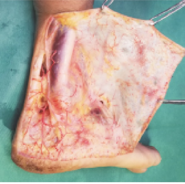
图(4)
计量
- 文章访问数: 5035
- HTML全文浏览量: 2854
- PDF下载量: 26
- 被引次数: 0
