

-
双侧后交通动脉(posterior communicating artery,PCoA)管径粗细不一[1],其中粗者与大脑后动脉相当甚至略粗,看上去宛如PCoA延续为大脑后动脉,称之为胚胎型大脑后动脉,被认为是颈内动脉系血管变异中发生率最高的类型[2]。存在胚胎型大脑后循环病人由于颅底动脉缺少潜在的侧支循环途径,因此大脑前后部发生缺血损伤的概率较大,增加了脑卒中发生的概率[3-4]。本文应用磁共振动脉成像(MRA)技术探讨有无胚胎型大脑后动脉与基底动脉管径及其缺血事件发生概率的关系,为预防和治疗病人缺血性脑卒中提供指导。
-
病例来自2011年10月至2012年1月在河北省沧州中西医结合医院神经内科就诊并行MRA检查的脑血管病病人148例,其中有胚胎型大脑后动脉病人48例,男20例,女28例,年龄(61.3±13.4)岁;无胚胎型大脑后动脉病人100例,男53例,女47例,年龄(57.5±12.4)岁。2组病人一般资料比较具有可比性。
-
采用SIEMENS Avanto 1.5T高场强MR扫描仪,标准头部8通道线圈。摆正受试者体位,以前后连合连线为轴位图像扫描基线。扫描范围包括脑前后循环的颅内段,采用3D-TOF(3 dimensional-time of flight)法, 常规横断面、矢状面、T1WI、T2WI扫描TR 32 ms,TE 3.85 ms,层厚0.8 mm。MRI包括TSE T2WI、SE T1WI、FLAIR T2WI及DWI序列。扫描参数,FSE T2WI:TR 3 510 ms,TE 96 ms,激励次数为1,层厚5.0 mm,间隔2.0 mm;SE T1WI:TR 450 ms,TE 9.9 ms,激励次数为1,层厚5.0 mm,间隔2.0 mm;DWI:TR 3 400 ms,TE 93 ms,激励次数为1,层厚5.0 mm,间隔2.0 mm;轴位FLAIR T2WI:TR 8 500 ms,TE 130 ms,TI 2 500 ms,激励次数为2,层厚5.0 mm,间隔2.0 mm。
-
(1) 根据PCoA变异情况,可分为6个等级,Ⅰ型:双侧PCoA管径基本一致;Ⅱ型:单侧PCoA管径粗于另一侧;Ⅲ型:单侧PCoA管径粗于另一侧,并延续为同侧的大脑后动脉;Ⅳ型:双侧PCoA均各自延续为同侧的大脑后动脉;Ⅴ型:单侧PCoA缺失;Ⅵ型:双侧PCoA均缺失(见图 1)。(2)基底动脉血管内径[5]:使用EPSON PERFEC-TIO NI660 PHOTO扫描仪对T1、T2加权像在基底动脉中段的横截面由留空效应形成的无信号圆形黑影放大20倍,动脉黑影每旋转60°测量1次直径,测量3次取平均值;若黑影呈椭圆形则取最小径为动脉内径。(3)后循环缺血事件:英国牛津郡社区脑卒中项目(OCSP)的Bamford分型明确为后循环供血区缺损症状,经MRI证实基底动脉狭窄、闭塞、纤细、弯曲,血流速度改变。
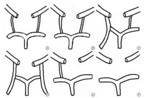
图 1 PCoA变异分型示意图
-
采用t检验和χ2检验。
-
本研究148例病例中Ⅰ型4例、Ⅱ型5例、Ⅴ型14例、Ⅵ型77例,共100例,为无胚胎型大脑后动脉组;Ⅲ型38例、Ⅳ型10例,共48例,为有胚胎型大脑后动脉组。
-
有胚胎型大脑后动脉组基底动脉上、中、下三段血管内径均小于无胚胎型大脑后动脉组(P < 0.01)(见表 1),即有胚胎型大脑后动脉的基底动脉内径较无胚胎型大脑后动脉更加纤细。
胚胎型大脑后动脉 n 上段管径 中段管径 下段管径 有 48 0.191±0.050 0.208±0.055 0.236±0.061 无 100 0.228±0.047 0.245±0.057 0.278±0.061 t — 4.31 3.86 3.89 P — < 0.01 < 0.01 < 0.01 表 1 2组病人基底动脉血管内径比较(x±s;cm)
-
后循环缺血事件患病组基底动脉上、中、下三段血管内径均小于不患病组(P < 0.05)(见表 2),即基底动脉血管纤细增加了后循环缺血事件事件的发病概率。
后循环缺血事件 n 上段管径 中段管径 下段管径 患病 11 0.206±0.045 0.219±0.051 0.252±0.053 不患病 37 0.223±0.044 0.240±0.056 0.274±0.063 t — 2.14 2.31 2.14 P — < 0.05 < 0.05 < 0.05 表 2 后循环缺血事件与基底动脉血管内径相关性分析(x±s;cm)
-
脑血管的基本构型在胚胎发育28~40 d形成。从胚胎发生角度来看,大脑后动脉源于PCoA。但从血流分布角度看,发育完好的大脑后动脉的血液来源于基底动脉。以往的经验告诉我们,当病人一侧颈内动脉闭塞时,较粗的PCoA可能会起代偿保护作用,防止发生血管梗死。相反,同侧PCoA较细会增加这些病人发生脑卒中的风险[6]。本研究利用MRA对148例脑血管病病人的血管结构异常进行分析,证实了一侧或双侧胚胎型大脑后动脉存在的情况下,基底动脉管径较为纤细,而基底动脉血管纤细增加了后循环缺血事件的发病率。可见有胚胎型大脑后动脉的情况下基底动脉缺血事件患病率明显增高,这与以往对胚胎大脑后动脉的认识不符。分析导致这一结果的原因有以下几个方面:(1)当一侧或双侧PCoA增粗并延续为胚胎型大脑后动脉时,由于颈内动脉不但供应同侧的大脑前动脉、大脑中动脉,还要供应同侧的大脑后动脉,所以同侧颈内动脉血流量增加,而基底动脉血流量则减低[7]。HONG等[8]的研究也证明双侧胚胎型大脑后动脉会导致后循环脑血流量降低。(2)长期低血流量的基底动脉会渐渐变得纤细,进而基底动脉小的分支可能减少或闭塞。亦有文献[9]报道,胚胎型大脑后动脉的病人,基底动脉变得纤细。(3)从生物血流动力学分析:当一侧或双侧胚胎型大脑后动脉存在时,前后循环血流发生了重新分配,颈内动脉系统的血流量增多,而血流量降低使得纤细的基底动脉清除栓子的能力下降[10-11],进一步增高了缺血事件的发生率。
近年来脑血管病病因的研究较多,但脑血管本身形态对颅内血流的影响,以及这种生物流体力学改变在脑血管疾病发生发展过程中的作用较少报道。认识并逐渐重视脑血管本身形态变异及生物流体力学变化与脑血管病发生、发展之间关系,对于临床脑血管病防治有极其重要的意义。
胚胎型大脑后动脉与基底动脉形态及其缺血事件之间的关系
Relationship between the embryonic posterior cerebral artery, and basilar artery morphology and its ischemic events
-
摘要:
目的应用磁共振动脉成像(MRA)技术探讨胚胎型大脑后动脉的存在与基底动脉形态及基底动脉缺血事件发生概率的关系。 方法随机选择行MRA检查的脑血管病病人148例。其中有胚胎型大脑后动脉病人48例,无胚胎型大脑后动脉病人100例。观察胚胎型大脑后动脉的开放情况,测量基底动脉上、中、下三段处的内径,及后循环缺血事件发生情况,进行统计学分析。 结果有胚胎型大脑后动脉组基底动脉上、中、下三段血管内径均小于无胚胎型大脑后动脉组(P < 0.01)。后循环缺血事件患病组基底动脉上、中、下三段血管内径均小于不患病组(P < 0.05)。 结论胚胎型大脑后动脉存在的情况下,基底动脉内径较小且缺血事件发生率增高。 Abstract:ObjectiveTo investigate the relationship between the embryonic cerebral artery, and morphology of basilar artery and probability of basilar artery ischemic events using magnetic resonance angiography. MethodsA total of 148 patients with cerebrovascular disease diagnosed by magnetic resonance angiography examination were randomly selected.There were 48 patients with embryonic posterior cerebral artery and 100 patients without embryonic posterior cerebral artery.The openness of the embryonic cerebral artery was observed.The inner diameters of the upper, middle and lower basal artery, and occurrence of posterior circulation ischemic events were measured and analyzed. ResultsThe diameters of the upper, middle and lower basilar artery in embryonic posterior cerebral artery group were smaller than those in non-embryonic posterior cerebral artery group(P < 0.01).In the posterior circulation ischemic event, the diameters of the upper, middle and lower basilar artery were smaller than those in non-disease group(P < 0.05). ConclusionsUnder the presence of the embryonic cerebral artery, the inner diameter of basilar artery is smaller, and the incidence rate of ischemic event increases. -
表 1 2组病人基底动脉血管内径比较(x±s;cm)
胚胎型大脑后动脉 n 上段管径 中段管径 下段管径 有 48 0.191±0.050 0.208±0.055 0.236±0.061 无 100 0.228±0.047 0.245±0.057 0.278±0.061 t — 4.31 3.86 3.89 P — < 0.01 < 0.01 < 0.01  下载: 导出CSV
表 2 后循环缺血事件与基底动脉血管内径相关性分析(x±s;cm)
后循环缺血事件 n 上段管径 中段管径 下段管径 患病 11 0.206±0.045 0.219±0.051 0.252±0.053 不患病 37 0.223±0.044 0.240±0.056 0.274±0.063 t — 2.14 2.31 2.14 P — < 0.05 < 0.05 < 0.05
下载: 导出CSV
-
[1] 兰慧.后交通动脉容积CT数字减影血管成像研究[J].中国临床解剖学杂志, 2017, 35(5):28. [2] 汪文胜, 胡春洪.颅脑与头颈部影像图解[M].北京:人民军医出版社, 2011:11. [3] 高旭萍, 闫荣.单纯性眩晕与卒中后头晕的椎基底动脉磁共振造影特征的差异对比研究[J].中国医师杂志, 2017, 19(5):675. doi: 10.3760/cma.j.issn.1008-1372.2017.05.011 [4] 王学廷, 董致成, 王涛, 等.椎动脉优势与基底动脉走行形态变化关系研究[J].人民军医, 2015(5):555. [5] 薛武荣, 韩宏明.颅内动脉变异的颅脑磁共振血管成像特征分析[J].医学综述, 2017, 23(3):601. doi: 10.3969/j.issn.1006-2084.2017.03.044 [6] CAN A, HO AL, EMMER BJ, et al.Association between vascular anatomy and posterior communicating artery aneurysms[J]. World Neurosurg, 2015, 84(5):125. [7] ZHANG C, LI S, PU F, et al.The effect of anatomic variations of circle of Willis on cerebral blood distribution during posture change from supination to standing:a model study[J]. Biomed Mater Eng, 2014, 24(6):2371. [8] HONG JM, LEE JS, SHIN DH, et al.Hemodynamic impact of fetal-variant Willisian circle on cerebral circulation:a duplex ultrasonography study[J]. Eur Neurol, 2011, 65(6):340. doi: 10.1159/000327213 [9] 陆琰琦.椎动脉多普勒超声和MRA对后循环缺血的诊断价值评估[J].现代诊断与治疗, 2017, 28(7):74. [10] 隋滨滨, 高培毅, 林燕, 等.4D血流MR成像评估颅内动脉血流动力学状态的实验研究[J].国际医学放射学杂志, 2017, 40(6):641. [11] CAPLAN LR, WONG KS, GAO S, et al.Is hypoperfusion an important cause of strokes? If so, how?[J]. Cerebrovasc Dis, 2006, 21(3):145. doi: 10.1159/000090791 -

 点击查看大图

图(1)表(2)
计量
- 文章访问数: 5050
- HTML全文浏览量: 2753
- PDF下载量: 7
- 被引次数: 0
