


-
脑在发育过程中皮质凹陷、折叠形成了脑沟,脑沟的存在增加了灰质的面积和体积,使得在有限的空间内可以容纳更多的神经元。一些学者曾强调这些脑的凹陷、折叠发生在两种细胞构筑有差异的皮质区域的边界,于是脑沟便被认为是脑功能分区的天然标志。因而,研究脑沟的侧别、性别和位置等差异可以在一定程度上反映脑功能的差异性。神经影像技术与立体定向仪的结合推动脑肿瘤外科切除、立体定向放疗术进入脑、神经解剖图像引导下直接对脑肿瘤进行精确定位,科学制定治疗计划及安全,准确实施瘤内近距离放疗时代,这些治疗过程的准确实施有赖于对病灶和局部解剖结构的准确定位。为此,本文探讨距状沟在容易识别和测量的旁正中矢状面上(X=±7mm)是否有侧别、性别及位置的差异性,构建该层面立体定位数据集并进行回归分析。
-
30名正常成年志愿者,男15名,女15名;年龄23~60岁。门诊体检及MRI检查无神经、精神病史及相关家族病史。选择自旋回波序列,TR 400 ms,TE 15 ms,以AC-PC线为扫描基线,获取3 mm层间距横断层和7 mm层间距矢状断层图像,Dicom3.0格式。
-
Signa 1.5T超导磁共振扫描仪及头颅正交线圈(美国GE公司);微型计算机(A8 S,华硕公司)图形图像工作站:efilm Workstation2.1(美国Merge eMed公司),以及数据测量Adobe Photoshop CS8.0软件包。3D-Doctor软件,版本1.2.0.1,Able Software Co.USA。
-
在3D-Doctor内打开图像,选择“Edit”-“calibrations”,打开“Image calibration Parameters”对话框,调整像素大小至实际数值,测量旁正中矢状断层上距状沟的长度。距状沟的弯曲代表相应的视觉皮层面积的增加,应沿着其实际形态测量沟长。距状沟前后端点的测量包含于下面旁正中矢状断层上距状沟立体定位数据集。
-
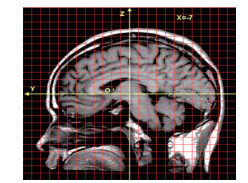
用eFilm2.1打开Dicom3.0格式的MRI数据。选中图像,“File”-“export”-“as images”,选择保存类型为“JPEG”。Photoshop8.0打开转换后的图像,将宽度和高度均调整为实际扫描的240 mm。测量时,首先在正中矢状断层上确立大脑原点(Y0,Z0),建立标准Y-Z二维坐标系(见图 1),旁正中矢状断层重复操作,距状沟在Y轴方向上每隔3mm定一个取样点测量坐标值(Y,Z)。
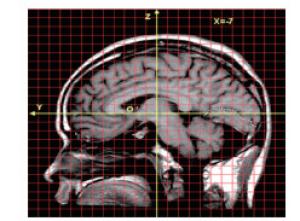
图 1 标准二维坐标系内,右侧旁正中矢状面上距状沟例图(X=-7mm)
-
采用t检验和曲线回归分析。
-
30例样本旁正中矢状断层上,左侧沟长(57.08±6.93)mm,与右侧沟长(55.81±7.63)mm比较差异无统计学意义(t=0.88,P>0.05),男性沟长(56.01±7.70)mm,与女性沟长(57.20±6.51)mm比较差异无统计学意义(t=0.46,P>0.05);距状沟前端的Y值侧别差异无统计学意义(P>0.05)(见表 1),前端左侧较右侧偏后0.86 mm;距状沟后端的Y值侧别差异有统计学意义(P < 0.05)(见表 1),左侧较右侧偏后4.34 mm。
部位 n 左侧/mm 右侧/mm t P 前端Y值 30 -35.45±3.37 -34.59±4.36 1.35 > 0.05 后端Y值 30 -81.37±4.58 -77.03±6.49 3.35 < 0.05 表 1 旁正中矢状断层上距状沟前后端Y值的侧别比较(x±s)
-
选取距状沟出现最完整的旁正中矢状断层上分析其在Z轴方向的变异性。距状沟位于连合间径(AC-PC)平面附近略偏上,中部Z值较高,两端Z值偏低,前端点的平均Z值大于0 mm,距状沟末端有一小段平均Z值略小于0 mm。从标准差来判断,距状沟由前向后变异性有增大趋势(见表 2)。
Y值 左侧 右侧 -30.00 1.24±5.94 4.75±5.52 -33.00 3.48±3.97 3.63±4.25 -36.00 5.23±3.36 3.61±4.44 -39.00 6.40±4.20 4.44±4.65 -42.00 7.62±4.21 5.75±4.41 -45.00 8.19±4.02 6.03±5.39 -48.00 9.06±3.82 5.60±6.01 -51.00 9.37±4.34 4.93±6.47 -54.00 9.24±4.71 5.00±6.19 -57.00 9.10±5.09 5.07±6.44 -60.00 8.47±5.77 4.03±7.22 -63.00 7.53±5.58 3.65±8.21 -66.00 5.99±5.56 2.44±8.87 -69.00 4.21±6.71 3.30±5.54 -72.00 1.96±6.94 1.64±6.52 -75.00 -0.37±7.04 -0.18±7.12 -78.00 -2.72±6.76 -0.42±7.71 -81.00 -1.34±5.15 -2.25±10.23 -84.00 -5.75±6.35 -0.15±10.07 -87.00 — 0.046±10.66 注:距状沟最前端取样点P5位于Y=-30.00 mm,最后端P95取样点位于Y=-78.00 mm 表 2 X=±7mm旁正中矢状断层上距状沟取样点的平均坐标值(n=30;x±s;mm)
-
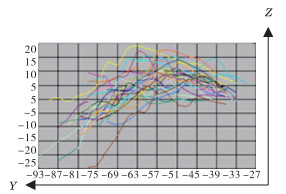
旁正中矢状断层(X=±7 mm)上,双侧距状沟在Z轴的分布范围(-25.0,20.0),左侧距状沟分布范围(-25.0,20.0),右侧距状沟分布范围(-20.0,20.0),呈现距离大脑原点越近距状沟的位置变异性越小的规律(见图 2、3)。旁正中矢状断层上,距状沟Z值对Y值的二次曲线回归方程:左侧:Z=-32.717-1.587Y- 0.016Y2(R=0.683,F=191.12,P < 0.01), 右侧:Z=-30.390-1.537Y-0.015Y2(R=0.565,F=166.72, P < 0.01)。

图 2 30例左侧距状沟在旁正中矢状断层(X=7mm)的分布图(左图)
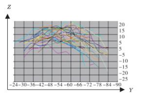
图 3 30例右侧距状沟在旁正中矢状断层(X=-7mm)的分布图(右图)
-
关于大脑半球不对称性的研究已较为深入,结构和功能具有一体性,功能的不对称性应该有对应的神经生物学基础[1-3]。王跃等[4]报道,后语言区的不对称性主要反映在枕叶长的不对称性上,用手习惯与半球不对称的关系极可能反映在枕叶宽和侧脑室后角长的不对称性上。这些不对称性会不会使得距状沟也存在侧别或性别差异,本研究的结果显示旁正中矢状面上距状沟沟长在侧别和性别方面均不具有统计学差异,与一些学者[5-6]报道距状沟沟深、沟长和视觉区的表面积没有明显侧别差异的结果吻合。有研究[7]显示侧脑室枕角左侧比右侧向后扩展5.1 mm,Brodmann 17、18区中心位置左侧较右侧偏内、偏后[8],与本研究结果距状沟后端的位置在Y轴方向上左侧较右侧偏后具有一致性,枕极处距状沟后端的位置左侧较右侧偏后4.34 mm。可能的解释是,手偏利和语言优势半球的存在导致大脑顺钟向转位,左枕叶扩张并超过中线,左枕极较右枕极位置偏后,即后语言区的不对称和运动皮质的不对称是影响枕叶形态不对称的主要原因之一。GILISSEN等[9-10]认为枕叶内侧面的视觉投射面和距状沟长度相关,纹状皮质体积及沟内面积和距状沟的深度相关。我们的结果可以为视觉投射面、纹状皮质沟内面积及纹状皮质体积的研究提供参照。
脑、神经影像辅助技术广泛应用于神经外科领域[11-12],如脑内肿瘤的切除、颅内血肿、脓肿的定向排空、囊性肿瘤的CT/MRI引导下穿刺引流、放射性核素内放射治疗等,准确实施上述手术的关键是对局部血管、大脑沟回、病灶等的准确定位。JUNG等[13]通过对420例脑部肿瘤病人的研究发现,使用影像导航技术定位的平均误差为1.15 mm,不仅可以清楚显示解剖结构和降低手术的损伤性,更是极大的增加了手术的可信度。在三维空间内,距状沟的准确定位可以在一定程度上帮助初级视皮质附近病灶手术的顺利实施,本研究构建了距状沟在旁正中矢状断层上的立体定位数据集,求得Z值对Y值的二次曲线回归方程,以期为初级视皮质附近区域病灶的准确定位提供参照。距状沟由前向后变异性逐渐增大,在空间位置上愈临近低级中枢脑沟的变异性越小,此可能是大脑进化在形态学上的反映,同时也进一步印证了在大脑立体定位研究中,选择AC-PC中点作为大脑原点定位脑内结构更具稳定性。
旁正中矢状断层上大脑距状沟立体定位数据集的构建及差异性分析
The building of the stereotactic data set of calcarine sulcus and difference analysis of the paramedian sagittal plane
-
摘要:
目的构建大脑半球旁正中矢状断层上(X=±7 mm)距状沟的立体定位数据集,探讨距状沟是否具有侧别、性别和位置差异性。 方法以连合间径中点为原点建立三维立体坐标系,30名健康成人大脑旁正中矢状断层T1MRI上,3D-Doctor软件测量距状沟沟长,该断层上Y值为3 mm的倍数处选取测量点构建数据集,求Z值对Y值的二次曲线回归方程。 结果旁正中矢状断层上距状沟沟长不具有侧别和性别差异(P>0.05),Y轴方向上距状沟后端左侧较右侧位置偏后(P < 0.05)。旁正中矢状断层上距状沟呈现距离大脑原点越近位置变异性越小的规律,旁正中矢状断层上距状沟Z值对Y值的二次曲线回归方程:左侧Z=-32.717-1.587Y-0.016Y2(P < 0.01),右侧Z=-30.390-1.537Y-0.015Y2(P < 0.01)。 结论构建的距状沟立体定位数据集预期对该沟附近的视皮质功能区定位、病灶定位及外科治疗具有指导意义。 Abstract:ObjectiveTo build the stereotactic data set of calcarine sulcus(CS) on the paramedian sagittal plane(X=±7 mm), and explore the CS variability among different lateral, gender and position. MethodsThe three-dimensional coordinate system was established with the midpoint of AC-PC line as the origin.The length of CS on paramedian sagittal T1MRI planes of the brain of 30 healthy adults were measured using 3D-Doctor workstation, the measurement points were selected at a multiple of 3 mm on this section to construct the data set, and the quadratic regression equation of Z value to Y value was calculated. ResultsThe differences of the length of CS in different laterals and genders were not statistically significant(P>0.05).In the direction of Y axis, the left side of the back end of the CS was behind the right side.On paramedian sagittal planes, the smaller the positional variability of CS was, the closer the CS to the origin of the brain was.On paramedian sagittal plane, the quadratic regression equations of Z-value to Y-value of the CS in the left side and right side were Z=-32.717-1.587Y-0.016Y2 and Z=-30.390-1.537Y-0.015Y2, respectively(P < 0.01). ConclusionsThe building stereotaxic data set in this study is expected to be of guiding significance for the localizating the visual cortical functional areas, lesion area and surgical treatment in the vicinity of the calcarine sulcus. -
Key words:
- calcarine sulcus /
- magnetic resonance imaging /
- regression analysis
-
表 1 旁正中矢状断层上距状沟前后端Y值的侧别比较(x±s)
部位 n 左侧/mm 右侧/mm t P 前端Y值 30 -35.45±3.37 -34.59±4.36 1.35 > 0.05 后端Y值 30 -81.37±4.58 -77.03±6.49 3.35 < 0.05  下载: 导出CSV
表 2 X=±7mm旁正中矢状断层上距状沟取样点的平均坐标值(n=30;x±s;mm)
Y值 左侧 右侧 -30.00 1.24±5.94 4.75±5.52 -33.00 3.48±3.97 3.63±4.25 -36.00 5.23±3.36 3.61±4.44 -39.00 6.40±4.20 4.44±4.65 -42.00 7.62±4.21 5.75±4.41 -45.00 8.19±4.02 6.03±5.39 -48.00 9.06±3.82 5.60±6.01 -51.00 9.37±4.34 4.93±6.47 -54.00 9.24±4.71 5.00±6.19 -57.00 9.10±5.09 5.07±6.44 -60.00 8.47±5.77 4.03±7.22 -63.00 7.53±5.58 3.65±8.21 -66.00 5.99±5.56 2.44±8.87 -69.00 4.21±6.71 3.30±5.54 -72.00 1.96±6.94 1.64±6.52 -75.00 -0.37±7.04 -0.18±7.12 -78.00 -2.72±6.76 -0.42±7.71 -81.00 -1.34±5.15 -2.25±10.23 -84.00 -5.75±6.35 -0.15±10.07 -87.00 — 0.046±10.66 注:距状沟最前端取样点P5位于Y=-30.00 mm,最后端P95取样点位于Y=-78.00 mm
下载: 导出CSV
-
[1] 隋东莉, 李成, 陈刘成, 等.大脑扣带沟分支的影像解剖学研究及意义[J].蚌埠医学院学报, 2015, 40(1):4. [2] 向春风, 周牧野, 李成, 等.大脑额下沟断层解剖学相关性研究[J].蚌埠医学院学报, 2019, 44(2):235. [3] 申素珍, 沈龙山, 王震寰.大脑中央后回前壁三维空间定位及非对称性研究[J].蚌埠医学院学报, 2018, 43(4):425. [4] 王跃, 姜均本.大脑语言区不对称性与半球不对称性的定量分析[J].重庆医科大学学报, 1992, 17(1):19. [5] 王海青, 丁娟, 孙博, 等.国人女性距状沟形态及侧差的三维MRI研究[J].解剖学报, 2010, 41(2):285. [6] AHMAD RM, LAURENT T, THIERRY M, et al.Main anatomical features of the calcarine sulcus:a 3D magnetic resonance imaging at 3T study[J].Surg Radiol Anat, 2019, 41(2):181. doi: 10.1007/s00276-018-2118-x [7] TOGA AW, THOMPSON PM.Mapping brain asymmetry[J].Nat Rev Neurosci, 2003, 4(1):37. doi: 10.1038/nrn1009 [8] HASNAIN MK, FOX PT, WOLDORFF MG.Structure-function spatial covariance in the human visual cortex[J].Cereb Cortex, 2001, 11(8):702. doi: 10.1093/cercor/11.8.702 [9] GILISSEN E, IBA-ZIZEN MT, STIEVENART JL, et al.Is the length of the calcarine sulcus associated with the size of the human visual cortex? A morphometric study with magnetic resonance tomography[J].J Hirnforsch, 1995, 36(4):451. [10] GILISSEN E, ZILLES K.The calcarine sulcus as an estimate of the total volume of human striate cortex:a morphometric study of reliability and intersubject variability[J].J Hirnforsch, 1996, 37(1):57. [11] 孙崇璟, 谢涛, 张晓彪, 等.后纵裂入路切除侧脑室三角区肿瘤的解剖学[J].解剖学报, 2018, 49(2):217. [12] THOMAS M.Stereotactic MR-guided focused ultrasound deep brain lesioning:the resurrection of posteroventral pallidotomy and thalamotomy for Parkinson's disease?[J].Acta Neurochirurgica, 2017, 159(7):1367. doi: 10.1007/s00701-017-3161-9 [13] JUNG TY, JUNG S, KIM IY, et al.Application of neuronavigation system to brain tumor surgery with clinical experience of 420 cases[J].Minim Invasive Neurosurg, 2006, 49(4):210. doi: 10.1055/s-2006-948305 -

 点击查看大图
图(3)表(2)
计量
- 文章访问数: 4088
- HTML全文浏览量: 1983
- PDF下载量: 37
- 被引次数: 0
