


-
准确的大脑皮层损伤的脑沟定位对于改善医生之间的沟通,预测和预防术后损伤是非常有必要的。这一过程有赖于对皮质解剖和图像的系统分析有很好的了解,然而,在神经和神经外科训练中常被忽视[1-3]。涉及大脑运动中枢的肿瘤、缺血性疾病等精准诊疗,精确定位和测定大脑中央沟空间三维坐标方法学至关重要[3]。本研究以大脑AC-PC为扫描定位基线,将31名健康成年人大脑横断位、冠状位、矢状位MRI的Dicom3.0格式文件转化为JPG格式后导入Photoshop软件,利用该软件的图像旋转、坐标轴平移、坐标值读取等功能,建立以AC-PC中点为原点、以AC-PC为Y轴的大脑立体定位坐标系,实现大脑结构的准确三维立体定位,并制作大脑中央沟的定位图谱。现作报道。
-
本实验选用31例头颅连续MRI断层扫描数据,横断层MRI扫描基线为前连合后缘中点到后连合前缘中点的连线,即连合间线(AC-PC);冠状面以AC-PC的中垂线为基线;矢状面以头颅正中矢状面为零层面。扫描野FOV 24.0 mm×24.0 mm,分辨率512×512,层厚6 mm。扫描过程中严格保持头颅固定。由于扫描野为大小不变的正方形,头颅断层影像域相对于正方形的四个顶点位置固定,图像配准通过正方形四个顶点自动完成,亦即图像的大小、配准信息包含Dicom3.0格式的信息中,无需人工干预。MRI扫描数据由eFilm Workstation工作站读取[4-6]。
-
任何具有一定体积和三维形态的、被置入X-Y-Z三维坐标系中的物体可以视为由一定数目连续的平行于X-Y平面、具有一定厚度的二维断层,并沿着Z轴方向叠加而成的结构。在每一个平行于X-Y平面的二维断层平面上,当确定了X-Y轴在该平面的投影线后,以其为该平面的X-Y轴建立二维平面坐标系,则平面上任意点在该平面坐标系中的X、Y坐标值与原三维坐标系中的X、Y坐标值相等。Z值的大小由所在平面与X-Y零层面的距离确定,正负则由该层面与零层面的上下方位关系决定。本实验中,定义AC-PC面为零层面,近颅侧的上方层面的Z值为正值,近尾侧的为负值。
基于上述数学原理,利用MRI断层扫描技术将三维形态的大脑“断层分割”为连续的近似二维的断层图像;确保图像的大小、点距与MRI扫描时设定的完全一致,将Dicom3.0格式MRI扫描数据转化为JPG格式后导入Photoshop软件,利用该软件的图像旋转、坐标轴平移、坐标值读取等功能,可便捷、精确测得断层面上中央沟在大脑外侧面的X、Y值。Z值等于所在层面与双连合层面间相间隔的层面数和层间距的乘积。中央沟位于双连合层面上方,故Z值一般均大于零。
-
各方位断层的中央沟的识别均采用经典形态识别法[7-11]结合计算机辅助连续结构追踪技术、计算机辅助三维定标法等识别,并参考空间三维形态[4-6]。记录当前MRI的年龄、性别、磁共振检查号、扫描野大小、点距等信息。于双连和平面测量连合间径并记录。导出该图像为JPG格式保存。导出的图片再导入Photoshop软件。选择双连合层面,选择“图像” →“图像大小”菜单栏,调整图像大小为24.0 cm×24.0 cm,与原Dicom图像的扫描野一致。在双连合平面,再次测量连合间径大小,与前次eFilm Workstation中测量一致,印证并确保图像大小、点距等几何信息与原始Dicom3.0格式的图像信息是完全一致的(见图 1)。
图 1 eFilm与Photoshop测量同一样本的连合间径值一致,均为25.Omm
-
Photoshop软件内自带坐标系是以图片左上角为坐标原点,同时坐标轴本身不能旋转,原点通常不与大脑原点重叠;原始坐标的Y轴也通常不与连合间径或其投影线重合或者平行。为避免繁杂的坐标转换计算,利用软件的画布旋转和坐标轴平移等功能,建立与脑立体定位一致的标准化的二维X-Y坐标系。具体方法:在双连合平面,旋转画布使Y轴与连合间径平行。测得连合间径及其倾斜角,测得该连合间径倾斜角-92.8°, 需要逆时针旋转2.8°实现连合间径与Y轴平行.菜单栏“图像” →“旋转画布” →“任意角度”,选择逆时针旋转2.8°。旋转后此时连合间径倾斜角变为“-90°”,与纵轴实现平行。前、后连合中点坐标的X值相等,设为X0。设定垂直、水平参考线及大脑原点。鼠标分别移至前连合中点、后连合中点,于信息面板显示两点坐标(X0, Y1),(X0, Y2),设Y0=(Y1+Y2)/2计算连合间径中点,即大脑原点在该坐标系的坐标为:(X0, Y0)。菜单栏点击命令“视图” →“新参考线”,设定垂直的参考线,方框内填写数值X0,此参考线为标准坐标轴Y轴所在位置;再设定水平参考线,依次选择“视图” →“新参考线”,选择“水平”,方框内填写Y0值,设定水平的参考线,此参考线为标准坐标轴X轴所在位置。两参考线交点即为大脑原点(连合间径中点)所在位置。平行移动坐标轴使Y轴与连合间径重合且原点与大脑原点重合。菜单栏点击命令“视图” →“标尺”,显示标尺。鼠标左键移至标尺左上角小方框内,拖拽十字坐标线,使其中心点与参考线的交点准确重合。移动后坐标轴Y轴与连合间径重叠,坐标原点也实现与大脑原点的重合,完成标准X-Y二维坐标系的建立。此时连合间径中点坐标值显示为(0,0),连合间径长度依然为240.0 mm,与前面所测一致,保证图像内解剖结构之间的位置关系没有改变(见图 2)。
图 2 标准坐标系建立,大脑原点处坐标值为(0, 0),且Y轴与连合间径重合
其他层面标准X-Y二维坐标系的建立。由于原始图像之间边界是自动配准的,其他层面图像经过同样的处理:画布旋转同样角度、在原始坐标系内点(X0, Y0)处设置水平及垂直参考线、平行移动原始坐标原点至参考线交点,此时该层面图像依然与双连合层面的图像严格配准,且该层面上的X-Y轴相当于双连合层面上的X-Y轴的投影。如此即完成其他层面的标准X-Y二维坐标系的建立。由数学坐标值的定义,该坐标系中图像上任一点的X、Y值与定义的、以大脑原点为坐标原点的标准X-Y-Z三维坐标系中X、Y值一致。
-
颅脑断层MR上中央沟的识别采用典型层面形态学并结合计算机辅助技术完成[4-7],同时参考中央后回等中央沟邻近结构形态学特征[8]。中央沟的在X-Y二维标准坐标系,利用软件的坐标直接读取功能,中央沟上取样点的坐标值可以通过信息面板直接显示。记录中央沟外侧面起始点X、Y坐标值,Z值由所在层面与双连合层面的间隔层数与层间距乘积确定,同一层面上任一点的Z值相同。实际测量中从中央沟易于辨识的较高横断层面开始。
-
采用上述方法,在30例头颅MRI上分别测量前、后连合、连合间径长度;测量大脑中央沟外侧缘三维立体定位坐标;测量中央沟三维立体定位坐标等,可构建中央沟立体定位数据集。
-
大脑前、后上下径普遍大于前后径,前、后联合在断面上呈长椭圆形,而非圆形。连合间径最大值均值23.651 3±1.675 53 mm,最大值25.330 mm,最小值21.976 mm,数值相对稳定(见表 1)。
结构 均值(x±s) 范围 前连合上下径 4.040 1±1.432 1 2.234 5~5.023 4 前连合前后径 2.661 2±0.703 1 2.126 2~4.367 4 后连合上下径 2.263 9±0.554 1 1.678 2~3.345 0 后连合前后径 1.798 9±0.452 0 1.467 1~2.234 1 连合间径 23.651 3±1.675 53 1.903 1~25.634 2 表 1 大脑前、后连合及连合间径的MRI测量(mm)
-
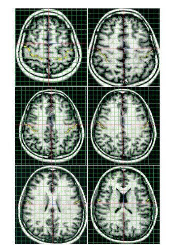
中央沟横断层MR定位方格例图,每方格大小10 mm×10 mm,红色实线相当于坐标轴X(横轴),Y(纵轴),为Photoshop产生,其交叉点即相当于坐标原点(0, 0)。黄色虚线为识别后手工标记的中央沟。中央沟的各取样点的X,Y值可以由定位方格直观给出,也可以由软件精确给出。Z轴数值则由所在层面距离经连合间径层面确定。中央沟横断层定位例图显示中央沟在断层上的中心位置相对逐渐前移,愈向下愈接近坐标轴横轴,同时长度减小,经胼胝体干层面以上层面均位于横轴前侧,即取样点Y>0(见图 3)。
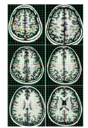
图 3 中央沟横断层MR定位方格例图
-
现代影像学技术的进步,成像分辨率及精度大幅提高,研究者广泛采用MRI进行活体脑的解剖研究,取得较丰富成果,并因此产生了基于大量影像学资料的大数据与计算医学研究热点[8-9]。大脑形态学研究目前仍然是生物医学工程、大脑功能研究、临床脑外科等多领域共同的活跃而重要的研究课题,脑解剖区域的可视化定位在脑形态学研究中具有非常重要的作用[1-3, 8-10]。
-
应用断层影像学资料,在微型计算机上进行三维立体定位的研究目前尚没有专门、通用的软件可资应用。包括3D-Doctor、eFilm等在内的Dicom3.0格式影像数据读取软件功能丰富,能够进行三维重建等,但一般也缺少能提供三维立体定位坐标值方便的测量或直接读取功能;少数软件理论上具备这一功能,但存在着步骤繁杂、坐标原点固定、不能进行坐标轴任意的旋转和平移等不足,难以方便地应用于三维立体定位研究或需要进行繁杂的坐标转换计算。Photoshop软件是由Adobe公司开发的功能强大的通用图形图像处理软件,可进行包括坐标轴旋转(通过画布旋转等效实现)、坐标轴平移、坐标值直接显示等。但Photoshop软件不能直接读取Dicom3.0格式的影像数据,需要编制、安装特定的接口程序。另一种可行的办法就是本研究采用的通过eFilm等Dicom3.0格式影像数据读取软件,将原数据格式转化为Photoshop软件能够直接读取的JPG等格式。但需要注意格式转化后可能存在图像大小、分辨率等的改变,这将使图像内包含的解剖结构间位置、距离等信息失真,严重影响测量的精确性。解决此问题的方法是:记录原始影像设备扫描时设定的扫描野大小、分辨率等信息;选取两个或者多个特征解剖学结构,本研究中选取前、后连合中点,在eFilm软件中测量其距离。图象格式转化后,在Photoshop中通过修改图像大小、分辨率等信息,使其与影像设备扫描时扫描野的设定值一致即可(本组设定的扫描野大小均为24.0 cm×24.0 cm);同时通过测量选定的特征解剖学结构间距离等信息,印证图像格式转化前后图像的大小、分辨率等信息完全一致,保证图像内解剖结构间位置关系的真实性,确保测量结果准确可靠。此外值得指出的是,恰当运用Photoshop的批处理功能及将重复的操作储存为“动作序列”,将大大减少重复操作,提高自动化水平,可显著提升图像处理及三维定位测量的效率。
头颅MRI立体定位坐标系及中央沟定位图谱的构建
Study on MRI stereotactic coordinate system of head and localization map of central sulcus
-
摘要:
目的探讨基于标准头颅MRI的立体定位方法,验证定位精度。 方法31例头颅连续MRI断层扫描数据,以Dicom 3.0格式导入eFlim 2.1工作站并以JPG格式导出,后者再导入Photoshop软件。分别通过图像大小校正、图像旋转、坐标轴平移等步骤使系统坐标原点与大脑原点重合、系统Y轴与大脑连合间线重合,建立标准坐标系。通过软件坐标直接读取功能获得断层上X、Y坐标值。Z值由所在层面距离双连合层面层数及层厚乘积决定。选取前连合后缘中点、后连合前缘中点的坐标值、两点距离等作为验证,在建立的立体定位坐标系中,计算连合间径长度并与eFilm软件直接测量结果对照。 结果31例头颅连续MRI断层扫描数据均实现准确三维立体定位,制定定位图谱。2种方法测得的连合间径长度一致。 结论基于标准头颅MRI的立体定位法可以应用于大脑中央沟三维立体定位的研究。 Abstract:ObjectiveTo study the stereotactic localization method based on standard head MRI, and verify the localization accuracy. MethodsThe continuous head MRI scan data with Dicom 3.0 format of 31 cases were imported into eFlim 2.1 workstation, exported as JPEG format, then imported into Photoshop.The standard coordinate system was established by image size correction, image rotation and coordinate axis translation to make the system coordinate origin coinciding with brain origin point, and system Y-axis coincide with the intersynaptic wiring of brain.The coordinate values(X, Y) of the fault were obtained through the coordinate direct reading function of software, and the Z value was determined by product of the number of layers and thickness beyween two connected layers.During establishing stereotactic coordinate system, the coordinate value of the midpoint of posterior margin of anterior commissure, midpoint of leading edge of the posterior commissure, and distance between two points were used to calculate the length of interstitial diameter, which was compared with eFilm software direct measuring. ResultsThe accurate three-dimensional stereotactic localization could be established using continuous MRI scan data in 31 cases, and the localization map was formulated.The interstitial diameter length detected by two methods was the same. ConclusionsThe stereotactic localization method based on standard MRI can be used in studying the rolandic fissure. -
表 1 大脑前、后连合及连合间径的MRI测量(mm)
结构 均值(x±s) 范围 前连合上下径 4.040 1±1.432 1 2.234 5~5.023 4 前连合前后径 2.661 2±0.703 1 2.126 2~4.367 4 后连合上下径 2.263 9±0.554 1 1.678 2~3.345 0 后连合前后径 1.798 9±0.452 0 1.467 1~2.234 1 连合间径 23.651 3±1.675 53 1.903 1~25.634 2  下载: 导出CSV
-
[1] DESTRIEUX C, TERRIER LM, ANDERSSON F, et al.A practical guide for the identification of major sulcogyral structures of the human cortex[J].Brain Struct Funct, 2017, 222(4):2001. doi: 10.1007/s00429-016-1320-z [2] FANG S, LIANG J, QIAN T, et al.Anatomic location of tumor predicts the accuracy of motor function localization in diffuse lower-grade gliomas involving the hand knob area[J].Am J Neuroradiol, 2017, 38(10):1990. doi: 10.3174/ajnr.A5342 [3] STEPHAN-OTTO C, NÚÑEZ C, ARCA G, et al.Three-dimensional map of neonatal arterial ischemic stroke distribution from early multimodal brain imaging[J].Stroke, 2017, 48(2):482. doi: 10.1161/STROKEAHA.116.014186 [4] 沈龙山, 王震寰, 张磊, 等.计算机辅助连续结构追踪技术对大脑中央沟精确定位的方法学研究[J].蚌埠医学院学报, 2008, 33(6):712. doi: 10.3969/j.issn.1000-2200.2008.06.024 [5] 沈龙山, 王震寰, 张磊, 等.计算机辅助三维定标法在冠、矢状位MRI脑结构定位的研究[J].蚌埠医学院学报, 2008, 33(6):716. doi: 10.3969/j.issn.1000-2200.2008.06.025 [6] 沈龙山, 王震寰, 张磊, 等.Rolando裂的三维重建与可视化[J].蚌埠医学院学报, 2008, 33(6):719. doi: 10.3969/j.issn.1000-2200.2008.06.026 [7] KANEKO OF, FISCHBEIN NJ, ROSENBERG J, et al.The "White Gray Sign" identifies the central sulcus on 3t high-resolution t1-weighted images[J].AJNR Am J Neuroradiol, 2017, 38(2):276. doi: 10.3174/ajnr.A5014 [8] 申素珍, 沈龙山, 王震寰.大脑中央后回前壁三维空间定位及非对称性研究[J].蚌埠医学院学报, 2018, 43(4):425. [9] 王震寰.计算医学——应对大数据的挑战向临床转化[J].蚌埠医学院学报, 2014, 39(1):1. [10] 袁现坤, 李淑宇.基于FreeSurfer面数据的网格点脑分区定位及其可视化[J].北京生物医学工程, 2015, 34(2):146. doi: 10.3969/j.issn.1002-3208.2015.02.06 -

 点击查看大图

图(3)表(1)
计量
- 文章访问数: 5398
- HTML全文浏览量: 2778
- PDF下载量: 18
- 被引次数: 0
