

 下载:
下载:

-
近年来,随着医学影像技术的飞速发展,三维重建技术无论是在CT、MRI还是超声诊断等方面,已被广泛应用于临床。扣带回是边缘系统和Papez环路的重要组成部分,具有调控情绪、感觉、运动、内脏活动、注意、认知等功能,无论是在形态还是功能上均与其他脑区存在紧密联系[1]。扣带回皮层及其周边脑部病变由于所处的脑部特殊位置,使治疗变得繁琐。例如针对侧脑室侧壁肿瘤,TOMINAGA等[2]提出经扣带回手术入路,并认为切开一侧扣带回不会导致严重的神经功能障碍。扣带回皮层的三维重建及可视化可以全面展示其整体及与侧脑室等邻近结构的形态特征及关系,能够将大脑扣带回皮层的病变区域更直观全面地展现,为指导侧脑室及额顶叶等各邻近脑部立体精准定位手术、脑部放射治疗精准剂量估算和微创外科等提供了影像学结构模型,同时也为系统解剖学及断层解剖学的脑部教学提供了三维立体数字模型。
-
1名成年健康志愿者(知情同意),女,20岁,右利手。临床检查及颅脑MRI检查,无神经和精神系统及家族遗传疾病。以AC-PC为扫描基线,获取冠状面、横、矢面断层T1 MRI图像。图像为Dicom3.0格式,自选回波序列,TR:1 830 ms,TE:22 ms,扫描野FOV:24.0 cm×24.0 cm,分辨率512×512,层厚2 mm,层隔:0 mm。扫描过程中严格限制头颅固定,要求志愿者闭眼,戴耳塞,防止外界干扰。
-
Singal 3.0超导磁共振扫描仪及头颅正交线圈(General Electric Co., USA);HP Z820 Workstation(显卡:NVIDIA Quadro4000, CPU主频:1.8 GHz);Lenovo V480型计算机,联想公司。
-
Able Software 3D-Doctor,V1.2.0.1 (Able Software Co.USA);RadiAnt DICOM Viewer 3.4.2;eFilm Workstation,版本2.1.2.352,(Merge eMed Co.USA)。
-
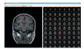
将冠状面头颅MRI T1WI图像数据直接以Dicom3.0格式导入3D-Doctor软件中。扫描野为大小不变的正方形,头颅断层影像域相对于正方形的四个定点位置固定,图像配准由软件自动完成。手工分割、边界提取大脑扣带回皮层(红色图线)、侧脑室(蓝色)、大脑纵裂(绿色)和大脑外表面轮廓(灰色)(见图 1)。

图 1 图像分割与边界提取(红色示大脑扣带回皮层,蓝色示侧脑室)
-
通过大脑原点(AC-PC连线的中点)建立三维坐标系,定X轴为左右横轴,Y轴为前后纵轴,Z轴为上下竖轴。对上述分割提取的断层进行三维重建。采用充分复杂面绘制法,首先在3D-Doctor软件任务栏中设定好扫描层面厚度及单位,其后依次选择“3D Rendering”→“Surface Rendering”→“Complex Surface”,完成三维重建。
-
点击3D-Doctor软件任务栏中“Tools”→“Smoothing surface models”→“是”,即可对重建后结构模型的后处理,使视觉效果显示更好。
-
在任务栏中点击“3D Object Setting”,在二级菜单中选择“Display”“Opacity”“Transparency”,设置目标模型的显示、透明度、透明效果。
-
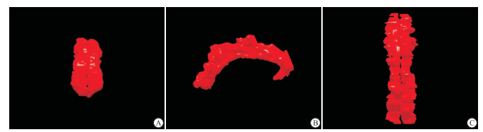
成功构建活体状态的大脑扣带回皮层三维可视化模型,获得了扣带回皮层的立体整体形态。正面观呈两侧基本对称的由下到上的长方形扁条带,位于大脑半球纵裂两侧,侧脑室上部中间;侧面观均类似平放的“新月形”,前后走形,前后稍低,中部上凸,表面凹凸不平,位于侧脑室上前方;顶面观类似左右基本对称的不规则长条形,表面依旧凹凸不平。重建出的结构可以在三维空间以任意角度放大、缩小旋转观察,可计算其长度、角度、体积等(见图 2)。
图 2 扣带回皮层三维模型
-
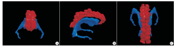
成功显示侧脑室与大脑扣带回皮层的空间位置关系。侧脑室位于大脑中央部,扣带回下方。正面观扣带回位于侧脑室中上部,与侧脑室前角、三角区中间部投影重叠,侧脑室基本呈镜像对称的“C”字形,位于大脑半球间裂两侧,下角略细长,形状不规则,表面不光整;侧面观扣带回皮层与正上方包饶侧脑室,但与其上下仍旧有空间距离,侧脑室依旧呈不规则大“C”字形;顶面观侧脑室后角及三角区显示清楚。三面观可见扣带回与侧脑室前角、中央部和后角基本平行走形(见图 3)。

图 3 扣带回皮层与侧脑室三维模型(红色示扣带回皮层,蓝色示侧脑室)
-
成功构建大脑扣带回皮层、侧脑室、大脑纵裂及大脑表面三维模型,成功显示它们之间的空间位置和毗邻关系。扣带回位于大脑半球内侧中央部,侧脑室位于其正下方,大脑纵裂分割这两部分结构,使其基本呈镜像对称关系。正面观大脑纵裂位于中央部,余三部分结构基本呈左右对称;侧面观大脑纵裂约占半球上3/5,侧脑室与扣带回约占中间部1/3;顶面观可见大脑表面的沟回结构,纵裂居中,余两部左右对称;底面观与顶面观类似,但大脑半球底部脑回显示清楚且对称,侧脑室前、下及三角区显示尤其清楚(见图 4)。
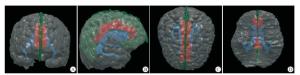
图 4 扣带回皮层、侧脑室、大脑纵裂、大脑表面三维模型(红色示扣带回皮层,蓝色示侧脑室,绿色示大脑纵裂,灰色示大脑表面)
-
近代随着科学技术的飞速发展,计算机及其衍生技术广泛应用于医学诊断及治疗的方方面面。但是这些技术在断层影像方面呈现的大多都是二维数据信息,尤其是在脑部断层影像方面,无论是CT、MRI、PET还是超声检查等各种影像检查技术中,运用最多的依旧是二维图像信息。没有三维立体的图像数据信息作为基础支撑,会使得一些脑部疾病在诊断中不能及时有效地被发现,从而对后续的治疗产生影响。
汤煜春[3]于2009年研究证实,中国人和ICBM152公认的Caucasian大脑存在形态结构区别。因此,对于中国人脑部数字化可视化的研究就显得很有必要。构建国人脑部三维立体数据集及可视化模型是疾病诊断和治疗的重要基础,其在临床解剖学和断层影像解剖学教学方面也有重要作用。而构建三维可视化模型,必然少不了计算机技术的应用,其构建的结构立体感强、形态逼真,能够很好地呈现兴趣结构的空间与位置关系,是作为可视化模型成功构建的技术支撑基础。本课题组正着力于该方面研究,可对国人基于MRI数据的脑部沟回数字化数据作为补充。而基于MRI数据的国人正常扣带回皮层的三维可视化模型目前并未见过多相关报道。本文的扣带回皮层研究正是在本课题组实验技术路径基础上开展。
扣带回皮层,在人类的情绪、记忆、内脏感觉、疼痛等发面发挥着非常重要的作用,近年来有关扣带回皮层的分部及功能研究更是成为热点问题。研究[4-7]显示,阿尔兹海默病、精神疾病、抑郁症、言语记忆及认知等均与扣带回皮层有关。本文研究结果显示,大脑扣带回皮层位于胼胝体上方两侧,紧贴大脑纵裂,呈前后走形的“新月形”。孙博[8]研究表明,女性中扣带回的灰质体积结构明显右侧化较多,说明扣带回皮层在性别和侧别上存在很大程度的差异,此结果可为与扣带回相关的临床神经系统疾病的影像学诊断和功能研究提供可视化的形态依据。顶面及侧面三维可视化模型可以看出其在灰白质交界处走形扭曲,这更增加对扣带回皮层边界识别和功能理解上的困难。从扣带回皮层与侧脑室及大脑纵裂的三维重建模型可以看出,其形态比较复杂,彼此位置关系紧密。
综上,建立扣带回皮层可视化三维模型,对扣带回皮层部位疾病的诊断和治疗研究有重要意义;三维可视化技术在组织结构及解剖关系复杂的疾病的诊治中已经广泛开展并运用[9],对与扣带回皮层结构区域有关的侧脑室肿瘤的微创手术入路和三维立体定向精准放疗治疗,精准医学个性化治疗方案的制定方面,也具有重要的指导意义。
大脑扣带回皮层的三维重建及可视化
Three-dimensional reconstruction and visualization of cerebral cingulate cortex
-
摘要:
目的建立健康成人活体MR图像的大脑扣带回皮层三维可视化模型,为脑立体定向微创手术提供扣带回皮层及邻近区域解剖学模型。 方法选取1名健康成年女性颅脑薄层冠状位MR图像数据,将其导入3D-Doctor软件,并使用手工分割方法建立大脑扣带回皮层、侧脑室、大脑纵裂和大脑外表面轮廓的三维重建模型。 结果成功构建大脑扣带回皮层三维可视化图像,成功显示大脑扣带皮层、侧脑室、大脑纵裂和大脑外表面的立体结构形态及与邻近脑组织结构关系。 结论大脑扣带回皮层的三维可视化模型的构建对扣带回解剖结构的识别、脑立体定向手术设计、扣带回皮层区域病变定位诊断和功能研究有重要价值。 Abstract:ObjectiveTo establish a three-dimensional visualization model of brain cingulate cortex in healthy adult MR image, and provide an anatomical model of cingulate cortex and adjacent structure for stereotactic minimally invasive surgery of brain. MethodsThe MR image data of a cranial thin layer coronal plane in a healthy adult woman were imported into 3D-Doctor software, and the three-dimensional reconstruction of the cingulate cortex, lateral ventricle, cerebral longitudinal fissure and outer contour of brain were established using the manual segmentation method. ResultsA three-dimensional visual image of the cingulate cortex of brain was successfully constructed, and could successfully show the three-dimensional structure of the cingulate cortex, lateral ventricle, cerebral longitudinal fissure and outer surface of brain, and its relationship with adjacent brain structure. ConclusionsThe construction of three-dimensional visualization model of the cingulate cortex of brain has important value in the recognition of the anatomical structure of cingulate gyrus, design of the stereotactic surgery of brain, and positioning diagnosis and functional study of cingulate cortex area. -
[1] 王微微, 吴逊.扣带回的解剖、生理及扣带回癫痫[J].中国现代神经疾病杂志, 2018, 18(5):315. doi: 10.3969/j.issn.1672-6731.2018.05.004 [2] TOMINAGA T, KAYAMA T, KUMABE T, et al.Transcingulate approach to lateral ventricle tumors.Technical casereport[J].Neurosurg Rev, 1996, 19(2):105. doi: 10.1007/BF00418079 [3] 汤煜春.中国人数字化标准脑图谱的建立[D].济南: 山东大学, 2009. [4] 覃媛媛, 张顺, 郭林英, 等.阿尔茨海默病患者扣带束和后扣带回的改变[J].神经损伤与功能重建, 2016, 11(1):27. [5] LOCKWOOD PL, WITTMANN MK.Ventral anterior cingulate cortex in social decision-making[J].Neurosci Biobehav Rev, 2018, 92(5):187. [6] WAGNER S, SEBASTIAN A, LIEB K, et al.A coordinate-based ALE functional MRI meta-analysis of brain activation during verbalfluency tasks in healthy control subjects[J].BMC Neurosci, 2014, 16(15):19. [7] MUNDY P.A review of joint attention and social-cognitive brain systems in typical development and autism spectrum disorder[J].Eur J Neurosci, 2018, 47(6):497. doi: 10.1111/ejn.13720 [8] 孙博.中国人大脑皮质性别差异的影像学研究[D].济南: 山东大学, 2010. [9] KAGAMI K, SHINMYO Y, ONO M, et al.Three-dimensional visualization of intrauterine conceptus through the uterine wall by tissue clearing method[J].Sci Rep, 2017, 7(1):5964. doi: 10.1038/s41598-017-06549-6 -

 点击查看大图
图(4)
计量
- 文章访问数: 4937
- HTML全文浏览量: 2650
- PDF下载量: 13
- 被引次数: 0
