

-
颌骨囊肿是发生于颌骨的含有液体或半固体物质的病理性(或异常的)囊性肿物,常内衬上皮,并以牙源性颌骨囊肿(odontogenic cyst,OC)最多见[1]。颌骨囊肿摘除术是治疗OC的常用方法,且是大多数骨内、骨外囊肿的首选方法,术后遗留的无效腔,常致创口延期愈合[2]。OC摘除术为完全清除病灶,常将暴露于囊腔内的牙根尖切除,这减少了受累牙的骨内根长度,不利于患牙的稳固及后期修复。Bio-oss骨粉及海奥生物膜已广泛用于各种颌骨缺损的引导骨组织再生(guided bone regeneration,GBR)[3-4]。本研究拟在术中保留囊肿累及牙根尖,放置Bio-oss骨粉包埋骨腔内牙根面并联合海奥生物膜阻挡上皮组织干扰植骨区的愈合,旨在保留松动患牙、加速松动牙的稳固并为后期种植等修复工作打下基础。
-
选取我科2017年10月至2018年6月收治的OC病人55例作为研究对象,年龄15~45岁。纳入标准:(1)颌骨囊性病变,囊腔长径1.5~4.0cm,未穿通鼻底、上颌窦底;(2)病历、临床症状、体格检查、全景片或口腔颌面锥体束CT(CBCT)等术前相关资料完整;(3)病人对本研究知情并签署同意书。排除标准:(1)造釉细胞瘤等实体病变,复发率较高者;(2)伴有系统性疾病影响骨愈合和再生效果者;(3)临床资料不全,难以配合随访术后1年以上者。按手术方法将病人分成2组,其中A组15例,男8例,女7例,行根面刮治保留根尖,用骨粉包埋牙根暴露于颌骨缺损的部分并使用生物膜覆盖;B组40例,男24例,女16例,行OC摘除术+根尖切除术。2组病人年龄、性别差异均具有可比性。
-
海奥(Heal-All)口腔修复膜(烟台正海生物技术有限公司,中国),将小牛皮采用组织工程技术制成的异种功能性天然脱细胞真皮基质胶原膜。Bio-oss骨粉(Geistlich AG,瑞士),源自牛骨的天然无机磷灰石结晶体。
-
常规拍摄口腔科CBCT及曲面体层摄影片(全景片)评估OC位置范围,术前行全口洁治+根管治疗术以利后续的根尖周刮治;病灶感染者控制炎症后择期手术。
-
A组病人手术切口采用病灶邻近牙龈缘切口、前庭沟切口或角形切口,翻瓣暴露术野,彻底刮净内容物及囊壁,牙科钻磨除粗糙骨质、骨壁;根据囊肿内容物判断性质,排除其他可疑病变后术中,采用微创器械如小刮匙,牙周刮治器彻底清除囊腔内病灶牙根周围残留囊壁、病变牙周膜,高频电刀烫除超充牙胶尖;根面平整后,冲洗创口,采用适量自体血与Bio-oss骨粉相混合覆盖于根尖区,至少覆盖根面2~3 mm以补偿骨质流失和吸收,使骨粉与周围骨壁相延续形成一个稳定整体以提供良好的血供;生物膜的放置注意UP面朝向软组织面,缝合时注意松弛度,必要时在黏骨膜瓣下做减张切口以防止骨粉移位。对于囊腔剩余部分,任由自体血充盈机化改建,同时方便自体比较术后成骨及改建情况;未见明显渗血,对位缝合创缘;口外加压包扎。B组病人行OC摘除术+根尖切除术。所有病人术后3、6、12个月进行随访,检查松动患牙恢复稳固情况且拍摄CBCT、全景片评价术后缺损区骨再生和改建效果。
-
(1) 咀嚼时无疼痛等自主不适反应。(2)门诊检查松动患牙:牙周无炎症,根尖区黏膜无瘘管,叩诊阴性,松动度Ⅰ度以下。
-
采用t(或t′)检验和方差分析。
-
术后2组病人伤口均无感染且愈合良好,A组未出现生物膜暴露,骨粉流失等症状;术后6个月,2组病例均无复发症状且移位膨隆软硬组织基本恢复至正常解剖结构。术前2组受累牙数和囊肿所致丧失正常功能牙数差异均无统计学意义(P>0.05)(见表 1)。2组病人术后3、6、12个月复查丧失功能患牙恢复情况差异均有统计学意义(P<0.01),术后6个月及12个月后恢复情况均优于与术后3个月(P<0.01);且A组丧失功能患牙恢复数及恢复百分比均优于B组(P<0.01)(见表 2)。
分组 n 受累牙数 丧失正常功能牙数 A组 15 3.00±0.93 2.07±0.70 B组 40 3.60±1.08 2.40±0.90 t — 1.90 1.67 P — >0.05 >0.05 表 1 2组受累牙数和丧失正常功能牙数比较(x±s)
分组 n 术后3个月 术后6个月 术后12个月 F P MS组内 恢复正常数 A组 45 1.53±0.64 1.87±0.52* 2.00±0.67** 7.04 <0.01 0.376 B组 144 1.05±0.45 1.56±0.60** 1.68±0.57** 54.47 <0.01 0.296 t — 4.68# 3.12 3.15 — — — P — <0.01 <0.01 <0.01 — — — 恢复正常百分比 A组 45 0.79±0.28 0.93±0.14** 0.98±0.09** 12.34 <0.01 0.035 B组 144 0.47±0.23 0.67±0.23** 0.73±0.22** 16.23 <0.01 0.051 t — 7.72 9.18# 11.00# — — — P — <0.01 <0.01 <0.01 — — — 与术后3个月比较*P<0.05,**P<0.01;#示t′值 表 2 术后3、6、12个月2组丧失功能牙恢复正常功能情况比较(x±s)
-
A组术后3个月,骨腔内自体血愈合区骨密度增高不明显,成骨较慢;根尖植骨区骨密度明显高于术前,已形成部分新骨,与周围正常骨组织密度差异尚大。术后6个月植骨区骨纹理逐渐形成,可见不甚明显骨腔边界,骨整合有序进行,与自体骨密度尚不契合;自发愈合区成骨开始,但未见明显骨小梁结构,仍可见骨腔透射影像。术后12个月根尖植骨材料基本吸收,骨纹理清晰可见,与周围骨密度接近,基本无明显界限,自发愈合区骨密度较6个月时稍增高,但成骨质量明显差于根尖部,局部仍可见缺损区骨腔与周围骨界限。典型病例见图 1。
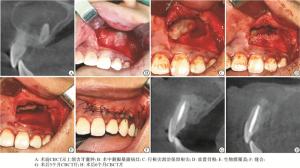
图 1 病人,男,16岁,诉上颌前牙区肿胀半年余
-
颌骨囊肿的治疗目标在于彻底的清除病变组织,降低复发率且尽最大的可能恢复受损区域的解剖和功能,囊肿摘除术中常行根尖切除术以绝复发。随着牙保存理念及微创操作观念的不断深入和口腔微创器械及修复材料的不断发展,学者们倾向于在术中尽量保留牙体组织。袁慧娟等[5]在根尖周外科术中彻底清除根尖病灶并保留患牙根尖,在术后随访中,根尖切除组与根尖保留组牙齿松动及瘘管发生情况差异无统计学意义,说明根尖保留与病灶复发无相关性,且保留根尖有利于后期桩核冠义齿修复和患牙的远期留存。在国内外文献中,尚未发现有学者采用根尖保留的方法刺激OC术后的根尖区骨再生,本研究创新性地将根尖保留应用于术中,探讨保留根尖对保存患牙和颌骨缺损修复的意义。本研究病人经过术后12个月随访,残留骨腔均不同程度的缩小且根尖部均未见复发症状,这说明OC术中,彻底清除病灶,保留很尖对OC的复发率无明显影响,与上述学者的研究结果一致。保留根尖即保留了OC术后近牙槽突缺损区的骨再生支架,有利于颊侧缺如骨板骨再生和维持牙槽骨丰满度。保留根尖对因龋坏、外伤所致牙冠较短患牙有重要意义,此类患牙多采用桩核冠修复,而桩核冠修复成功的关键在于至少4 mm的根尖封闭和足够的骨内桩长度。鉴于本研究中未单独设置根尖保留组,保留根尖与根尖截除的复发关系有待于后期研究。
颌骨囊肿术后所遗留无效腔的修复是口腔功能重建的一个重要课题,为解决术后形成的死腔,多采用蝶形手术、血块充填法、植骨术、生物材料移植、囊肿减压成形术等方法[2]。以自体血充满骨腔,小的颌骨囊肿骨缺损的平均愈合时间要1年,而中大型颌骨囊肿则需要2~5年,单纯的血凝块难以机化成理想的生理形态和良好骨结构,常导致义齿修复及种植的困难[6]。多数学者认为骨移植促进了颌骨缺损区域的再生,可以加速骨愈合、防止软组织塌陷、提高骨骼强度,在较大的颌骨缺损中值得应用[3-4, 7-11]。Bio-oss的三维多孔结构利于液体浸润和快速血管化,可为成骨细胞提供良好的生长通道。Bio-oss已广泛用于牙槽嵴的扩展/重建、拔牙窝的位点保存、种植手术、牙周手术、颌骨囊肿填充等口腔操作中;在骨再生中,Bio-oss起到一个成骨支架的作用,利用骨传导性为成骨细胞提供一个有利的生长环境,刺激成骨细胞的生长发育[9]。Bio-oss不会被天然骨完全吸收和替代,但也不会被纤维结缔组织所替代,因其骨传导性及空间维持能力,可被认为是一种永久性的移植物留存于宿主体内[10-11]。WILTFANG等[8]研究发现,将一层薄薄的Bio-oss覆盖于移植的髂骨骨松质表面可减少移植骨的吸收,巩固了自体骨增量的远期疗效;联合Bio-Gide覆盖植骨区效果更明显。PAPPALARD等[12]利用Bio-oss充填OC术后遗留骨腔后成骨速率更快,刺激了新骨的形成,加快了种植等后期修复的等待时间。本研究影像学检查发现根尖植骨区在术后较早的发生了骨再生,其成骨效果优于自体血愈合区,印证了以上学者的观点,应用GBR刺激了颌骨缺损区骨再生。
GBR是利用屏障膜阻挡快速增值的上皮细胞和结缔组织,促进生长缓慢的细胞如成骨细胞的生长进而形成骨骼[9]。海奥生物膜为来源于小牛皮的可吸收胶原蛋白膜,保留了天然的胶原纤维空间结构,已被广泛用于GBR、牙周组织引导再生和预防味觉出汗(Frey′s)综合征[13]。海奥膜一层为致密面(UP面),起到有效的屏障作用,防止软组织长入缺损部位,并可引导和支持软组织生长;另一层为疏松面,具有细胞外基质的三维空间结构,可调节、引导细胞长入,促进成骨细胞黏附与沉积;它去除了诱发宿主免疫排斥反应的成分,在体内降解时间维持在6个月左右且代谢产物没有细胞毒性。周俊波等[6]采用Bio-oss/海奥膜修复OC缺损骨腔发现其加速了成骨,方法可行、疗效确切。徐明等[14]将GBR技术应用于小型颌骨囊肿(长径1.5~3.0 cm)术后缺损骨腔,有效的修复了OC导致的局部骨缺损,恢复了颌骨的生理功能,方便后期义齿修复。本研究A组术后6个月植骨区骨纹理逐渐形成,骨整合有序进行,与自体骨密度尚不契合;自发愈合区成骨虽开始,但未见明显骨小梁结构,仍可见骨腔透射影像;与以上学者研究结果一致,GBR应用于OC摘除术中骨再生疗效肯定。保留根尖及采用GBR的意义还在于,即使部分松动牙因术后愈合不佳拔除,植骨可以发挥类似上颌窦提升术的效果,实现骨增量,为后期的种植义齿修复打下基础。
本研究证实,只要彻底清除病灶,保留根尖未明显增加OC的复发率;联合GBR有利于囊肿累及牙齿的保留,加速了松动患牙的稳固;刺激了术后骨腔根尖部的骨再生,有利于恢复良好的软硬组织解剖形态,为修复颌骨囊肿导致的骨缺损打下了坚实的骨质基础,有利于术后的修复重建工作的开展。手术方法可行,疗效肯定。
根尖保留联合引导骨组织再生在颌骨囊肿术中的应用研究
Application study of apical preservation combined with guided bone regeneration in jaw cyst surgery
-
摘要:
目的探讨颌骨囊肿术中采用根尖刮治保留根尖联合应用Bio-oss骨粉/海奥人工生物膜的临床疗效。 方法选取颌骨囊肿病人55例,分为2组,A组(15例)术中行根尖周刮治术保留根尖,用骨粉覆盖牙根暴露于颌骨缺损的部分,并应用生物膜覆盖;B组(40例)行单纯囊肿摘除术+根尖切除术。所有病人术后均随访12个月以上,比较2组病人术前和术后颌骨囊肿累及牙功能恢复、颌骨缺损骨再生及改建情况。 结果A组术后囊肿累及牙恢复正常功能优于B组(P < 0.01),A组根尖植骨区成骨效果优于自体愈合区。 结论颌骨囊肿摘除术中保留根尖联合引导骨再生术疗效较好,效果可靠。 Abstract:ObjectiveTo explore the clinical effects of periapical curettage preservation combined with Bio-oss bone meal/Haiao artificial biomembrane in jaw cyst surgery. MethodsFifty-five patients with jaw cyst were divided into the group A(15 cases) and group B(40 cases).The group A was treated with the periapical curettage preserving the root tip, bone meal covering the lack of part of the root exposed to jaw and biofilm covering.The group B was treated with simple cyst excision combined with apicoectomy.All patients were followed up for more than 12 months.The recovery of tooth function, bone regeneration and reconstruction of jaw defects between two groups before and after operation were compared. ResultsAfter operation, the normal function recovery in cyst involved teeth in group A was faster than that in group B(P < 0.01).The osteogenic effect in bone graft area in group A was better than that in autogenous healing area. ConclusionsThe efficacy of apical preservation combined with guiding bone regeneration in jaw cyst excision is good and reliable. -
Key words:
- jaw cyst /
- apical preservation /
- guided bone regeneration /
- excision
-
表 1 2组受累牙数和丧失正常功能牙数比较(x±s)
分组 n 受累牙数 丧失正常功能牙数 A组 15 3.00±0.93 2.07±0.70 B组 40 3.60±1.08 2.40±0.90 t — 1.90 1.67 P — >0.05 >0.05  下载: 导出CSV
表 2 术后3、6、12个月2组丧失功能牙恢复正常功能情况比较(x±s)
分组 n 术后3个月 术后6个月 术后12个月 F P MS组内 恢复正常数 A组 45 1.53±0.64 1.87±0.52* 2.00±0.67** 7.04 <0.01 0.376 B组 144 1.05±0.45 1.56±0.60** 1.68±0.57** 54.47 <0.01 0.296 t — 4.68# 3.12 3.15 — — — P — <0.01 <0.01 <0.01 — — — 恢复正常百分比 A组 45 0.79±0.28 0.93±0.14** 0.98±0.09** 12.34 <0.01 0.035 B组 144 0.47±0.23 0.67±0.23** 0.73±0.22** 16.23 <0.01 0.051 t — 7.72 9.18# 11.00# — — — P — <0.01 <0.01 <0.01 — — — 与术后3个月比较*P<0.05,**P<0.01;#示t′值
下载: 导出CSV
-
[1] HILL CM, RENTON T.Oral surgery Ⅱ:Part 3.Cysts of the mouth and jaws and their management[J].Bri Dent J, 2017, 223(8):573. doi: 10.1038/sj.bdj.2017.916 [2] 张志愿.口腔颌面外科学[J].7版.北京: 人民卫生出版社, 2012: 306. [3] GÜVEN O, TEKIN US.Healing of bone defects by an osteopromotion technique using solvent-dehydrated cortical bone plate:a clinical and radiological study[J].J Craniofac Surg, 2006, 17(6):1105. doi: 10.1097/01.scs.0000244922.15006.6e [4] LALABONOVA H, DASKALOV H.Jaw cysts and guided bone regeneration (a late complication after enucleation)[J].J IMAB, 2013, 19(4):401. doi: 10.5272/jimab. [5] 袁慧娟, 段昌华, 张佩芬, 等.根尖截除与保留治疗牙根尖周病疗效比较分析[J].中国实用口腔科杂志, 2014, 7(7):416. [6] 周俊波, 马露, 陈洁, 等.牛骨粉(Bio-Oss)/人工生物膜(海奥)联合治疗下颌骨缺损骨腔的临床观察[J].口腔医学, 2015, 35(10):830. [7] JIE L, KERNS DG.Mechanisms of guided bone regeneration:a review[J].Open Dent J, 2014, 8(1):56. doi: 10.2174/1874210601408010056 [8] WILTFANG J, JÄTSCHMANN N, HEDDERICH J, et al.Effect of deproteinized bovine bone matrix coverage on the resorption of iliac cortico-spongeous bone grafts-a prospective study of two cohorts[J].Clin Oral Implants Res, 2014, 25(2):e127. doi: 10.1111/clr.2014.25.issue-2 [9] ELGALI I, OMAR O, DAHLIN C, et al.Guided bone regeneration:materials and biological mechanisms revisited[J].Eur J Oral Sci, 2017, 125(5):315. doi: 10.1111/eos.2017.125.issue-5 [10] LIM HK, KIM JW, LEE UL, et al.Risk Factor Analysis of Graft Failure With Concomitant Cyst Enucleation of the Jaw Bone:A Retrospective Multicenter Study[J].J Oral Maxillofac Surg, 2017, 75(8):1688. [11] ETTL T, GOSAU M, SADER R, et al.Jaw cysts-filling or no filling after enucleation A review[J].J Cranio-Maxillofac Surg, 2012, 40(6):485. doi: 10.1016/j.jcms.2011.07.023 [12] PAPPALARDO S, CORONELLA M, LANZA ML, et al.Multidetector CT Dentascan evaluation of bone regeneration obtained with deproteinised bovine graft in residual cavity after mandibular cyst enucleation[J].Radiol Med, 2013, 118(4):523. doi: 10.1007/s11547-012-0911-x [13] 马士卿, 张旭, 孙迎春, 等.引导骨组织再生膜的研究进展[J].口腔医学研究, 2016, 32(3):308. [14] 徐明, 宋治锋, 王秦宁.GBR技术在颌骨囊肿手术中的临床应用观察[J].口腔颌面外科杂志, 2015, 25(6):440. doi: 10.3969/j.issn.1005-4979.2015.06.010 -

 点击查看大图

图(1)表(2)
计量
- 文章访问数: 5017
- HTML全文浏览量: 3251
- PDF下载量: 18
- 被引次数: 0
