


-
动脉粥样硬化是一种全身性、脂质驱动的动脉壁炎症性疾病[1],是心血管疾病和引起进一步的急性并发症的主要原因。尽管心血管医学已经取得了巨大进展,动脉粥样硬化疾病依旧是导致发展中国家的病人死亡主要原因[2]。颈动脉是人体的重要大血管,是观察整体血管动脉粥样病变发展情况的重要部位,可以反映人体整体的动脉硬化的负荷情况[3]。
超微血管成像技术(SMI)是一种新兴的可以专门观测低速血流状态的微小血管的新的血管成像模式,它不需要对比介质, 运用独特的算法显示出具有明显较高图像质量的微血管血流信号。
本实验利用SMI模式检测颈动脉斑块中新生血管的数量,并与超声造影(CEUS)模式作对比,旨在探究SMI评价颈动脉斑块中新生血管的价值。
-
选取2017年1月至2018年6月我院常规彩色多普勒颈动脉超声检查中发现的62例颈动脉斑块病人,其中男50例,女12例,年龄39~79岁,经SMI筛查出以低回声、低回声为主的混合回声及中等偏低回声的颈动脉斑块中有血流信号的共70个斑块,70个斑块平均长度(13.8±5.25)mm, 平均厚度(3.2±0.71)mm。所有病人均进行常规二维超声检查、SMI检查,经SMI检查筛查后部分病人进行CEUS检查。本研究经过本院伦理研究会通过,被检病人均知情同意,并签署知情同意书。
-
纳入标准:(1)病人基本情况良好,凝血功能均正常。(2)病人于双侧颈动脉发现可疑易损性斑块,斑块厚度大于2.0 mm。排除标准:(1)急性心内膜炎,瓣膜修复,急性全身感染,败血症,高凝状态,近期的血栓栓塞以及晚期肝肾系统疾病。(2)严重脑血管病病人、不合作的病人和不能耐受完整超声颈动脉检查的病人。(3)颈部手术后伤口有敷料的病人。(4)对选用的六氟化硫造影剂或其他组成成分过敏,临床近期发现的急性冠脉综合征或缺血性心脏病不稳定的病人,包括正逐渐变为或者进行性的心肌梗塞。(5)心脏症状于过去7 d内明显恶化,出现典型的心绞痛症状;心脏的症状明显恶化;心脏冠状动脉介入史或其他因素表明临床不稳定的因素在近3个月内发生(例如,最近的心电图、实验室或临床恶化的迹象);急性心力衰竭,Ⅲ/Ⅳ级的心功能衰竭和严重心律失常。(6)心脏病病人、重度肺高压病人(肺动脉压>90 mmHg)、系统高血压未控制的病人和呼吸窘迫综合征等无法耐受造影检查的病人。
-
采用东芝Aplio500彩色多普勒超声诊断仪,频率为5~10 MHz线阵探头,检查病人颈部血管。被检病人取仰卧位,枕头放在颈背部,颈部放松状态,头向后仰完全暴露颈部同时偏向被检查的对侧。扫查方法:检查医师将探头水平平移于甲状腺横切面的水平,并向外侧扫查,然后可以探测到颈总动脉的横截面,探头向下移动到颈总动脉起始处开始探查,并连续不断的观察颈总动脉,当探查到甲状软骨上边缘水平时,在横截面图像上出现血管的膨大区域(即颈动脉分叉处),并且当膨大区域向上接近下颌角后方时出现两条血管横截面,即为颈内动脉和颈外动脉,仔细观察上述颈内和颈外血管,在常规二维灰度图像模式下和彩色多普勒血流显像结合观察斑块大小、位置,数目、形态和回声特征等。由一名检查医师操作,两名高级职称医师进行双盲评估,对评分差异较大的斑块重新协商评定。
-
检查医师稳定探头,将探头顺沿着血管走行方向从病人颈根部开始扫查至颈总动脉,横扫至颈总动脉的分叉处后,检查颈内动脉和颈外动脉,尽可能的完全扫查达颈部的最高位置。检查医师选定好颈动脉内目标斑块,用标尺准确测量选定斑块的长度和厚度,并记录数据以及斑块所在的位置,于二维超声图像上最清晰显示被检斑块的位置稳定探头,启动SMI,选择灰阶模式SMI模式,常规二维图像与SMI图像将于屏幕上双幅显示出来。调整机器的增益以及深度,仔细观察所示斑块内有无点状、线状强回声,即新生血管的形成,记录数据。
-
选定经SMI检查的有微血管信号的斑块进一步行CEUS检查, 病人平躺,放松状态,头向后仰并偏向被检查对侧,检查医师将斑块清晰的扫查显示出来,稳定探头,并将仪器预设值调为屏幕双屏显示的的造影条件,机械指数设为0.07,聚焦点位于斑块的后缘,按下CEUS模式按钮,使用Bracco公司的超声造影剂Sonovo(注射用六氟化硫微泡),助手医生使用注射器将5.0 mL 0.9%氯化钠溶液注入六氟化硫造影剂瓶中,充分震荡和混匀,以获得乳白色六氟化硫悬浮液,并分2次抽取1.2 mL、0.8 mL六氟化硫悬浮液, 通过病人左肘的正中静脉以团注方式注入,在每次注入六氟化硫悬浮液后立即再次注射5.0 mL 0.9%氯化钠注射液,第一次注入造影剂同时开始TimeA计时并且按下RAW键进行数据储存,造影剂注入15 s后观察颈动脉的造影剂灌注情况,观察时间3 min,待第一次观察结束后,第二次注入造影剂,同时开始TimeB计时并进行RAW模式下数据储存,继续观察颈动脉造影剂的灌注情况以及斑块内是否有少量点状、线状新生血管的形成,记录斑块内检测到新生血管信号的数量、位置和形状。血管造影完成后,释放病人的静脉通路,审查医师观察病人的反应,15 min后,如无不良反应,则返回病房。
-
观察统计病人颈动脉斑块的数量、类型、位置、大小,新生血管在斑块内的位置和形态,进行SMI与CEUS检查新生血管分布一致性的比较。
-
SMI:0级显示图像上斑块内没有强回声,未见新生血管的形成;1级为在图像上斑块内显示1个或数个点状强回声,并且仅在斑块的肩部或基底部;2级为图像上显示斑块内可见斑点状、线状或树突状的新生血管。
CEUS:0级为在图像上斑块中不存在可见的造影剂微气泡;1级为图像上斑块内可见点状造影剂微气泡增强;2级为图像上斑块内存在可见造影剂微气泡点线状的增强,可以贯穿斑块或大部分贯穿斑块,或可见血液流动征。
-
采用t检验和Kappa一致性检验。Kappa≥0.75表示SMI与CEUS检查一致性很好,0.40<Kappa<0.75表示两者之间有较好的一致性,Kappa≤0.4表示两者之间差异很大。
-
62例病人经颈动脉常规彩色多普勒超声检查后,共计发现可疑易损性斑块(低回声、低回声为主的混合回声及中等偏低回声)103个(见图 1),103个可疑易损性斑块均进行SMI检查,筛选出有血流信号的斑块共计70个,70个斑块的平均长度(13.8±5.25)mm,平均厚度(3.2±0.71)mm,其中38个为低回声,17个为低回声为主的混合回声,15个为中等偏低回声。SMI检查的103个受检斑块中,微血流信号显示率约67.9%,其中斑块部位位于颈动脉分叉处38个,颈总动脉中段24个,颈内动脉8个,低回声斑块38个,低回声为主的混合回声斑块17个,中等偏低回声斑块15个(见表 1)。
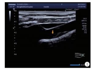
图 1 病人右侧颈总动脉后壁低回声斑块
厚度 长度 低回声斑块 混合回声斑块 等回声斑块 低回声斑块 混合回声斑块 等回声斑块 颈内动脉 左 2.60* (2)4.25±0.07 — 10.80* 17.08±0.55 — (1) (2) (0) (1) (2) (0) 右 3.45±0.92 4.30*- 2.70±0.28 12.35±0.50 13.00* 11.70±1.27 (2) (1) (2) (2) (1) (2) t — — — — — — P — — — — — — 颈总动脉分叉处 左 3.39±1.18 3.70±0.42 3.20±0.42 10.67±3.54 18.80±5.37 11.10±1.27 (16) (2) (2) (16) (2) (2) 右 2.98±1.35 3.26±0.50 4.04±0.56 11.72±5.33 12.97±0.75 13.48±2.47 (10) (3) (5) (10) (3) (5) t 0.82 1.01 1.88 0.61 1.53 1.25 P >0.05 >0.05 >0.05 >0.05 >0.05 >0.05 颈总动脉中段 左 2.78±0.47 3.90±0.17 3.32±0.93 12.14±2.11 22.70±2.06 15.70±6.33 (5) (3) (14) (15) (3) (4) 右 3.15±0.65 3.06±0.50 3.05±0.35 14.22±5.63 19.13±6.57 12.10±3.11 (4) (6) (2) (4) (6) (2) t 0.99 2.75 0.39 0.73 0.89 0.73 P >0.05 >0.05 >0.05 >0.05 >0.05 >0.05 ()内为例数;*示例数为1的原始数据 表 1 病人颈动脉斑块的大小、部位和类型(x±s;mm)
-
在颈动脉SMI模式中,检查结果显示血流信号的70个斑块中,在斑块近心端的肩部检测到18个有新生血管信号;29个斑块基底部检测到29个,斑块远心端肩部检测出18个,斑块顶部8个(见表 2)。Kappa分析显示,SMI与CEUS检测颈动脉斑块内新生血管的一致性很好(Kappa=0.769,P < 0.01)(见图 2~3、表 3)。
部位 斑块近心端肩部 斑块基底部 斑块远心端肩部 斑块顶部 合计 左 右 左 右 左 右 左 右 低回声 6 5 7 6 5 4 4 1 38 混合回声 1 2 3 3 2 4 1 1 17 中等回声 2 2 3 4 1 2 0 1 15 合计 9 9 13 16 8 10 5 3 70 表 2 SMI显示斑块内血流信号分布情况(n)

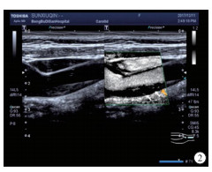
图 2 SMI检查右侧颈动脉低回声斑块内基底部可见点状及细线状新生血管血流信号

图 3 SMI检查病人右侧颈动脉低回声斑块内基底部可见点状及细线状新生血管血流信号
CEUS SMI 合计 0级 1级 2级 0级 10 5 0 15 1级 0 30 5 35 2级 0 0 20 20 合计 10 35 25 70 表 3 SMI与CEUS一致性比较(n)
-
动脉粥样硬化病变的过程是炎症诱导的紊乱,晚期动脉粥样硬化病变,通过诱导斑块的破裂,可能引起急性临床事件,进而引起血栓的形成。动脉粥样硬化斑块中存在活化的T淋巴细胞、巨噬细胞以及肥大细胞等炎性细胞,T细胞会产生干扰素,干扰素发挥作用后会降解细胞外基质,从而影响斑块的形态变化。另外,血管内皮细胞中黏附分子的上调加速了炎性细胞的黏附,斑块将继续不断产生炎症反应,导致斑块不稳定,最终使斑块成为易损性斑块[4],主要表现为大的坏死核心和纤薄的纤维帽,胆固醇沉积,炎症细胞浸润和钙化[5]。越来越多的研究[6]表明,斑块血管和斑块内出血是导致不稳定病变的重要因素,易损性斑块内的新生血管形成是不成熟、不规则和脆弱的,因为它们以不连续的基底膜和数量很少的紧密连接的内皮细胞为特征,结构完整性受损,大多数动脉血管内皮生长因子来自外膜,逐渐侵袭进入冠状动脉粥样硬化斑块,新生血管在冠状动脉粥样硬化斑块的进展和易感性中起着重要作用,此外,这些早期的新生血管的周细胞覆盖比较差,很容易导致斑块内出血,诱发斑块破裂和闭塞性血栓形成,导致管腔狭窄或闭塞[7],最终发展为临床疾病,包括有缺血性心脏病、中风和周围血管疾病[8]。
目前,超声多普勒作为检查斑块内血管分布的重要技术,为临床提供了经济、简便、快捷的方法,有多种超声模式可以完成检测,虽然传统的彩色多普勒超声可以根据斑块回声、形态提供有效信息,粗略评估斑块的组成,但它不能准确量化斑块易损性。
CEUS具有高时间分辨率和空间分辨率,可用于研究斑块新生血管和大血管[9]。使用这种成像技术检查,不规则的小血管壁、低回声斑块、溃疡斑块的描绘比二维超声更标准[10],是从形态学成像到功能性成像的进阶[11]。CEUS有助于增强斑块形态和识别新生血管的变化。由于其在辐射暴露和低成本的基础上的方便、安全和便携性,常被用于医学诊断成像。近年来CEUS技术的快速发展大大提高了超声诊断的质量,丰富了超声图像的评价参数,广泛应用于观察斑块内新生血管。CEUS可以在体内的斑块内检测到新生血管,据报道,在CEUS中观察到的斑块内的增强可归因于斑块新生血管形成,有助于临床分类颈动脉斑块。
SMI技术检测的是低速的血流信号,它拥有高灵敏度和高分辨率,其中的剪影模式SMI技术则能更敏感地捕捉低速血流信号[12-13],抑制二维组织信息使血流信息凸显,因其减去了背景信号而只显示低流速的血管,因此,可以清楚地观察颈动脉易损性斑块中的微小新血管形成,比彩色多普勒超声更直观地显示血管的走行、数量以及难以发现的低速新生血管,为临床早期诊断、尽早干预、治疗提供新的技术手段,改善病人的预后情况,SMI通过抑制背景信号来增强血流信号,侧重于血流的显示SMI,通过SMI可以获得保持了高帧率和高分辨率的图像[14],相比于彩色多普勒血流显像和能量多普勒显像,SMI提供了对血管分支细节更好的描述[12]。SMI在超声检查过程中对检查医师的要求更为严格,探头使用时要尽量保持平稳,探头的移动速度要尽量缓慢、轻柔,SMI的标尺选择较为严谨,一般情况下选择1.3~1.5 cm/s,当选择的标尺过高时,部分低速血流不能显示,而选择太低时,则会增加运动伪影[15]。
本研究中,利用新型超声技术SMI评估颈动脉斑块中的新生血管,并对其进行分级,结果显示SMI评估斑块内新生血管化和CEUS具有很好的一致性,可以认为SMI是作为评价颈动脉斑块内新生血管简单易行的方法,可以通过图像上强回声数量等指标进行分级反映颈动脉斑块的新生血管数量,对评价颈动脉粥样硬化斑块的稳定性具有较高的临床应用价值,对临床并发症的防治具有积极的指导意义。
然而以CEUS结果为标准,SMI检查结果低估了颈动脉新生血管5处,高估了颈动脉新生血管5处,可能是由于SMI技术易受到操作者的影响,而CEUS显示新生血管更为敏感。此外,有点状钙化的易损性斑块容易与mSMI状态下的血流信号混淆,两者在图像上均表现为强回声,所以操作者在检查过程中应多角度扫查。
本实验存在一定的局限:(1)实验样本量较少;(2)受实验条件限制,SMI检测出斑块中的新生血管仅与CEUS做对比;(3)虽然选择了统一的分级标准,但是人为评级仍然存在误差。
综上所述,SMI技术可以检测颈动脉斑块内的新生血管,且与CEUS存在很好的一致性。因其高分辨率、高灵敏度以及无需借用造影介质等特点,在颈动脉不稳定性斑块检测中具有较高的研究价值,可作为检测颈动斑块新生血管的重要影像学方法。
超微血管成像技术检测颈动脉斑块新生血管价值探讨
Value of superb micro-vascular imaging in detecting neovascularization of carotid plaque
-
摘要:
目的探讨超微血管成像技术(SMI)在检测颈动脉斑块新生血管中的优缺点。 方法选取进行常规彩色多普勒颈动脉超声检查中发现的62例颈动脉粥样硬化病人,进行常规彩色多普勒超声检查后继续SMI筛查,查出有新生血管信号的颈动脉斑块70个,进一步进行超声造影(CEUS)检查,比较两种方法检测颈动脉斑块中新生血管数量的一致性。 结果将SMI检测出新生血管的斑块根据新生血管的数量、形态分布情况进行分级,经颈动脉SMI检查后评分为0级的斑块10例,1级的斑块35例,2级的斑块25例。经颈动脉CEUS检查,评分为0级的斑块15例,1级的斑块35例,2级的斑块20例。两种模式检测颈动脉斑块内新生血管的一致性很好(Kappa=0.769,P < 0.05)。 结论SMI与CEUS检测颈动脉斑块内新生血管有很好的一致性,且SMI的安全性高,操作简便,可重复性强,在评价颈动脉斑块新生血管中有重要的参考价值,可作为检测颈动脉斑块新生血管的重要方法。 Abstract:ObjectiveTo investigate the clinical significnace of superb micro-vascular imaging(SMI) in the detection of neovascularization of carotid plaque. MethodsSixty-two patients with carotid atherosclerosis were identified using the routine color Doppler, and 70 carotid plaque cases with new blood vessel signal were screened using SMI, and further examined using contrast-enhanced ultrasound(CEUS).The consistency of two methods in detecting the number of neovascularization in carotid plaque was compared. ResultsThe plaques were graded according to the distribution and number of neovascularization detected by SMI, grade 0 in 10 cases, grade 1 in 35 cases and grade 2 in 25 cases were identified according to the score of SMI.The grade 0 in 15 cases, grade 1 in 35 cases and grade 2 in 20 cases were identified according to the score of carotid CEUS.The consistency of SMI CEUS in detecting the neovascularization of carotid plaque was good(Kappa=0.769, P < 0.05). ConclusionsSMI has good coincidence with CEUS in the diagnosis of neovascularization of carotid plaque.SMI has high safety, easy operation and high reproducibility.SMI has important reference value in evaluating the neovascularization of carotid plaque, and can be used as an important method in the diagnosis of neovascularization of carotid plaque. -
表 1 病人颈动脉斑块的大小、部位和类型(x±s;mm)
厚度 长度 低回声斑块 混合回声斑块 等回声斑块 低回声斑块 混合回声斑块 等回声斑块 颈内动脉 左 2.60* (2)4.25±0.07 — 10.80* 17.08±0.55 — (1) (2) (0) (1) (2) (0) 右 3.45±0.92 4.30*- 2.70±0.28 12.35±0.50 13.00* 11.70±1.27 (2) (1) (2) (2) (1) (2) t — — — — — — P — — — — — — 颈总动脉分叉处 左 3.39±1.18 3.70±0.42 3.20±0.42 10.67±3.54 18.80±5.37 11.10±1.27 (16) (2) (2) (16) (2) (2) 右 2.98±1.35 3.26±0.50 4.04±0.56 11.72±5.33 12.97±0.75 13.48±2.47 (10) (3) (5) (10) (3) (5) t 0.82 1.01 1.88 0.61 1.53 1.25 P >0.05 >0.05 >0.05 >0.05 >0.05 >0.05 颈总动脉中段 左 2.78±0.47 3.90±0.17 3.32±0.93 12.14±2.11 22.70±2.06 15.70±6.33 (5) (3) (14) (15) (3) (4) 右 3.15±0.65 3.06±0.50 3.05±0.35 14.22±5.63 19.13±6.57 12.10±3.11 (4) (6) (2) (4) (6) (2) t 0.99 2.75 0.39 0.73 0.89 0.73 P >0.05 >0.05 >0.05 >0.05 >0.05 >0.05 ()内为例数;*示例数为1的原始数据  下载: 导出CSV
表 2 SMI显示斑块内血流信号分布情况(n)
部位 斑块近心端肩部 斑块基底部 斑块远心端肩部 斑块顶部 合计 左 右 左 右 左 右 左 右 低回声 6 5 7 6 5 4 4 1 38 混合回声 1 2 3 3 2 4 1 1 17 中等回声 2 2 3 4 1 2 0 1 15 合计 9 9 13 16 8 10 5 3 70
下载: 导出CSV
表 3 SMI与CEUS一致性比较(n)
CEUS SMI 合计 0级 1级 2级 0级 10 5 0 15 1级 0 30 5 35 2级 0 0 20 20 合计 10 35 25 70
下载: 导出CSV
-
[1] SANNINO A, BREVETTI L, GIUGLIANO G, et al.Non-invasive vulnerable plaque imaging:how do we know that treatment works?[J]. Eur Heart J Cardiovasc Imaging, 2014, 15(11):1194. [2] GO AS, MOZAFFARIAN D, ROGER VL, et al.Heart disease and stroke statistics-2014 update:a report from the American Heart Association[J]. Circulation, 2014, 129(3):e28. [3] 栗静, 石正洪.颈动脉易损性斑块的研究进展[J].中风与神经疾病杂志, 2017, 34(1):88. [4] 刘国荣, 高素玲, 李旗, 等.炎症因子对缺血性脑卒中患者颈动脉易损性斑块的影响[J].中国煤炭工业医学杂志, 2013, 16(6):914. [5] YAHAGI K, KOLODGIE FD, OTSUKA F, et al.Pathophysiology of native coronary, vein graft, and in-stent atherosclerosis[J]. Nat Rev Cardiol, 2016, 13(2):79. [6] HAASDIJK RA, DEN DEKKER WK, CHENG C, et al.THSD1 preserves vascular integrity and protects against intraplaque haemorrhaging in ApoE-/- mice[J]. Cardiovasc Res, 2016, 110(1):129. [7] KUME T, OKURA H, FUKUHARA K, et al.In vivo detection of vasa vasorum neovascularization using intravascular ultrasound:a comparison between acute coronary syndrome and stable angina pectoris[J]. J Cardiol, 2017, 69(4):601. [8] XU S, BENDECK M, GOTLIEB AI.Vascular pathobiology: atherosclerosis and large vessel disease[M]. Cardiovascular Pathology(Fourth Edition), 2016: 85. [9] CLEVERT DA, SOMMER WH, HELCK A, et al.Improved carotid atherosclerotic plaques imaging with contrast-enhanced ultrasound(CEUS)[J]. Clin Hemorheol Microcirc, 2011, 48(1):141. [10] SCHINKEL AFL, KASPAR M, STAUB D.Contrast-enhanced ultrasound:clinical applications in patients with atherosclerosis[J]. Int J Cardiovasc Imaging, 2016, 32(1):35. [11] 商宁, 杜辉, 李军, 等.超声造影联合弹性成像在TI-RADS 4类甲状腺结节良恶性鉴别诊断中的应用价值研究[J].中国医学装备, 2017, 14(1):74. [12] MACHADO P, SEGAL S, LYSHCHIK A, et al.A novel microvascular flow technique:initial results in thyroids[J]. Ultrasound Q, 2016, 32(1):67. [13] 李响, 康姝, 王学梅, 等.超微血管成像与彩色多普勒血流成像在乳腺肿瘤诊断中的应用[J].中国医学影像技术, 2015, 31(5):663. [14] ARTUL S, NSEIR W, ARMALY Z, et al.Superb microvascular imaging:Added value and novel applications[J]. J Clin Imaging Sci, 2017, 7(1):45. [15] 程令刚, 何文, 张红霞, 等.超微血管成像评价颈动脉斑块内新生血管[J].中国医学影像技术, 2015, 31(5):647. -

 点击查看大图

图(3)表(3)
计量
- 文章访问数: 5445
- HTML全文浏览量: 2664
- PDF下载量: 16
- 被引次数: 0
