

-
Graves病(GD)在原发性甲亢中最为常见,可累及全身,主要侵犯心血管系统。目前,临床多为甲亢与心功能关系的相关研究,其与血管弹性关系的研究尚少,且颈动脉结构和功能改变并不完全一致[1-2],因此,两者应该有效结合进行评价。根据《血管和浅表超声检查指南》内中膜增厚诊断标准,将其定义为内-中膜厚度(intima-medium thickness,IMT)≥1.0 mm[3]。本研究应用超声彩色脉搏波技术(UFPWV)定量评价内中膜尚未增厚GD病人早期颈动脉弹性参数的变化,并分析该变化与危险因素之间的关系。现作报道。
-
选取2018年12月至2019年6月蚌埠医学院第一附属医院初诊、内分泌科或核医学科确诊的IMT < 1 mm的GD病人76例,均未接受过抗甲状腺药物治疗,其中男22例,女54例,年龄12~76岁,病程1~24个月。排除高血压、颈动脉增厚及斑块、糖尿病、严重心脏疾病及妊娠哺乳期病人。另选取同期来我院健康体检者76名作为对照组,与GD组年龄和性别相匹配,其中女53例,男23例,年龄14~74岁,均经询问病史、体格检查、甲状腺超声及功能检查无异常。
-
采用法国声科Aixplorer超声诊断仪。病人取仰卧位,头适度后仰,选择频率4~15 MHz高频线阵探头,切换至Thyroid模式,测量2组受试者甲状腺两侧叶的最大上下径(a)、左右径(b)和前后径(c),单位为cm,根据V=0.479abc(mL)计算每侧叶甲状腺容积并记录。切换至Carotid-PWV模式,选择颈总动脉长轴切面距分叉处近端1~1.5 cm处平直段与内中膜贴合度(Fit)高的节段,取样框宽度约1.5 cm,系统自动测量颈总动脉后壁IMT,测量3次取平均值并记录;病人屏气,图像稳定后进入UFPWV模式,2 s后恢复呼吸,5 s后系统自动测得收缩期开始时脉搏波传导速度(beginning of systole,BS)和收缩期结束脉搏波传导速度(end of systole,ES),记录每条颈总动脉3次测量的平均值,其中“Δ±”为BS与ES的标准差,反映的是图像采集的稳定性,Δ < 10%视为可信数据[4],较以往研究以标准差Δ < 20%视为可信数据,标准更为严格(见图 1)。并采用化学发光法测定2组受试者血清甲状腺激素水平,包括血清游离三碘甲状原氨酸(FT3)、游离甲状腺素(FT4)和促甲状腺激素(TSH)水平。
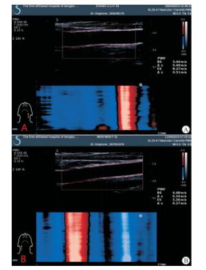
图 1 UFPWV技术测量颈总动脉弹性参数
-
采用t检验、χ2检验和Pearson线性相关分析。
-
2组性别、年龄差异均无统计学意义(P>0.05);GD组病人收缩压、脉压和心率均明显高于对照组(P < 0.01),舒张压低于对照组(P < 0.05);GD组病人FT3、FT4均明显高于对照组,TSH明显低于对照组(P < 0.01)(见表 1)。
分组 n 男 女 年龄/岁 心率/(次/分) 收缩压/mmHg 舒张压/mmHg 脉压/mmHg FT3/(pmol/L) FT4/(pmol/L) TSH/(mIU/L) GD组 76 22 54 40.58±12.88 94.20±18.06 123.68±10.68 74.65±7.41 48.96±6.82 14.72±6.33 100.21±60.57 0.03±0.05 对照组 76 23 53 40.70±13.56 70.59±7.51 114.67±8.73 77.08±7.47 37.59±7.48 4.23±1.07 25.81±5.67 2.25±1.02 t — 0.03* 0.06 -10.52* -5.70 2.02 -9.74 -14.25 -10.66 19.01 P — >0.05 >0.05 < 0.01 < 0.01 < 0.05 < 0.01 < 0.01 < 0.01 < 0.01 *示χ2值 表 1 2组受试者一般资料比较(x±s)
-
GD组颈总动脉IMT与对照组差异无统计学意义(P>0.05),BS及ES均明显高于对照组(P < 0.01)(见表 2)。
分组 IMT/mm BS/(m/s) ES/(m/s) GD组 0.55±0.09 6.58±0.75 8.62±1.38 对照组 0.54±0.09 5.64±0.76 6.49±1.20 t -0.48 -7.64 -10.18 P >0.05 < 0.01 < 0.01 表 2 2组颈总动脉IMT、BS及ES比较(x±s)
-
Pearson相关分析结果显示,GD病人血管弹性参数BS、ES与IMT均呈正相关关系(P < 0.01);BS、ES与甲状腺容积均无明显相关关系(P>0.05);ES与FT3水平呈正相关关系(r=0.467,P < 0.01),BS与FT3、FT4、TSH,ES与FT4、TSH均无明显相关关系(P>0.05)。
-
动脉结构与功能的改变是心血管疾病的病因,GD的主要靶器官是心血管系统,其结构及功能的损害对临床延缓和控制甲亢继发的心血管病变非常重要。颈动脉硬化改变常早于其他动脉,所以在临床上常被作为检查窗口。动脉弹性参数可在动脉结构发生改变之前较好地反映动脉功能的变化,PWV已被公认为是可靠的动脉弹性参数[5],UFPWV参考BS与ES,能更好地反映颈动脉功能变化,较传统的Complior SP法测量速度快,精准度及稳定性更高[6-9],在血管内中膜增厚以前就可以超早期预测心血管疾病的发生,现已开始应用于临床[10-11]。
本研究结果显示,GD组颈动脉IMT较对照组无明显增厚,BS及ES均明显高于对照组,提示GD病人在动脉结构发生改变以前弹性就已经减低,这与FANG等[12]研究结果相符。高水平甲状腺激素损伤血管内皮细胞,使动脉弹性减低[13],但在较短时内内中膜的变化并不明显。本研究中,GD组病人收缩压及脉压均明显高于对照组,舒张压低于对照组,这与杨寒凝等[11]研究结果一致。可能原因为随着血压增高,颈总动脉壁张力增大,内皮细胞功能受损,血管弹性减低。因此,控制血压对GD病人意义重大。
相关分析结果显示,GD病人血管弹性参数与甲状腺容积无明显相关性,提示肿大的甲状腺可以推挤颈动脉外移,甚至产生外压性形变,但不会损伤血管内皮功能。而BS及ES与IMT均呈正相关关系,这与滕飞等[14]观点相同,可见颈动脉功能与结构密切相关。此外,GD病人ES与FT3水平呈明显正相关关系,DELITALA等[15]也得出同样研究结果,这可能与FT3较FT4及TSH生物活性更强有关。
综上所述,在GD病人病程早期,颈动脉IMT尚未增厚以前,其动脉弹性已经发生早期变化。UFPWV技术通过精准测量颈动脉PWV值,可较敏感地发现这种早期变化,从而超早期预测甲亢继发的心血管病变。
彩色脉搏波技术定量评价Graves病早期颈动脉结构及功能变化
Application value of UFPWV in quantitative evaluating the early changes of carotid artery structure and function in patients with Graves disease
-
摘要:
目的应用彩色脉搏波技术(ultrafast pulse wave velocity,UFPWV)定量评价Graves病(Graves disease,GD)病人早期颈动脉结构和功能变化。 方法选取内-中膜厚度(intima-medium thickness,IMT)未增厚GD病人76例(GD组),同期健康体检成人76名作为对照组,测量2组受试者颈动脉IMT,采用UFPWV采集脉搏波传导速度,包括收缩期开始时脉搏波传导速度(beginning of systole,BS)和收缩期结束脉搏波传导速度(end of systole,ES),比较相关参数,分析BS、ES与IMT、甲状腺容积及血清甲状腺激素水平的相关性。 结果GD组颈总动脉IMT与对照组差异无统计学意义(P>0.05),BS及ES均明显高于对照组(P < 0.01)。GD病人血管弹性参数BS、ES与甲状腺体积均无明显相关性(P>0.05),但均与IMT呈明显正相关关系(P < 0.01),ES与游离三碘甲状原氨酸水平亦呈正相关关系(P < 0.01)。 结论UFPWV可发现GD病人颈动脉IMT增厚以前的早期弹性变化,可作为评价GD病人动脉弹性的新型有效手段。 Abstract:ObjectiveTo explore the application value of ultrafast pulse wave velocity(UFPWV) in quantitative evaluating the early changes of carotid artery structure and function in patients with Graves disease(GD). MethodsSeventy-six GD patients without intima-media thickness(IMT) of carotid artery thickening and 76 healthy people were divided into the GD group and control group, respectively.The carotid IMT in two groups was measured.The pulse wave conduction velocity was recorded using the UFPWV technology, and which included the pulse wave conduction velocity at the beginning and end of systole(BS and ES).The related parameters were compared, and the correlation of BS and ES with IMT, thyroid volume and serum thyroid hormone level were analyzed. ResultsThere was no statistical significance in IMT between GD group and control group(P>0.05), and the BS and ES in GD group were significantly higher than those in control group(P < 0.01).The BS and ES of vascular elasticity parameters were not correlated with the thyroid volume(P>0.05), but were positively correlated with IMT(P < 0.01).The ES was positively correlated with the level of free triiodothyronine. ConclusionsThe early changes of carotid artery elasticity in patients with GD before IMT thickening can be detected by UFPWV, which can be used as a new effective method to evaluate the artery elasticity in patients with GD. -
Key words:
- Graves disease /
- carotid artery /
- ultrafast pulse wave velocity /
- intima-media thickness
-
表 1 2组受试者一般资料比较(x±s)
分组 n 男 女 年龄/岁 心率/(次/分) 收缩压/mmHg 舒张压/mmHg 脉压/mmHg FT3/(pmol/L) FT4/(pmol/L) TSH/(mIU/L) GD组 76 22 54 40.58±12.88 94.20±18.06 123.68±10.68 74.65±7.41 48.96±6.82 14.72±6.33 100.21±60.57 0.03±0.05 对照组 76 23 53 40.70±13.56 70.59±7.51 114.67±8.73 77.08±7.47 37.59±7.48 4.23±1.07 25.81±5.67 2.25±1.02 t — 0.03* 0.06 -10.52* -5.70 2.02 -9.74 -14.25 -10.66 19.01 P — >0.05 >0.05 < 0.01 < 0.01 < 0.05 < 0.01 < 0.01 < 0.01 < 0.01 *示χ2值  下载: 导出CSV
表 2 2组颈总动脉IMT、BS及ES比较(x±s)
分组 IMT/mm BS/(m/s) ES/(m/s) GD组 0.55±0.09 6.58±0.75 8.62±1.38 对照组 0.54±0.09 5.64±0.76 6.49±1.20 t -0.48 -7.64 -10.18 P >0.05 < 0.01 < 0.01
下载: 导出CSV
-
[1] 张光华, 安静, 洪林巍.极速成像技术检测脉搏传导速度早期评价原发性高血压患者颈动脉弹性的研究[J].中国临床医学影像杂志, 2016, 27(4):297. [2] 张红, 周琦, 姜珏.极速脉搏波技术定量评价老年高血压患者颈动脉弹性的应用研究[J].临床内科杂志, 2015, 32(6):380. doi: 10.3969/j.issn.1001-9057.2015.06.006 [3] 何文, 金占强.超声新技术在浅表器官中的应用[J].中国医学影像技术, 2016, 32(5):643. [4] 邱兰燕, 钱林学, 刘冬, 等.极速成像技术检测的脉搏波传导速度与颈动脉硬化相关性的研究[J].中华超声影像学杂志, 2014, 23(3):203. doi: 10.3760/cma.j.issn.1004-4477.2014.03.008 [5] 杨谧, 杨寒凝.实时剪切波及彩色脉搏波成像对正常颈动脉硬度的研究[J].中国临床医学影像杂志, 2018, 29(7):468. [6] WEIRMCCALL JR, KAMALASANAN A, CASSIDY DB, et al.Assessment of the effects of technique on pulmonary arterial pulse wave velocity measurement[J]. Heart, 2016, 102(Suppl 3):A18. doi: 10.1136/heartjnl-2016-309668.24 [7] SALLES S, CHEE AJ, GARCIA D, et al.2-D arterial wall motion imaging using ultrafast ultrasound and transverse oscillations[J]. IEEE Trans Ultrason Ferroelectr Freq Control, 2015, 62(6):1047. doi: 10.1109/TUFFC.2014.006910 [8] MIRAULT T, PERNOT M, FRANK M, et al.Carotid stiffness change over the cardiac cycle by ultrafast ultrasound imaging in healthy volunteers and vascular Ehlers-Danlos syndrome[J]. J Hypertens, 2015, 33(9):1890. doi: 10.1097/HJH.0000000000000617 [9] TANTER M, FINK M.Ultrafast imaging in biomedical ultrasound[J]. IEEE Trans Ultrason Ferroelectr Freq Control, 2014, 61(1):102. doi: 10.1109/TUFFC.2014.2882 [10] 栾云, 殷立平, 黄辉, 等.脉搏波速度技术对原发性高血压患者颈动脉弹性功能的初步探讨[J].东南国防医药, 2016, 18(5):451. doi: 10.3969/j.issn.1672-271X.2016.05.001 [11] 杨寒凝, 杨谧, 孙月, 等.实时剪切波弹性成像及彩色脉搏波成像技术评估不同级别高血压患者颈总动脉弹性[J/CD].中华医学超声杂志: 电子版, 2018, 15(9): 679. [12] FANG J, KANG C, LI Z, et al.Correlation research of carotid arterial sclerosis and left ventricular diastolic function in hyperthyroidism[J]. Minerva Cardioangiol, 2014, 62(5):379. [13] DEYNELI O, AKPNAR IN, MERILILER OS, et al.Effects of levothyroxine treatment on insulin sensitivity, endothelial function and risk factors of atherosclerosis in hypothyroid women[J]. Ann Endocrinol, 2014, 75(4):220. [14] 滕飞, 吴长君.彩色脉搏波新技术评价2型糖尿病患者早期颈动脉功能变化的初步研究[J].医学研究杂志, 2015, 44(11):132. doi: 10.11969/j.issn.1673-548X.2015.11.037 [15] DELITALA AP, ORRÙ M, FILIGHEDDU F, et al.Serum free thyroxine levels are positively associated with arterial stiffness in the SardiNIA study[J]. Clin Endocrinol, 2015, 82(4):592. doi: 10.1111/cen.12532 -

 点击查看大图

图(1)表(2)
计量
- 文章访问数: 4041
- HTML全文浏览量: 2089
- PDF下载量: 23
- 被引次数: 0
