

-
生物组织软硬度和组织生物学特性密切关联,弹性成像通过采集组织的软硬度信息,有助于评估组织病变的良恶性。近年来,弹性成像技术在乳腺疾病诊断方面的应用发展较为成熟,包括静态弹性成像、声脉冲辐射力弹性成像(acoustic radiationforce impulse, ARFI)以及实时剪切波弹性成像等不同的成像方法[1],其中ARFI技术包括声触诊组织成像(virtual touch tissue imaging,VTI)和声触诊组织定量(virtual touch tissue quantification,VTQ)[2],VTQ有效避免了传统弹性成像技术压力不稳的弊端。然而,VTQ在技术上也存在一些缺点,如只能单点测量、取样框面积大、需重复多次测量、时有出现无测值情况等,因此,在使用中颇为受限。最近,在此基础上发展的声触诊组织成像定量测量技术(virtual touch tissue imaging quantification,VTIQ)有效改进了以上缺点。本研究旨在探讨VTQ和VTIQ在乳腺良恶性结节鉴别诊断中的应用价值。现作报道。
-
收集2017年4月至2018年4月来我院诊疗的乳腺疾病病人58例共58个乳腺实性结节,所有病人均为女性,年龄16~73岁,所有病例均经手术治疗或穿刺活检证实,共计良性结节34个,恶性结节24个,结节最大径7.3~56 mm。排除标准:病变同侧乳腺有手术、微创、穿刺活检病史者;乳腺假体植入者;单纯囊性病变。
-
使用德国西门子AcusonS3000型彩色多普勒超声诊断仪,配有ARFI及VTIQ成像软件,线阵探头9L4,频率4~9 MHz,成像宽度范围37 mm。
-
病人仰卧位,必要时半侧卧位,充分暴露双侧胸部及腋窝,选择仪器预设置的乳腺条件,根据不同情况灵活调节仪器参数(如聚焦深度、聚焦部位、增益等),以便清晰显示正常乳腺各层解剖结构及病变声像图。先行常规超声检查,以乳头为中心,以顺时针或逆时针辐射状扫查,或从上到下,从左到右逐一切面扫查,详细观察病灶及其周围腺体组织的声像图改变。
-
检查时将探头轻轻置于病人皮肤表面,涂抹适量耦合剂,切忌人为施压,调整放大图像,取病变最大切面,激活VTQ技术,将取样框置于病变同一位置保持探头不变,嘱病人屏气5 s,点击“Update键”,重复测量6~10次剪切波速度值(SWV), 去除最大值和最小值,计算平均值,测量时应尽量避开病变内囊性部分及钙化部分,出现X.XX m/s时记为8.4 m/s[3]。
-
调节图像大小适中,使病变区及周边腺体组织清晰显示,将取样框覆盖病灶,依次显示质量模式和速度模式图,质量模式可控制图像弹性质量分布,质量由低到高为红色→黄色→绿色,选取有效测量区域即质量模式下均匀绿色分布区域,使感兴趣区(region of interest,ROI)处于病灶内,ROI大小为1 mm×1 mm,嘱病人屏气,获取速度模式测量图像,图像中SWV值自低至高分别为蓝色→绿色→黄色→红色。以病变周围腺体组织表现为浅蓝色或淡绿色且分布均匀、病变内表现为红色或黄色为标准,调整大小适宜的SWV量程(最大10 m/s),获取供最终测量的VTIQ速度模式图。测量时,根据速度模式图中不同色彩,分别将ROI置于病变SWV最高区、最低区、周边及中央区域。对病变重复定位测量6~10次,获取SWV平均值并记录。将所有病例的常规超声图像及VTQ、VTIQ图像储存在超声仪器硬盘内,以备脱机分析。
-
采用t(或t′)检验和受试者工作特征(ROC)曲线分析。
-
58个乳腺结节中包括恶性结节24个,良性结节34个,其中恶性组包括浸润性导管癌21例,导管内癌3例;良性组包括乳腺纤维腺瘤14例,乳腺腺病7例,肉芽肿性乳腺炎5例,良性分叶状肿瘤4例,导管内乳头状瘤4例。
-
VTQ和VTIQ检查乳腺恶性结节的SWV值均明显高于良性结节(P < 0.01);对于乳腺良性和恶性结节,VTIQ测得SWV值与VTQ测得差异均无统计学意义(P>0.05)(见图 1~2、表 1)。

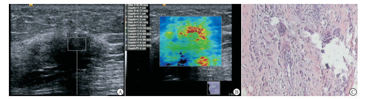
图 1 乳腺纤维腺瘤的VTQ、VTIQ及病理检查
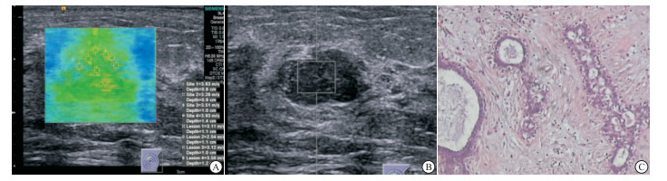
图 2 乳腺浸润性导管癌的VTQ、VTIQ及病理检查
分组 n VTQ VTIQ t P 良性结节 34 3.45±0.95 3.70±0.66 1.26Δ >0.05 恶性结节 24 4.42±0.99 4.79±0.76 1.45 >0.05 t — 3.76 5.82 — — P — <0.01 <0.01 — — Δ示t′值 表 1 VTQ、VTIQ测得乳腺良、恶性结节SWV值比较(x±s;m/s)
-
VTQ和VTIQ诊断结果见表 2。VTIQ技术ROC曲线下面积为0.820,高于VTQ技术的0.702(Z=2.72,P < 0.05)。ROC曲线显示,VTQ技术诊断良恶性结节最佳阈值为SWV值3.97 m/s,以3.97 m/s为截断点,其鉴别诊断良恶性结节灵敏度、特异度、准确度依次为66.7%、70.6%、69.0%,AUC为0.702;VTIQ最佳阈值为SWV值4.09 m/s,以4.09 m/s为截断点,其灵敏度、特异度、准确度依次为79.2%、85.3%、82.8%,AUC为0.820。
检查技术 检查结果 病理结果 良性 恶性 VTQ 良性 24 8 恶性 10 16 VTIQ 良性 29 5 恶性 5 19 表 2 VTQ和VTIQ技术诊断结果
-
乳腺良恶性结节灰阶超声表现存在一定程度交叉重叠,单纯依靠常规超声常常难以对乳腺结节良恶性进行准确定性。2013版BI-RADS分类把病变弹性评估纳入伴随征象[4-5],并且已有研究[6-7]表明乳腺恶性肿瘤内部组织硬度显著高于周边或者对侧相应区域正常腺体组织。
VTQ技术是一项横向振动技术,横向振动是以剪切波的形式向四周传播,VTQ即通过检测剪切波的传播信号,显示并计算SWV,定量评估组织软硬度,组织越软,剪切波速度值越低,相反,组织越硬,剪切波速度值越高。VTQ使弹性成像技术由定性评价和半定量评价迈入了定量评价模式,较为广泛地应用于鉴别诊断乳腺良恶性肿块[8-9],但是它也存在一些缺陷,使得VTQ在应用中受到限制。
要改进上述缺陷,VTIQ技术应运而生,它是在VTI和VTQ技术基础上进行的改革创新,克服了以往技术的不足,对乳腺肿块的诊断可兼有定性和定量分析模式[10-12],且研究[12-13]表明VTIQ技术提高了操作者的可重复性和独立性。VTIQ技术具有质量、速度、时间、位移四种模式,测量的ROI范围为1 mm×1 mm(VTQ测量ROI:5 mm×6 mm),取样框较小,对于较小病灶测量更精确,可多次同时选取ROI进行重复测量,测得SWV值更能反映出整体病灶的软硬度,更具有可靠性。此外,VTIQ技术SWV测值范围为0~10 m/s,较VTQ技术测值范围更宽,且可手动调节,避免了VTQ技术测量时因被测组织存在高硬度区域出现无法测值(X.XX m/s)的情况[14]。
本研究结果显示,采用VTQ、VTIQ两种技术所测得乳腺恶性结节的SWV值均高于良性结节,说明恶性结节硬度显著高于良性结节,测定乳腺结节SWV值有助于鉴别乳腺结节良恶性,分析原因可能为恶性肿瘤内细胞含量丰富,细胞增生异型性显著,细胞排列较为紧密,呈索条状、巢状,浸润性生长,多伴有间质纤维化、钙化,因此肿瘤硬度增加,剪切波在其内传播速度增快,SWV测值增高。此外,本研究结果显示,VTQ技术AUC为0.702,VTIQ技术AUC为0.820,VTIQ诊断效能高于VTQ技术,与既往研究[15-16]相似。分析其可能原因为VTIQ技术具有质量控制模式,可以有效选择质量分布均匀一致呈绿色的区域进行测量,同时VTIQ速度模式图直观显示病变内部软硬度信息,指引观察者选择高硬度区域放置ROI进行测量,提高测量的准确性。而VTQ只能单点测量,不能直观显示病变内硬度分布情况。一些乳腺良性病变富含纤维组织及瘢痕增生,使得剪切波在其内传播增快,导致SWV测值增高,出现假阳性病例。本研究中,有10个乳腺良性结节VTQ技术测得SWV值>3.97 m/s,判为恶性结节;使用VTIQ技术,仍有5个良性结节误判为恶性病变,分析原因可能为部分乳腺良性结节内部含有较多钙化或者纤维成分,尽管在检查时观察者已经选择二维超声图像上非钙化或者非纤维化部分进行测量,但是钙化区或者纤维化区域周围病变组织硬度与病变内远离钙化处的组织硬度相比显著增高,使得SWV值增高,导致假阳性出现。一些恶性肿瘤由于细胞含量多,间质纤维化成分少,或者由于生长过快,肿块内部出现出血坏死液化区域,导致病变组织内部硬度降低,SWV值降低,出现假阴性情况,本研究中有8例恶性肿瘤VTQ技术测得被判为良性肿瘤,采用VTIQ技术有5例恶性肿瘤误诊为良性。因此,尽管VTIQ技术较VTQ技术诊断效能有所提高,但是临床应用中还应结合其它超声新技术或者钼靶等提高乳腺恶性肿瘤的诊断准确率。
本研究亦具有一定局限性,如本次研究没有纳入乳腺常规超声检查信息,仅以VTIQ技术或者VTQ技术作为唯一诊断指标;研究病例较少,病理类型较为单一,尤其是乳腺恶性病变,以浸润性导管癌居多。
综上,相较于VTQ技术,VTIQ技术易于操作,在一定程度上改进了VTQ定量测量存在的问题,在乳腺良恶性结节鉴别诊断中VTIQ技术诊断效能优于VTQ技术,在临床应用中可增加诊断信息,具有较好临床应用前景。
不同声触诊组织成像定量技术在乳腺良恶性结节鉴别诊断中的应用价值
Application value of different virtual touch tissues imaging quantification techniques in the differential diagnosis of benign and malignant breast nodules
-
摘要:
目的探讨声触诊组织定量技术(VTQ)与声触诊组织成像定量技术(VTIQ)在乳腺良恶性结节鉴别诊断中的应用价值。 方法收集乳腺疾病病人58例共58个乳腺实性结节,术前或者穿刺活检前分别应用VTQ、VTIQ技术测量病灶内部剪切波速度值(SWV),与病理结果对照,分别绘制VTQ、VTIQ技术诊断乳腺良恶性结节的ROC曲线,比较2种技术诊断效能。 结果58个乳腺结节中包括恶性结节24个,良性结节34个。VTQ和VTIQ测得乳腺恶性结节的SWV值均明显高于良性结节(P < 0.01);对于乳腺良性和恶性结节,VTIQ测得SWV值与VTQ测得差异均无统计学意义(P>0.05)。VTIQ技术ROC曲线下面积为0.820,高于VTQ技术的0.702(P < 0.05)。VTQ技术诊断良恶性结节最佳阈值为SWV值3.97 m/s,以3.97 m/s为截断点,其鉴别诊断良恶性结节灵敏度、特异度、准确度依次为66.7%、70.6%、69.0%,AUC为0.702;VTIQ最佳阈值为SWV值4.09 m/s,以4.09 m/s为截断点,其灵敏度、特异度、准确度依次为79.2%、85.3%、82.8%,AUC为0.820。 结论VTIQ技术鉴别诊断乳腺良恶性结节的效能优于VTQ技术,具有较好临床应用前景。 -
关键词:
- 声触诊组织定量技术 /
- 声触诊组织成像定量技术 /
- 乳腺结节
Abstract:ObjectiveTo explore the application value of virtual touch tissue quantification(VTQ) and virtual touch tissue imaging quantification(VTIQ) in the differential diagnosis of benign and malignant breast nodules. MethodsFifty-eight breast solid nodules in 58 patients with breast disease were collected.The internal shear wave velocity value (SWV) of the nodules were measured using VTQ and VTIQ techniques before operation or before biopsy, and the results of which were compared with the pathological results.The ROC curve of benign and malignant breast nodules diagnosed by VTQ and VTIQ techniques were drew to compare the diagnostic efficacy of two techniques. ResultsFifty-eight breast nodules included 24 malignant nodules and 34 benign nodules.The mean value of SWV measured by VTQ and VTIQ in malignant nodules was significantly higher than that in benign nodules(P < 0.01).For benign and malignant breast nodules, there was no statistical significance in the SWV value measured by VTIQ and VTQ(P>0.05).The area under ROC curve of VTIQ technique(0.820) was higher than that of VTQ technique(0.702)(P < 0.05).The best threshold of SWV value in benign and malignant nodules diagnosed by VTQ technique was 3.97 m/s.With 3.97 m/s as cut-off point, the sensitivity, specificity and accuracy of differential diagnosis of benign and malignant nodules were 66.7%, 70.6% and 69.0%, respectively, and the AUC was 0.702.The optimal threshold of SWV value diagnosed by VTIQ was 4.09 m/s.With 4.09 m/s as the cut-off point, and the sensitivity, specificity and accuracy were 79.2%, 85.3% and 82.8%, respectively, and the AUC was 0.820. ConclusionsThe efficacy of VTIQ technique is superior to VTQ in the differential diagnosis of benign and malignant breast nodules, and the VTIQ technique has a better clinical application prospect. -
表 1 VTQ、VTIQ测得乳腺良、恶性结节SWV值比较(x±s;m/s)
分组 n VTQ VTIQ t P 良性结节 34 3.45±0.95 3.70±0.66 1.26Δ >0.05 恶性结节 24 4.42±0.99 4.79±0.76 1.45 >0.05 t — 3.76 5.82 — — P — <0.01 <0.01 — — Δ示t′值  下载: 导出CSV
-
[1] SHⅡNA T, NIGHTINGALE KR, PALMERI ML, et al.WFUMB guidelines and recommendations for clinical use of ultrasound elastography:part 1:basic principles and terminology[J]. Ultrasound Med Biol, 2015, 41(5):1126. doi: 10.1016/j.ultrasmedbio.2015.03.009 [2] RICCI P, MAGGINI E, MANCUSO E, et al.Clinical application of breast elastography:state of the art[J]. Eur J Radiol, 2014, 83(3):429. [3] MENG W, ZHANG G, WU C, et al.Preliminary results of acoustic radiation force impulse (ARFI) ultrasound imaging of breast lesions[J]. Ultrasound Med Biol, 2011, 37(9):1436. doi: 10.1016/j.ultrasmedbio.2011.05.022 [4] 李小龙, 徐辉雄, 伯小皖, 等.声触诊组织成像和定量技术对BI-RADS 4乳腺病灶良恶性的诊断价值[J].影像诊断与介入放射学, 2015, 24(4):290. doi: 10.3969/j.issn.1005-8001.2015.04.006 [5] 朱庆莉, 姜玉新.乳腺影像报告与数据系统指南(第5版)超声内容更新介绍[J/CD].中华医学超声杂志: 电子版, 2016, 13(1): 5. [6] ASTERIA C, GIOVANARDI A, PIZZOCAR A.US-elastography in the differential diagnosis of benign and malignant thyroid nodules[J]. Thyroid, 2008, 18(5):523. doi: 10.1089/thy.2007.0323 [7] 赵晓虹, 丛淑珍, 李康, 等.乳腺黏液癌的声像图特征与病理结果对比分析[J].中国医学影像技术, 2008, 20(8):1212. doi: 10.3321/j.issn:1003-3289.2008.08.019 [8] 欧冰, 智慧, 杨海云, 等.比较声触诊组织量化与弹性应变率比值法诊断乳腺疾病[J].中国医学影像技术, 2012, 28(10):321. [9] 沈春云, 徐春燕, 张绪霞.ABVS联合ARFI技术对乳腺病灶鉴别诊断价值[J].中国超声医学杂志, 2015, 31(12):1835. [10] IANCULESCUA V, CIOLOVANA LM, DUNANTB A, et al.Added value of Virtual Touch IQ shear wave elastography in the ultrasound assessment of breast lesions[J]. Eur J Radiol, 2014, 83(5):773. [11] GOLATTA M, MARTINA MS, HARCOSA A, et al.Normal breast tissue stiffness measured by a new ultrasound technique:virtual touch tissue imaging quantification (VTIQ)[J]. Eur J Radiol, 2013, 82(11):676. doi: 10.1016/j.ejrad.2013.06.029 [12] GOLATTA M, SCHWEITZER-MARTIN M, HARCOS A, et al.Evaluation of virtual touch tissue imaging quantification, a new shear wave velocity imaging method, for breast lesion assessment by ultrasound[J]. Biomed Res Int, 2014, 2014:960262. [13] KAPETAS P, PINKER-DOMENIG K, WOITEK R, et al.Clinical application of Acoustic Radiation Force Impulse Imaging with Virtual Touch IQ in breast ultrasound:diagnostic performance and reproducibility of a new technique[J]. Acta Radiol, 2017, 58(2):140. doi: 10.1177/0284185116641347 [14] 张志勇, 吕志红.慢性乙型肝炎肝纤维化患者餐前餐后肝脏弹性指数的变化[J].临床内科杂志, 2016, 33(5):346. doi: 10.3969/j.issn.1001-9057.2016.05.020 [15] 詹嘉, 刘迎春, 陈悦, 等, 声触诊组织成像和定量技术鉴别诊断乳腺肿块良恶性的初步探讨[J].中国临床医学影像杂志, 2017, 28(6):405. doi: 10.3969/j.issn.1008-1062.2017.06.007 [16] MATSUZUKA T, SUZUKI M, SAIJO S, et al.Stiffness of salivary gland and tumor measured by new ultrasonic techniques:Virtual touch quantification and IQ[J]. Auris Nasus Larynx, 2015, 42(2):128. doi: 10.1016/j.anl.2014.08.021 -

 点击查看大图
图(2)表(2)
计量
- 文章访问数: 3982
- HTML全文浏览量: 2023
- PDF下载量: 7
- 被引次数: 0
