

-
肩袖损伤是引起肩关节疼痛及功能障碍的常见原因之一, 而冈上肌肌腱损伤又是其最常见的类型,研究[1]表明冈上肌肌腱损伤常伴随肌肉萎缩和脂肪的浸润,这些变化对治疗方案的选择和预后评估具有重要的临床意义。目前关于冈上肌脂肪变性程度的报道多为半定量研究,如磁共振波谱、化学位移成像等,同时关于肌肉萎缩程度的报道亦相对较少。本研究采用MRI最新的脂肪定量技术测量冈上肌脂肪分数(fat fraction,FF),同时测量冈上肌相对大小作为萎缩程度指标,进而评估它们与肌腱损伤程度之间的相关性。
-
选取2018年9月至2019年5月于我院骨科运动医学病区就诊的肩袖损伤病人,所有病人均伴有肩部不同程度疼痛。纳入标准:经临床及MRI诊断为冈上肌腱损伤。排除标准:有急性外伤史、关节炎病史、手术史及其他相关全身性及骨肌系统疾病者。最终入组病人40例,其中男26例,女14例,年龄42~71岁,平均年龄(56.27±7.51)岁。同时选取14名健康志愿者,男7名,女7名,年龄40~65岁,平均年龄(53.95±5.64)岁。所有研究对象均行常规肩关节MRI检查,同时进行脂肪定量扫描。
-
本研究采用飞利浦IngeniaCX 3.0 T MRI设备,行常规肩关节平扫及mDIXON-Quant检查。采用肩关节专用线圈。常规序列参数:斜冠状位、横轴位fs PDWI:TR3600ms, TE50 ms,层厚4 mm,层间距1 mm;斜冠状位、斜矢状位TIWI:TR650 ms, TE20 ms,层厚4 mm,层间距1 mm。脂肪定量序列采用mDIXON-Quant序列,扫描参数:TR/TE 9.4 ms/1.43 ms,ΔTE 1.3 ms,翻转角3°,回波数6,重建层厚3 mm,采用7峰值脂肪模型进行脂肪定量。
根据常规斜冠状位MRIfsPDWI显示冈上肌腱不同表现,将40例病人分为退变组、部分撕裂组和完全撕裂组。14名健康志愿者作为对照组。脂肪定量扫描后自动重建并生成FF图。
-
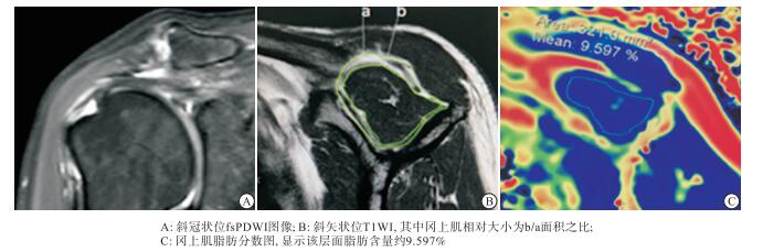
全部影像学图像传输至飞利浦后处理工作站进行分析。所获得MRI图像均由两名高年资影像医师独立完成分析,取其平均结果。依据常规MR影像评价冈上肌腱损伤严重程度:对照组,肌腱形态完整,呈均匀一致低信号;退变组,冈上肌腱在fs PDWI上呈中等或稍高信号,形态正常;部分撕裂组,冈上肌腱在fs PDWI上部分连续性中断,信号增高;完全撕裂组,肌腱信号强度明显增加,连续性中断,断端回缩(见图 1)。选择斜矢状肩胛骨显示为Y型层面作为研究层面,于该层面手动描画冈上肌轮廓作为兴趣区(ROI),即可直接得出冈上肌FF值,测量3次取平均值。同时于该层面T1WI像上手动描画冈上肌轮廓和冈上肌及其周围脂肪轮廓,得出其面积大小,将冈上肌与冈上肌及其周围脂肪面积之比作为冈上肌相对大小值用来评价其萎缩程度(见图 2)。
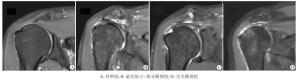
图 1 冈上肌斜冠状位fs PDWI图像

图 2 冈上肌部分撕裂病人矢状肩胛骨Y型层面MRI图像
-
采用方差分析、q检验、Spearman相关分析。
-
各组间FF值,对照组 < 退变组 < 完全撕裂组,对照组 < 部分撕裂组组 < 完全撕裂组,差异均有统计学意义(P < 0.01),退变组和部分撕裂组间差异无统计学意义(P>0.05);冈上肌相对大小,对照组>退变组>完全撕裂组,对照组>部分撕裂组组>完全撕裂组,差异均有统计学意义(P < 0.05~P < 0.01),退变组和部分撕裂组间差异无统计学意义(P>0.05)(见表 1)。
分组 n FF值 冈上肌相对大小 对照组 14 3.17±0.99 0.74±0.07 退变组 10 6.43±0.48* 0.66±0.07* 部分撕裂组 13 7.19±0.44** 0.61±0.08** 完全撕裂组 17 10.97±1.72**△△## 0.48±0.08**△△## F — 122.77 31.86 P — < 0.01 < 0.01 MS组内 — 1.289 0.006 q检验:与对照组比较*P < 0.05, **P < 0.01;与退变组比较△△P < 0.01;与部分撕裂组比较##P < 0.01 表 1 各组间FF及冈上肌相对大小比较(x±s)
-
FF值与冈上肌腱损伤程度呈正相关关系(rs=0.95,P < 0.01),冈上肌相对大小与冈上肌腱损伤程度呈负相关关系(rs=-0.80,P < 0.01);冈上肌相对大小与FF值呈负相关关系(r=-0.73,P < 0.01)。
-
肩袖是控制肩关节旋转、维持肱骨头稳定的重要结构,其中冈上肌腱是构成肩袖最为重要的,也是最易损伤的肌腱,其主要由水、胶原及蛋白聚糖构成。冈上肌腱损伤早期多属于可逆性,以组织生化改变为主;晚期则属于不可逆损伤。GOUTALLIER等[2]首先报道了肌腱损伤后由于肌腱对骨的附着力丧失,使得肌肉在横向及径向上可能均会发生一定程度的萎缩;同时肌小节长度和数量减少、肌原纤维溶解和变性,脂肪组织会累积于肌束内、外及肌腱内,从而引起不可逆性的肌肉内脂肪浸润[3-4]。这些改变不仅影响冈上肌腱的功能,同时影响治疗手段的选择和预后的恢复情况。目前临床研究已经证实冈上肌脂肪浸润过高可能是手术失败或预后不良的重要原因之一[5]。当冈上肌内脂肪浸润累及一半以上时则为外科手术的禁忌证。因此定量研究冈上肌脂肪浸润及肌肉萎缩程度对临床具有重要的指导意义。目前临床上常用的Goutallier分级是利用MRI斜矢状位T1WI来主观定性评价脂肪浸润程度,将肩袖肌腹内无脂肪组织定为0级,肩袖肌腹内见少量高信号脂肪组织为1级,肩袖肌腹内见脂肪组织但少于肌肉量为2级,肩袖肌腹内高信号的脂肪组织量与肌肉量相等为3级,肩袖肌腹内高信号的脂肪组织量大于肌肉量为4级。但该方法主观因素较强,不能准确评估肌肉脂肪含量。
随着现代医学影像的高速发展,现有的肌肉内脂肪定量方法通常包括定量CT以及MRI。定量CT目前国内外报道多为其在肝脏脂肪浸润方面的应用[6],而对于肌肉内脂肪浸润程度的评估仍处于研究阶段。MRI关于脂肪含量的研究略多,如化学位移成像、波谱分析等。国内包磊等[7]利用磁共振化学位移成像技术主要采用信号强度抑制率和信号强度指数等指标定量测量和评估冈上肌脂肪性退变程度对肌腱病变进行分级。KENN等[8]利用MRI波谱分析推断脂肪变性可以量化,但其空间分辨率低、扫描时间长且可重复性较差,因此较难推广。以上MRI技术均为间接评估肌肉内脂肪含量。mDIXON Quant是目前一种新的直接测量脂肪含量的MRI技术,该技术是3D梯度回波DIXON序列,采集6个回波,分析7个脂肪峰,且扫描速度快,同时可以计算出T2*值和R2*值。正常情况下脂肪在磁共振波谱中有9个脂肪峰,而脂肪的第8峰已经很接近水峰了,因此mDIXON Quant技术中分析7个脂肪峰值保证了脂肪定量的准确性。扫描结束后可以直接得到脂肪分数图,后处理时只需确定层面及感兴趣区即可直径得到ROI内面积的脂肪含量百分比,操作简单、可重复性高。
本研究中选择斜矢状肩胛骨显示为Y形层面作为研究层面,直接测量该层面内冈上肌脂肪含量,同时测量该层面冈上肌相对大小值。结果显示4组中随着损伤程度的增加,FF值逐渐增加,且完全撕裂组FF值明显高于其他组,系冈上肌脂肪浸润程度越重越容易引起肌腱的损伤。该结论与国内王翔等[9]报道结果一致。国外有学者研究[10]发现随着冈上肌萎缩百分比例的下降,肩袖损伤撕裂程度增加。本研究中冈上肌相对大小值随损伤程度逐渐减低,与其结果相符。同时FF值与冈上肌相对大小值呈负相关,说明随着脂肪浸润的加深肌肉萎缩度加深。
综上所述,3.0T MRI脂肪定量技术可以准确评估冈上肌脂肪浸润情况,且与肌腱损伤具有一定的相关性,同时可以间接反映肌肉萎缩情况,为临床干预提供重要的指导作用。但本研究中样本量较小,不能进一步得出不同类型肌腱损伤的脂肪浸润相对值。此外本研究未对肌肉脂肪含量对预后的影响进行阐述。笔者随后将进一步加大样本量,同时密切追踪随访治疗后变化。
MRI定量研究冈上肌腱损伤程度与脂肪浸润及肌肉萎缩程度的相关性
Correlations between the degree of supraspinatus tendon injury and degrees of fat infiltration and muscle atrophy using the quantitative study of MRI
-
摘要:
目的 测量冈上肌腱损伤病人冈上肌脂肪含量及肌肉萎缩程度,研究其与冈上肌腱损伤程度之间的相关性。 方法 选取40例肩关节冈上肌肌腱损伤病人及14名健康志愿者(对照组)作为研究对象,均采用3.0T MRI进行常规肩关节MRI检查及脂肪定量扫描(mDixon-Quant)。获取冈上肌脂肪分数图及冈上肌相对大小值。按冈上肌肌腱MRI不同表现将40例病人分为退变组、部分撕裂组和完全撕裂组。分析比较各组间冈上肌脂肪含量及相对大小值,冈上肌脂肪含量及相对大小值与冈上肌腱损伤程度间的相关性。 结果 各组间FF值,对照组 < 退变组 < 完全撕裂组,对照组 < 部分撕裂组组 < 完全撕裂组,差异均有统计学意义(P < 0.01),退变组和部分撕裂组差异无统计学意义(P>0.05);冈上肌相对大小,对照组>退变组>完全撕裂组,对照组>部分撕裂组组>完全撕裂组,差异均有统计学意义(P < 0.05~P < 0.01),退变组和部分撕裂组差异无统计学意义(P>0.05);FF值与冈上肌腱损伤程度呈正相关(rs=0.95,P < 0.01),冈上肌相对大小与冈上肌腱损伤程度呈负相关(rs=-0.80,P < 0.01),冈上肌相对大小与FF值呈负相关(r=-0.73,P < 0.01)。 结论 3.0T MRI可定量评价冈上肌脂肪浸润及萎缩程度,同时显示其与肌腱损伤程度的关系。 Abstract:Objective To measure the fat content and degree of muscle atrophy in patients with supraspinatus tendon injury, and study the correlations between the degree of supraspinatus tendon injury, and degrees of fat infiltration and muscle atrophy. Methods Forty patients with supraspinatus tendon injury of shoulder joint and 14 healthy examinees (control group) were investigated.The routine shoulder joint MRI examination and fat quantitative scan(mDixon-Quant) were performed.The fat fraction diagram of supraspinatus and relative size of supraspinatus were obtained.According to the different manifestations of MRI, the patients were divided into the degeneration group, partial tear group and complete tear group.The supraspinatus fat content and relative value, and its correlation with degree of supraspinatus tendon injury among four groups were analyzed and compared. Results Among four groups, the FF value in the control group, degeneration group and complete tear group, and in the control group, partial tear group and complete tear group gradually increased in turn, and the difference of which was statistically significant(P < 0.01).The difference of the FF value between degeneration group and partial complete tear group was not statistically significant(P>0.05).Among four groups, the supraspinatus volume in the control group, degeneration group and complete tear group, and in the control group, partial tear group and complete tear group gradually decreased in turn, and the difference of which was statistically significant(P < 0.05 to P < 0.01).The difference of the supraspinatus volume between degeneration group and partial complete tear group was not statistically significant(P>0.05).The FF value was positively correlated with the degree of supraspinatus tendon injury(rs=0.95, P < 0.01), the relative size of supraspinatus was negatively correlated with the degree of supraspinatus tendon injury(rs=-0.80, P < 0.01), and the relative size of supraspinatus was negatively correlated with the FF value (r=-0.73, P < 0.01). Conclusions 3.0T MRI can quantitatively evaluate the degree of fat infiltration and atrophy of supraspinatus and show the relationship between the degrees of fat infiltration and atrophy and degree of tendon injury. -
Key words:
- magnetic resonance imaging /
- supraspinatus tendon /
- muscular atrophy /
- fat infiltration
-
表 1 各组间FF及冈上肌相对大小比较(x±s)
分组 n FF值 冈上肌相对大小 对照组 14 3.17±0.99 0.74±0.07 退变组 10 6.43±0.48* 0.66±0.07* 部分撕裂组 13 7.19±0.44** 0.61±0.08** 完全撕裂组 17 10.97±1.72**△△## 0.48±0.08**△△## F — 122.77 31.86 P — < 0.01 < 0.01 MS组内 — 1.289 0.006 q检验:与对照组比较*P < 0.05, **P < 0.01;与退变组比较△△P < 0.01;与部分撕裂组比较##P < 0.01  下载: 导出CSV
-
[1] CHOI CH, KIM SS, KIM SJ, et al.Arthroscopic changes of the bicepspulley in rotator cuff tear and its clinical significance in relation totreatment[J].Clin Orthop Surg, 2015, 7(3):365. doi: 10.4055/cios.2015.7.3.365 [2] GOUTALLIER D, POSTEL JM, BERNAGEAU J, et al.Fattymuscle degeneration in cuff ruptures.Pre-and postoperative evaluationby CT scan[J].Clin Orthop Relat Res, 1994(304):78. [3] FUKUTA S, TSUTSUI T, RUI A, et al.Tendon retraction with rotator cufftear causes a decrease in cross-sectional area of the supraspinatusmuscle on magnetic resonance imaging[J].J Shoulder Elbow Surg, 2016, 25(7):1069. doi: 10.1016/j.jse.2015.11.008 [4] 刘少华, 陈疾忤, 李宏, 等.关节镜下单排与缝线桥技术修复中型肩袖撕裂——临床与核磁共振评价[J].中国运动医学杂志, 2017, 36(2):97. [5] 丁建, 陈艳, 姚建.肩周炎与肩袖损伤的MRI鉴别诊断在临床治疗中的意义[J].医学影像学杂志, 2017, 27(2):324. [6] 徐黎, 过哲, 程晓光, 等.定量CT与MR mDixon-quant测量肝脏脂肪含量的相关性研究[J].放射实践学杂志, 2017, 32(5):456. [7] 包磊, 姚伟武, 杨世埙.磁共振化学位移成像定量测量肩袖损伤后冈上肌脂肪性退变[J].放射学实践, 2012, 27(11):1246. [8] KENN W, BÖHM D, GOHLKE F, et al.2D SPLASH:a new method to determine the fatty infiltration of the rotator cuff muscles[J].Eur Radiol, 2004, 14(12):2331. doi: 10.1007/s00330-004-2410-5 [9] 王翔, 屈瑾, 雷新玮.慢性冈上肌腱损伤肌肉萎缩及脂肪浸润的定量研究[J].国际医学放射学杂志, 2017, 40(4):391. [10] SCHAEFER O, WINTERER J, LOHRMANN C, et al.Magnetic resonanceimaging for supraspinatus atrophy after rotator cuff repair[J].Clin Orthop Relat Res, 2002(403):93. -

 点击查看大图

图(2)表(1)
计量
- 文章访问数: 6104
- HTML全文浏览量: 3025
- PDF下载量: 30
- 被引次数: 0
