

 下载:
下载:
-
卵巢癌是常见且最致命的妇科恶性肿瘤,在女性生殖系统肿瘤中其发病率占第3位,治疗后复发率约70%,预后不良[1],早期诊断发现卵巢癌术后转移或复发对病人治疗方案有很重要的意义。临床常用血浆CA125检测指标对卵巢癌随访,判断卵巢癌术后中是否发生转移或复发,其灵敏度和特异度良好,但是当病人CA125水平高于正常值时,不能在解剖学上明确肿瘤的定位诊断。另外在良性肿瘤时,如腹膜炎性病变、子宫内膜异位症等,CA125水平也会升高[2]。其在发现卵巢癌术后转移或复发仅做常规筛查作用。18F-脱氧葡萄糖正电子发射断层-计算机断层扫描(18F-FDG PET/CT,PET/CT)为解剖形态与生理及病理功能成像的新的显像设备,其全身显像可以早期探查肿瘤的转移、复发情况。本研究探讨利用PET/CT检测血CA125升高的卵巢癌病人术后转移或复发诊断的价值。现作报道。
-
32例卵巢癌术后病人,术后规范化综合治疗(放疗、化疗及靶向治疗等),PET/CT检查近1月左右血CA125水平升高(>35.0 IU/mL),CA125水平(547.12±45.78)IU/mL,均疑为转移或复发;年龄42~76岁;病理分型:浆液性18例,黏液性9例,差分化癌4例,透明细胞癌1例。诊断肿瘤转移或复发的依据:再次手术或穿刺活检的病理结果或结合临床及其他影像学资料随访结果。
-
使用Siemens公司的Biograph PET/CT显像仪。18F-FDG(脱氧葡萄糖)购自(江原南京安迪科正电子研发有限公司),药品的放射化学纯度>95%。病人PET/CT检查前禁止饮食4~6 h,静脉注射18F-FDG,18F-FDG剂量按体质量7.4 MBq/kg给予。静脉注射18F-FDG正电子显像剂,安静休息60 min,行PET/CT显像前排空小便。一般6~7个床位,每个床位采集2~3 min,CT扫描条件120 kV,100 mA,层厚0.5 cm,采集完成后应用系统软件CT的数据将PET的图像衰减校正,利用迭代法(OSEM)重建获得病人的矢状、冠状和横断面影像以及PET和CT的融合图像。脑部图像的采集在注射显像剂40 min后应用3D模式,时间5~8 min。血CA125检测:静脉血2 mL左右,离心收集血清,测定血CA125。检测仪器为Axsm全自动免疫系统(美国雅培),试剂为原装配套产品,以血清CA125>35.0 U/mL为阳性。
-
病人的所有的PET图、CT平扫图像及PET/CT融合图像,使用融合软件进行帧对帧相互比较分析,利用感兴趣区(ROI)半定量技术分析法与肉眼观察法相互结合,半定量分析法是在病灶范围内18F-FDG代谢最高的地方勾ROI,避开视觉法可见的非肿瘤性病灶,如出血、坏死及囊变病灶等,尽可能勾画肿瘤病灶范围内实质,软件量取获得最大标准化摄取值SUVmax,以SUVmax≥2.5为阳性,SUVmax < 2.5为阴性。图像的观察由2位主治以上具临床诊断经验的核医学医师通过眼目测法读取PET/CT图像结果,根据病灶局部18F-FDG代谢情况和病变部位、数目多少、形态大小、密度及病变范围等诊断,诊断标准SUVmax≥2.5的病灶为18F-FDG代谢阳性图像的观察。
-
记录PET/CT和传统影像学(CT、超声)在检测卵巢癌术后血CA125升高病人中转移或复发的的敏感性、特异性以及准确性。敏感性=真阳性例数/(真阳性例数+假阴性例数)×100%;特异性=真阴性例数/(真阴性例数+假阳性例数)×100%;准确性=(真阳性例数+真阴性例数)/总例数×100%。
-
采用χ2检验。
-
32例卵巢癌病人中经临床和病理最后诊断26例发生转移,转移发生率约(81.3%)。转移或复发灶的分布位置:其中盆腔为最多见,有20例,其次为腹腔和胸腔12例;转移器官情况:淋巴结转移最多见,有25例;伴肺转移3例,伴肝脏、脾脏和骨转移各1例。未发现脑转移病人。病灶SUVmax:3.9~11.8,均高于2.5。PET/CT在本研究中的敏感性为92.3%、特异性为83.3%、准确性为90.6%。PET/CT误诊1例,其为肺部孤立性病灶,PET图、CT平扫图像及PET/CT融合图像均表现为阳性,且SUVmax=3.8,随访病理为炎性。PET/CT在炎性病变及肿瘤性病变有容易误诊,尤其是SUVmax≥2.5时,容易出现假阳性。
-
PET/CT的敏感度92.3%(24/26)、准确度90.6%(29/32)均高于传统影像学检查的65.4%(17/26)和62.5%(3/6)(χ2=5.65,P < 0.05和χ2=79.96,P < 0.01);PET/CT的特异度83.3%(5/6),与传统影像学(CT、超声)的特异度50.0%(3/6)比较差异无统计学意义(χ2=1.38,P>0.05)。
-
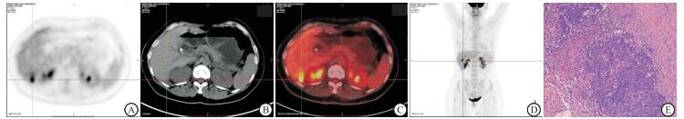
病人女,42岁,卵巢癌病人术后20余月,血CA125:77.40 IU/mL,肝脏CT三期增强示肝脏右后叶小结节影,无明显强化;PET、CT、MIP图和PET/CT融合图像十字交叉线位置显示:肝脏右后叶包膜下FDG代谢异常增高灶(见图 1A~D),考虑肝脏转移。肝脏右后叶术后病理示:肝脏转移性腺癌。免疫组织化学结果:瘤细胞ER(1+)、PR(-)、AFP(-)、P53(3+)、CA125(3+)、hetoatocvte(-)、Ki-67(+,80%),结合免疫组织化学标记,考虑为卵巢浆液性乳头状腺癌肝转移(见图 1E)。
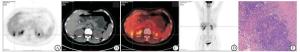
图 1 典型病例影像学检查和病理检查
-
卵巢癌是妇科常见的恶性肿瘤,即使得到了有效治疗达到治疗后的完全缓解,仍有50%~80%的病人可能会转移或复发[3]。本研究显示32例中最终证实26例为转移或复发,占81.3%,略高于80%,可能是因为我们选取的病人在随访过程中血CA125均高于正常参考值所致。卵巢癌转移或复发特征为多发部位或器官,可能会出现在盆腔局部,如阴道和盆腔;远处如腹主动脉旁淋巴结、肝脏、双肺和脑,其中以盆腔部位和淋巴结器官转移最多见。本研究中转移灶的分布位置:其中20例盆腔转移,为最多见转移部位,其次为腹腔和胸腔12例;25例以淋巴结转移,为最多见的转移器官;伴肺转移3例,伴肝脏、脾脏和骨转移各1例。未发现脑转移病人。多数病人在治疗后3年易于复发或转移,因此病人癌术后的定期随访是十分重要[4]。
血CA125检测可以作为卵巢癌术后病情随访监测指标,但血CA125异常升高时多数病人已经出现肿瘤转移,难以实现早期诊断,这也是卵巢癌复发或转移率高和死亡率高的因素之一[5]。本研究32例病人中,其中26例发生转移或复发,且血CA125也不能够对肿瘤进行定位的判断,临床常常用其进行早期的转移或复发的筛查,当血CA125水平异常时,可以积极采取进一步影像检查来定位及定性。PET/CT可检测多数肿瘤的发生,对于已知的肿瘤病人,可用于评估肿瘤的分期、监测治疗的疗效反应和监测肿瘤病人术后或治疗后复发或转移情况,特别是传统影像学(CT或超声)等检查仍无法明确缺定诊断的病人,有利于后期病情随访[6]。有研究[7-8]证明PET/CT作为一种以功能和代谢显像为特征的技术,在对肿瘤病情全面评估方面具有无创、敏感、准确性高等优点,对卵巢癌术后肿瘤转移或复发方面显示其独特的价值,PET/CT在检测肿瘤病人的转移或复发病灶的灵敏度、准确率高[9-10]。本研究显示,PET/CT对卵巢癌术后复发或转移灶的敏感性为92.3%、准确性是90.6%,高于传统影像学检查的65.4%、62.5%,与报道[11-12]相一致。卵巢癌转移途径有局部侵犯和淋巴道转移等,且部分病灶沿肠管壁生长,由于这些小病灶具有难辨别、分布广、位置移动等特点,普通影像学检查很难发现或低估病灶范围。使用PET/CT作为卵巢癌术后的随访,由于其是全身检查,可以检查肿瘤的原发部位、了解局部转移和远处转移的小病灶,给临床医生提供治疗方案的有效依据,有利于提升卵巢癌术后转移或复发病人的预后[13]。PET/CT对于肿瘤病人的随访复发或转移,可为临床医师为病人制定治疗方案提供依据,已得到临床医师的普遍认同。
综上所述,肿瘤标记物CA125是作为卵巢癌术后转移或复发的主要反映肿瘤存在的筛查手段;PET/CT不仅可以诊断原发肿瘤,因其为全身检查,可以检查病人全身主要的脏器,对卵巢癌的术后转移或复发的诊断亦起到很大的贡献,检查卵巢癌术后病人转移效能好,值得临床应用。
18F-FDG PET/CT检测卵巢癌术后CA125升高病人转移或复发的效果分析
Application value of 18F-FDG PET/CT in the detection of metastasis or recurrence of ovarian cancer patients with CA125 increasing after operation
-
摘要:
目的探讨18F-FDG PET/CT(PET/CT)在检测卵巢癌术后血CA125升高病人转移或复发的效果。 方法回顾32例行PET/CT检查的卵巢癌术后血CA125升高疑似转移或复发的病人,行PET/CT检查与传统影像学(CT或超声)结果比较,经再次手术或穿刺活检的病理结果或结合临床及其他影像学资料随访结果确诊。 结果PET/CT示转移或复发常见部位依次是盆腔、腹腔、胸腔;转移器官有淋巴结、肺、肝、脾脏和骨骼等;其中转移或复发病灶部位SUVmax均高于正常参考值。PET/CT的敏感度及准确度均高于传统影像学(P < 0.05和P < 0.01),特异度与传统影像学差异无统计学意义(P>0.05)。 结论PET/CT检查在卵巢癌术后转移或复发的检测中具有较高的诊断效能。 Abstract:ObjectiveTo explore the value of 18F-FDG PET/CT in detecting the metastasis or recurrence of ovarian cancer patients with serum CA125 increasing after surgery. MethodsThe clinical data of 32 suspected metastasis or recurrence ovarian cancer patients with postoperative CA125 level increasing were retrospectively analyzed.The results of 18F-FDG PET/CT were compared with traditional imaging(CT or ultrasound).The diagnosis results of patients were confirmed by the pathological examination of reoperation or puncture biopsy, and following-up data of clinical and imaging data. ResultsThe results of 18F-FDG PET/CT showed that the common sites of metastasis or recurrence were pelvis, abdominal cavity and thorax in turn, and the metastasis organs included the lymph node, lung, liver, spleen and bone.The SUVmax value of metastatic or recurrent lesions was higher than the normal reference value.The sensitivity and accuracy of 18F-FDG PET/CT were higher than those of traditional imaging(P < 0.05 and P < 0.01), but the difference of the specificity between two methods was not statistically significant(P>0.05). Conclusions18F-FDG PET/CT has a high diagnostic efficacy in the metastasis or recurrence of ovarian cancer after operation. -
Key words:
- computed tomography /
- ovarian cancer /
- metastasis or recurrence /
- traditional imaging
-
[1] PERRONE AM, DONDI G, LIMA GM, et al.Potential prognostic role of 18F-FDG PET/CT in invasive epithelial ovarian cancer relapse.A preliminary study[J].Cancers, 2019, 713(11):1. [2] 刘丽君, 杜丹丽.卵巢浆液性癌54例临床分析[J].蚌埠医学院学报, 2014, 39(5):609. [3] CENGIZ A, KO ZP, KARA P, et al.The role of 18F-FDG PET/CT in detecting ovarian cancer recurrence in patients with elevated CA-125 levels[J].Mol Imaging Radionucl Ther, 2019, 28(1):8. doi: 10.4274/mirt.galenos.2018.00710 [4] 龚静, 刘陶, 王雅琴, 等.CEA及CA125检测与PET/CT显像在诊断卵巢癌中的价值[J].实用妇产科杂志, 2016, 32(12):946. [5] 覃春霞, 韩娜, 张永学, 等.卵巢混合性生殖细胞瘤18F-FDG PET/CT影像诊断思路探讨[J].中华核医学与分子影像杂志, 2017, 37(4):234. [6] 张倩, 辛军, 曹礼, 等.影像表现与病理对照分析[J].中华核医学与分子影像杂志, 2017, 37(8):460. [7] CHUNG HH, KIM W, PARK NH, et al.Prognostic importance of peritoneal lesion-to-primary tumour standardized uptake value ratio in advanced serous epithelial ovarian cancer[J].Eur Radiol, 2018, 28(5):2107. doi: 10.1007/s00330-017-5194-0 [8] TAKAGI H, SAKAMOTO J, OSAKA Y, et al.Utility of 18F-fluorodeoxyglucose-positron emission tomography in the differential diagnosis of benign and malignant gynaecological tumours[J].J Med Imag Rad Oncol, 2018, 62(4):471. doi: 10.1111/1754-9485.12707 [9] 卓小丽, 李诗运, 戴儒奇, 等.18 F-FDG PET/CT显像与血清CA125水平的关系及在老年巢癌早期诊断、分期及术后随访中的价值[J].中国老年学杂志, 2017, 37(20):5070. [10] KIM CK, PARK BK, CHOI JY, et al.Detection of recurrent ovarian cancer at MRI:comparison with integrated PET/CT[J].Comput Assist Tomogr, 2007, 31(6):868. doi: 10.1097/rct.0b013e31803e8c45 [11] 伍日照, 黄斌豪, 邹伟强, 等.PET/CT显像在卵巢癌患者术后复发与转移中的应用效果[J].中国当代医药, 2019, 12(26):112. [12] AMIT A, HODES A, LAVIE O, et al.The role of F18-FDG PET/CT in predicting secondary optimal de-bulking in patients with recurrent ovarian cancer[J].Surg Oncol, 2017, 26(4):347. doi: 10.1016/j.suronc.2017.07.004 [13] 唐丽媛, 谢力, 王晓囡, 等.血清HE4、CA125和CA19-9检测在卵巢肿瘤诊断中的临床意义[J].中国妇幼保健, 2015, 30(15):2352. -

 点击查看大图
图(1)
计量
- 文章访问数: 4914
- HTML全文浏览量: 2191
- PDF下载量: 17
- 被引次数: 0
