


-
目前,新型冠状病毒疾病(coronavirus disease 2019, COVID-19)的爆发引起了全世界广泛关注[1]。由于新型冠状病毒(2019 novel coronavirus, 2019-nCov)可人传人进行传播[2-4],所以,早期快速筛查并将可疑或确诊的COVID-19病人进行隔离是目前公认的重要防控措施。诊断COVID-19的金标准为呼吸道或血液标本中2019-nCov核酸阳性[5]。由于部分病人早期可表现为多次2019-nCov核酸阴性[6],为早期快速诊断带来一定的困难,不利于2019-nCov传播的防控。多项研究[2, 7-9]表明,COVID-19病人出现发热、干咳、乏力,肺CT影像表现具有一定的特异性,为COVID-19早期诊断提供了可靠的依据。但是,并非每位病人的影像表现都比较典型,也有部分病人病灶局限累及1~2叶,单发或非胸膜下,对于这些非典型病人的影像在年龄分布上有无特点,是本研究关注的方向之一。本文就不同年龄组的临床和CT影像特点进行探讨。
-
收集蚌医一附院、安医一附院、浙江同德医院2020年1月24日至2月11确诊的符合新型冠状病毒感染的肺炎诊疗方案(试行第五版)COVID-19病人(含38例有疫区旅居史者)临床及CT影像资料,所有病人均评估初次CT影像资料。107例中男61例,女46岁,年龄7~89岁。入选标准:(1)呼吸道或血液标本中2019-nCov核酸阳性;(2)临床及初次CT影像资料完善。排除标准:(1)图像质量不佳(比如呼吸不配合病人),不能满足纹理分析要求;(2)肺CT未发现明显异常。收集临床基本资料(性别,年龄),临床症状(有无发热,咳嗽,乏力,头晕、头疼,胸闷、胸痛,呼吸困难,全身酸痛或其它),实验室检查[有无C-反应蛋白(CRP)增高,白细胞异常,淋巴细胞减低]及有无疫区旅居史[5]。根据不同年龄段将COVID-19病人分为3组(见图 1): < 30岁组,30~60岁组和>60岁组。将COVID-19分为轻或普通型和重或危重型[5],24例重型或危重型病人的年龄频率分布见图 2。
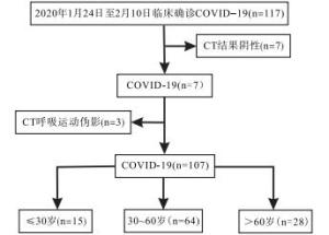
图 1 3组COVID-19的入组流程图

图 2 24例重型和危重型病人年龄频率分布图(柱形图代表相应年龄段和发生的频率,红色曲线图代表重型或危重型病人的分布趋势)
-
所有病人均使用美国Light Speed 64排螺旋CT(VCT)、日本东芝Aquilion 64层和德国西门子公司Emotion 16排扫描仪行高分辨CT扫描,扫描参数:120 kV,35 mAs,螺距1.2,视野320 mm×320 mm,采集矩阵512×512,重建层厚:0.625~1.25 mm。所有图像传送至PACS工作站(ADW4.6 GE),由2名胸组放射科医生解读,如有异议,经商议后取得一致意见。分别在肺窗(窗宽1 500~1 600,窗位-600~-800),纵隔窗(窗宽400~500,窗位20~40)进行观察病灶。首先,在肺窗观察病灶位置(是否为胸膜下),累及肺叶,形态(磨玻璃影,结节影),并观察是否合并晕征、反晕征、铺路石征、筛孔征、腔征(空泡或空腔)、树芽征、间质受累、血管扩张和支气管扩张。在纵隔窗观察肺内病灶是否存在实性成分、有无胸腔积液、胸膜增厚及淋巴结肿大。
-
采用方差分析和χ2检验。
-
轻或普通型83(77.6%)例,重或危重型24(22.4%)例,重型或危重型频率峰值出现在60~80岁。临床症状典型表现为发热105例(98.1%),咳嗽83例(77.6%),部分可表现为乏力25例(23.3%),头晕、头疼12例(11.2%),胸闷、胸痛23例(21.5%),呼吸困难14例(13.1%),全身酸痛10例(9.3%)。实验室检查CRP增高68例(63.6%,),WBC异常45例(42.1%)(其中3例升高,42例减低),淋巴细胞减低47例(43.9%)。
-
以多发病灶[71(66.4%)]、磨玻璃影[101(94.4%)]、胸膜下病灶[101(94.4%)]、晕征[84(78.5%)]及血管扩张[96(89.7%)]为典型表现,可表现为结节样[18(16.8%)]改变(见图 3、4);部分病灶内见实性成分[62(57.9%)]、反晕征[7(6.5%)]、铺路石征[37(34.6%)]、筛孔征[12(11.2%)]、腔征[11(10.3%)]、树芽征[3(2.8%)]、支气管扩张[47(43.9%)];23例(21.5%)见胸膜增厚,9例(8.4%)见少量胸腔积液,7例(6.5%)见淋巴结肿大。
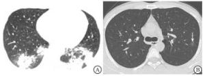
图 3 典型和非典型CT表现
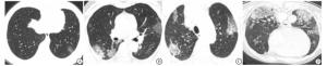
图 4 典型CT表现
-
107例COVID-19病人中,呼吸困难多见于>60岁组,差异有统计学意义(P < 0.01);白细胞异常病人中除3例表现为白细胞升高外,其余均减低。轻或普通型多见于 < 30岁组、30~60岁组,重或危重型多见于>60岁组,差异有统计学意义(P < 0.01)(见表 1)。
特征 n < 30岁组
(n=15)30~60岁组
(n=64)>60岁组
(n=28)χ2 P 临床表现 发热 105 14 63 28 2.45 >0.05 咳嗽 83 10 50 23 1.37 >0.05 乏力 25 3 14 8 0.60 >0.05 头晕、头疼 12 3 6 3 1.39 >0.05 胸闷、胸痛 23 2 12 9 2.76 >0.05 呼吸困难 14 0 5 9▲■■ 12.77 < 0.01 全身酸痛 10 1 6 3 0.19 >0.05 其它 11 1 7 3 0.25 >0.05 实验室检查 CRP增高 68 4 42 22 11.66 >0.05 白细胞异常 45 3 32 10 5.11 >0.05 淋巴细胞减低 47 4 31 12 2.36 >0.05 临床类型 轻或普通型 83 14 53 16▲■■ 9.87 < 0.01 重或危重型 24 1 11 12 疫区旅居史 38 8 24 6 4.62 >0.05 与 < 30岁组比较▲P < 0.05,▲▲P < 0.01;与30~60岁组比较■P < 0.05,■■P < 0.01 表 1 COVID-19病人临床资料比较(n)
-
3组COVID-19病人,年龄越大组更常见多肺叶累及,差异有统计学意义(P < 0.01);本文中胸膜增厚均为轻度,胸腔积液除1例中等量外,其余均为微量积液。单发非胸膜下病灶: < 30岁组3例(20%,3/15),30~60岁组1例,>60岁组0例。铺路石征、实性成分多见于>60岁组,差异有统计学意义(P < 0.01)(见表 2)。
特征 n < 30岁组
(n=15)30~60岁组
(n=64)>60岁组
(n=28)χ2 P CT表现(肺窗)肺叶分布 1~2叶 36 12 21▲▲ 3▲▲■■ 21.05 < 0.01 多叶 71 3 43 25 形态 磨玻璃影 101 13 60 28 3.40 >0.05 结节影 18 4 10 4 1.23 >0.05 胸膜下病灶 101 13 60 28 3.40 >0.05 征象 晕征 84 10 50 24 2.11 >0.05 反晕征 7 1 5 1 0.57 >0.05 铺路石征 37 1 20 16▲▲■ 11.78 < 0.01 筛孔征 12 1 6 5 1.77 >0.05 腔征 11 1 7 3 0.25 >0.05 树芽征 3 1 1 1 1.24 >0.05 间质受累 血管扩张 96 12 57 27 2.93 >0.05 支气管扩张 47 4 27 16 3.88 >0.05 CT表现(纵隔窗) 实性成分 62 6 32 24▲▲■■ 12.50 < 0.01 胸膜增厚** 23 1 14 8 2.79 >0.05 胸腔积液** 9 1 3 5 4.45 >0.05 淋巴结肿大 7 1 3 3 1.16 >0.05 与 < 30岁组比较▲P < 0.05,▲▲P < 0.01;与30~60岁组比较■P < 0.05,■■P < 0.01 表 2 COVID-19病人CT影像资料分析(n)
-
107例COVID-19病人中35.5%(38/107)有武汉旅居史,典型的临床表现为发热、咳嗽,少见的有乏力、头晕、头疼、胸闷、胸痛、呼吸困难等,除咳嗽较SONG等[8]报道比率高以外(77.6%,83/107;47%,27/51),其它临床表现与先前的文献[8, 10-11]报道相似。本文呼吸困难在>60岁组病人的发生率相对较高,与>60岁组病人年龄偏大,重型危重型病人偏多密切相关,也从侧面反映了老年人对2019-nCoV抵抗力差,肺部受累后换气功能下降,血氧含量减低,诱发组织器官缺氧,病人通过加快呼吸代偿,时间较久极易引发呼吸困难。
CT上COVID-19病人典型的表现为两肺散在的磨玻璃影,通常病灶累及多个肺叶,胸膜下最常见,病灶周围常伴有血管扩张和晕征,其次支气管扩张和铺路石征也较为常见,这与目前研究[7, 9, 12-15]基本相符。值得注意的是本组有4例单非胸膜下病灶,其中3例(20%,3/15)为 < 30岁组病人,影像学表现不典型,容易被误认为一般磨玻璃结节影,而影响病人早期诊断,同时也不利于疫情的防控,对年轻非典型表现病人应给予高度关注,建议隔离观察和密切随访。磨玻璃影的形成可能与病毒率先累及胸膜下血管支气管造成阻塞、扩张、渗出,出现片状炎症反应,炎症进展向周围不均匀性扩散致使病灶扩大,边缘密度增高形成晕征有关,随着时间延长病灶内部小叶间隔增厚形成铺路石征,若疾病继续进展则致使病灶实性成分增多。3组病人间的病灶累及范围随着不同组病人年龄的差异而明显不同,病灶局限于1~2叶者多见于年轻的 < 30岁组病人,病灶散在弥漫分布在>60岁组病人最为常见,出现这一现象的主要原因可能与不同组病人免疫力有关。>60岁组病人的铺路石征和实性成分最为多见,推测原因可能与不同年龄组病人对病毒的敏感性不同, < 30岁组病人年轻对外来病毒的刺激敏感性高、调动肌体的免疫应答机制迅速,再加上病人因年龄优势而抵抗力强,致使病毒容易被早期识别、鉴定、攻击和查杀,限制了疾病进展,容易使病灶局限于一或二叶内,也是这组病人多为轻型和普通型的原因之一,>60岁组病人年龄偏大,对外来异类病毒的识别、查杀能力弱,病毒的查杀时间长,容易出现扩散蔓延,而长时间不愈则会出现病灶内间质水肿、小叶间隔增厚和实性成分增多,可能也是铺路石征和实性占比在本组病人中比例较高的原因之一。
本研究存在以下局限性:(1)本研究样没有对COVID-19病人的疾病进展影像进行动态评估;(2)本研究着重对COVID-19病人不同年龄组的影像差异进行研究,未能探讨不同分型病人CT影像是否存在差异及研究哪些CT影像可以用来预测和评估病人预后,是后续研究方向;(3)本研究只是对2019-nCoV病毒感染的CT影像进行分析,没有和HN、SARS等病毒性肺炎进行CT影像鉴别分析。
综上所述,通过对107例COVID-19病人的临床和CT影像资料评估,发现典型的病人临床表现为发热、咳嗽;CT表现为两肺多发的胸膜下磨玻璃影,常伴晕征、血管和支气管扩张。不同年龄组间的临床和CT影像均具有一定特征性,病灶局限或单发多见,尤其是单发非胸膜下结节应引起高度重视。
不同年龄段新型冠状病毒肺炎的临床和CT影像特征分析
Clinical and CT findings of coronavirus disease 2019 in the different age groups
-
摘要:
目的分析不同年龄组间新型冠状病毒肺炎(coronavirus disease 2019,COVID-19)临床和CT影像特征的差异性。 方法回顾性分析107例经临床证实COVID-19病人的临床和CT资料,依据不同年龄段分为3组(< 30岁组,30~60岁组,>60岁组),对每组病人CT特点及临床资料进行分析。 结果107例COVID-19的典型临床表现为发热(98.1%,105/107)和咳嗽(77.6%,83/107),实验室检查C反应蛋白升高(63.6%,68/107),白细胞和淋巴细胞减低(42.1%,45/107;43.9%,47/107);典型CT表现为两肺多叶散在的胸膜下磨玻璃影(94.4%,101/107),最常伴有血管扩张(89.7%,96/107)和晕征(78.5,84/107)。呼吸困难和临床分型在 < 30岁组、>60岁组和30~60岁组、>60岁组之间差异有统计学意义(P < 0.05)。3组间的肺叶分布差异最为明显(P < 0.05)。铺路石征和实性成分在>60岁组和 < 30岁组、30~60岁组之间差异有统计学意义(P < 0.05)。 结论COVID-19的临床表现、实验室检查和CT表现具有一定特征性,但不同年龄组间又各具特点,掌握这些临床和CT影像特征、特点有助于早期明确诊断。 -
关键词:
- 新型冠状病毒肺炎 /
- 临床特征 /
- 体层摄影术,X线计算机
Abstract:ObjectiveTo analyze the differences of the clinical and CT imaging features of coronavirus disease 2019 (COVID-19) in the different age groups. MethodsThe clinical and CT data of 107 patients with clinically confirmed covid-19 were retrospectively analyzed and divided into three groups (< 30 years old group, 30-60 years old group, and >60 years old group) according to their ages.The CT findings and clinical data were analyzed in three groups. ResultsThe typical clinical symptoms of COVID-19 in 107 patients showed fever (98.1%, 105/107) and cough (77.6%, 83/107).Laboratory examination showed increased C-reactive protein (63.6%, 68/107), and decreased white blood cells and lymphocytes (42.1%, 45/107;43.9%, 47/107).The typical CT findings displayed scattered subpleural ground-glass opacity in two lungs (94.4%, 101/107), most commonly accompanied with vasodilation (89.7%, 96/107) and halo sign (78.5%, 84/107).Dyspnea and clinical type were statistically significant between < 30 years old group, >60 years old group and 30-60 years old group, >60 years old group (P < 0.05).The difference of distribution of lung lobes in three groups was significant (P < 0.05).There were significant differences in paving stone sign and solid components between >60 years old group, < 30 years old group and 30-60 years old group (P < 0.05). ConclusionsThe clinical manifestations, laboratory examination and CT findings of COVID-19 have some characteristics, but different age groups possess their own characteristics.Grasping characteristics of these clinical and CT imaging features is helpful for early definite diagnosis. -
Key words:
- coronavirus disease 2019 /
- clinical features /
- tomography, X-ray computed
-
表 1 COVID-19病人临床资料比较(n)
特征 n < 30岁组
(n=15)30~60岁组
(n=64)>60岁组
(n=28)χ2 P 临床表现 发热 105 14 63 28 2.45 >0.05 咳嗽 83 10 50 23 1.37 >0.05 乏力 25 3 14 8 0.60 >0.05 头晕、头疼 12 3 6 3 1.39 >0.05 胸闷、胸痛 23 2 12 9 2.76 >0.05 呼吸困难 14 0 5 9▲■■ 12.77 < 0.01 全身酸痛 10 1 6 3 0.19 >0.05 其它 11 1 7 3 0.25 >0.05 实验室检查 CRP增高 68 4 42 22 11.66 >0.05 白细胞异常 45 3 32 10 5.11 >0.05 淋巴细胞减低 47 4 31 12 2.36 >0.05 临床类型 轻或普通型 83 14 53 16▲■■ 9.87 < 0.01 重或危重型 24 1 11 12 疫区旅居史 38 8 24 6 4.62 >0.05 与 < 30岁组比较▲P < 0.05,▲▲P < 0.01;与30~60岁组比较■P < 0.05,■■P < 0.01  下载: 导出CSV
表 2 COVID-19病人CT影像资料分析(n)
特征 n < 30岁组
(n=15)30~60岁组
(n=64)>60岁组
(n=28)χ2 P CT表现(肺窗)肺叶分布 1~2叶 36 12 21▲▲ 3▲▲■■ 21.05 < 0.01 多叶 71 3 43 25 形态 磨玻璃影 101 13 60 28 3.40 >0.05 结节影 18 4 10 4 1.23 >0.05 胸膜下病灶 101 13 60 28 3.40 >0.05 征象 晕征 84 10 50 24 2.11 >0.05 反晕征 7 1 5 1 0.57 >0.05 铺路石征 37 1 20 16▲▲■ 11.78 < 0.01 筛孔征 12 1 6 5 1.77 >0.05 腔征 11 1 7 3 0.25 >0.05 树芽征 3 1 1 1 1.24 >0.05 间质受累 血管扩张 96 12 57 27 2.93 >0.05 支气管扩张 47 4 27 16 3.88 >0.05 CT表现(纵隔窗) 实性成分 62 6 32 24▲▲■■ 12.50 < 0.01 胸膜增厚** 23 1 14 8 2.79 >0.05 胸腔积液** 9 1 3 5 4.45 >0.05 淋巴结肿大 7 1 3 3 1.16 >0.05 与 < 30岁组比较▲P < 0.05,▲▲P < 0.01;与30~60岁组比较■P < 0.05,■■P < 0.01
下载: 导出CSV
-
[1] World Health Organization.WHO Director-General's remarks at the media briefing on 2019-nCoV on 11 February 2020[EB/OL].[2020-02-11].https: //www.who.int/dg/speeches/detail/who-director-general-s-remarks-at-the-media-briefing-on-2019-ncov-on-11-february-2020. [2] CHAN JFW, YUAN S, KOK KH, et al.A familial cluster of pneumonia associated with the 2019 novel coronavirus indicating person-to-person transmission:a study of a family cluster[J].The Lancet, 2020.DOI:10.1016/s0140-6736(20)30154-9. [3] LI Q, GUAN X, WU P, et al.Early transmission dynamics in Wuhan, China, of novel coronavirus-infected pneumonia[J].N Engl J Med, 2020.DOI:10.1056/NEJMoa2001316. [4] BENVENUTO D, GIOVANETTI M, CICCOZZI A, et al.The 2019-new coronavirus epidemic:evidence for virus evolution[J].J Med Virol, 2020.DOI:10.1002/jmv.25688. [5] 国家卫生健康委办公厅, 国家中医药管理局办公室.关于印发新型冠状病毒肺炎诊疗方案(试行第六版修正版)的通知[EB/OL].http://yzs.satcm.gov.cn/zhengcewenjian/2020-02-19/13222.html-2020-02-19. [6] 中华医学会放射学分会.新型冠状病毒肺炎的放射学诊断:中华医学会放射学分会专家推荐意见(第一版)[J].中华放射学杂志, 2020, 54:E001. [7] 高璐, 张静平, 杜永浩, 等.输入性新型冠状病毒肺炎的CT表现[J].西安交通大学学报(医学版), 2020:1. [8] SONG F, SHI N, SHAN F, et al.Emerging coronavirus 2019-nCoV pneumonia[J].Radiology, 2020:200274.DOI:10.1148/radiol.2020200274. [9] CHUNG M, BERNHEIM A, MEI X, et al.CT imaging features of 2019 novel coronavirus (2019-nCoV)[J].Radiology, 2020:200230.DOI:10.1148/radiol.2020200230. [10] WANG D, HU B, HU C, et al.Clinical characteristics of 138 hospitalized patients with 2019 novel coronavirus-infected pneumonia in Wuhan, China[J].JAMA, 2020.DOI:10.1001/jama.2020.1585. [11] 徐鹤, 李淑华, 赵灿灿, 等.新型冠状病毒肺炎32例临床表现及影像学特征初探[J].蚌埠医学院学报, 2020, 45(2):150. [12] KOO HJ, LIM S, CHOE J, et al.Radiographic and CT features of viral pneumonia[J].Radiographic, 2018, 38(3):719. doi: 10.1148/rg.2018170048 [13] XU YH, DONG JH, AN WM, et al.Clinical and computed tomographic imaging features of novel coronavirus pneumonia caused by SARS-CoV-2[J].J Infect, 2020, 80(4):394. [14] SHI H, HAN X, JIANG N, et al.Radiological findings from 81 patients with COVID-19 pneumonia in Wuhan, China:a descriptive study[J].Lancet Infect Dis, 2020, 20(4):425. [15] WANG J, XU Z, WANG J, et al.CT characteristics of patients infected with 2019 novel coronavirus:association with clinical type[J].Clin Radiol, 2020, 75(6):408. doi: 10.1016/j.crad.2020.04.001 -

 点击查看大图

图(4)表(2)
计量
- 文章访问数: 4501
- HTML全文浏览量: 2216
- PDF下载量: 15
- 被引次数: 0
