

 下载:
下载:
-
近几年来,随着产检的规范化以及产前超声筛查和诊断技术的不断进步,越来越多的胎儿泌尿系畸形在产前被发现,其中胎儿肾盂扩张占大部分,占80%~87%[1]。当产前系统超声检查提示胎儿肾盂扩张,但未见其他结构畸形或其他超声软指标异常时,则称为孤立性肾盂扩张,其临床预后及产前咨询值得重视和关注。2019年4月《中华医学遗传学》刊发了《低深度全基因组测序技术在产前诊断中的应用专家共识》[2],指出染色体拷贝数变异测序(CNV-seq)可以作为一线产前诊断技术对可能存在胎儿染色体异常的孕妇进行产前诊断。本研究回顾性分析76例孤立性肾盂扩张行羊水穿刺的胎儿,对羊水CNV-seq结果进行分析,以期为胎儿孤立性肾盂扩张提供重要的产前咨询依据。现作报道。
-
选择2016年11月至2018年12月在海南省人民医院经超声诊断的孤立性胎儿肾盂扩张病例76例,所有胎儿均为单胎,平均孕(22.0±4.0)周,孕妇平均年龄28岁。
-
肾盂前后径(anteroposterior diameter, APD)是肾盂扩张最敏感的指标。本文采用STOCKS等[3]提出的肾盂扩张诊断标准,并结合国内相关文献[4]:APD 15~20周≥0.4 cm,21~30周≥0.5 cm,31~40周≥0.7 cm,40周以上≥1.0 cm为肾盂扩张,若肾盂分离值≥1.5 cm可诊断肾盂积水。
-
采用美国GE公司的Voluson E8型彩色多普勒超声显像仪对胎儿各系统器官进行超声检查,如发现胎儿有肾盂扩张,在腹部横断面,显示双肾,测量APD,并观察双肾大小及肾皮质厚度。
-
所有入组孕妇根据妊娠周数,知情同意后在ALOKA5500SV型超声仪穿刺探讨引导下行羊膜腔穿刺术,抽取羊水20~30 mL进行染色体核型分析及低深度全基因组测序分析(染色体拷贝数变异测序技术,CNV-seq)。染色体核型分析:常规培养羊水细胞、制片、G显带,显微镜下检查20个核型。CNV-seq:通过离心法收集羊水细胞,将约50 ng羊水细胞DNA打碎,通过末端标记法、接头连接和PCR扩增的方式建立DNA文库,应用CNV-seq分析羊水细胞DNA,检测染色体拷贝数变异,分辨率为0.1 Mb。根据Decipher、DGV、千人基因组以及OMIM等公共数据库对多态性拷贝数变异(CNV)进行查询,然后根据美国医学遗传学学会制定的指南对致病性进行评估解读[5]。拷贝数变异分为致病性、可能致病性、致病性未知、可能良性或良性。CNV-seq分析由北京贝瑞和康生物技术有限公司完成
-
76例孤立性肾盂扩张胎儿的染色体核型分析仅1例唐氏综合征,并且该孕妇无创产前检测(non-invasive prenatal testing, NIPT)的结果为21-三体高风险;1例9号染色体倒位,为染色体多态。76例胎儿CNV-seq 1例为唐氏综合征(NIPT结果为21-三体高风险)(见图 1), 2例结果为CNV,其余均未发现致病性、可能致病性或致病性未知的CNV。
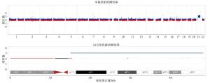
图 1 CNV-seq结果提示染色体21-三体
-
76例肾盂扩张胎儿均随访成功,其中男51例,女25例,男:女=2.04:1。2例引产,其中1例因唐氏综合征引产,另1例因超声复查提示胎儿双肾重度积水(APD>15 mm)引产;其余74例活产,平均出生孕周39.2周,剖宫产25例,顺产49例,所有新生儿均随访至出生后1个月,复查B超未见有肾盂扩张加重或出现肾积水。
-
随着产前超声的规范化,胎儿肾盂扩张成为产前较为常见的超声软指标异常,在所有胎儿中的发生率为1%~5%[6-7]。妊娠16周后超声检查即可以显示胎儿肾脏。胎儿肾盂扩张根据病因分为病理性和生理性,并且大部分为生理性。生理性肾盂扩张为一过性,可随着胎儿生长发育而自然消退,无需处理。一般认为与以下因素有关:胎儿泌尿系统发育不成熟、胎儿生理性尿量增多、孕妇在超声检查前大量饮水、妊娠期高孕激素状态抑制输尿管蠕动等;病理性肾盂扩张的病因包括泌尿道梗阻、后尿道瓣膜、异位输尿管疝、重复肾或全身多系统畸形等,常伴有肾积水、肾皮质改变、巨膀胱等异常。
产前筛查中通常将肾盂扩张作为一种超声软指标异常,可合并有其他超声软指标异常,或提示胎儿染色体非整倍体风险增高。关于胎儿肾盂扩张与染色体非整倍体的关系,此前已有多篇文献进行了探讨。HAVUTCU等[8]报道,301例孤立性胎儿肾盂扩张行产前诊断,结果未发现一例染色体非整倍体,因此认为孤立性胎儿肾盂扩张与染色体非整倍体关联性不大。COCO等[9]对366例肾盂扩张的胎儿进行产前诊断,发现在83.3%孤立性肾盂扩张和16.7%合并有其他结构畸形的胎儿中,各有1例唐氏综合征,并得出孤立性肾盂扩张筛查唐氏综合征的阴性预测值为99.9%,而阳性预测值仅为0.33%,因此该研究认为孤立性肾盂扩张不应该作为介入性产前诊断指征。国内的商梅娇等[10]对122例胎儿肾盂扩张进行介入性产前诊断分析,发现在67例孤立性肾盂扩张胎儿中,无一例染色体非整倍体,因此,同样认为孤立性肾盂扩张不应该直接作为产前诊断的指征。
然而,上述报道均仅进行了染色体核型分析,即G显带染色体核型分析,其分辨率为10 Mb。而随着分子生物技术的发展,近年来的介入性产前诊断除了进行染色体核型分析外,染色体的拷贝数变异检测亦成为常规,这其中就包括CNV-seq,基于高通量测序技术的全基因组染色体拷贝数变异检测,其分辨率达0.1 Mb,即可以检测0.1 Mb以上的染色体微缺失微重复。并且CNV-seq技术无需细胞培养,操作简单,所需羊水样本量低。WANG等[11]对3 249例羊水样本进行前瞻性的CNV-seq分析,认为与染色体核型分析相比,CNV-seq技术能额外检出1%的明确致病性CNV,并指出CNV-seq可以作为一线诊断技术对可能存在胎儿染色体异常的孕妇进行产前诊断。在《专家共识》中同样指出,CNV-seq可以作为一线产前诊断技术[2]。
那么,应用CNV-seq是否能额外检出除染色体非整倍体外的染色体微缺失微重复,此前鲜有报道。本研究回顾性分析了76例胎儿孤立性肾盂扩张的CNV-seq结果,结果显示,76例孤立性肾盂扩张胎儿,除1例因NIPT提示21-三体高风险行CNV-seq结果为唐氏综合征,其余75例CNV-seq结果均未见致病性、可能致病性或致病性未知的CNV,而且75例染色体核型分析除1例9号染色体倒位外,也未见染色体核型异常,因此,本研究认为如果唐筛或NIPT低风险,孤立性肾盂扩张不需要常规行产前CNV-seq分析,本研究支持孤立性肾盂扩张不应该作为产前诊断指征,除非孕妇唐氏筛查或NIPT提示高风险。另外,本研究76例孤立性肾盂扩张胎儿中,除1例21-三体、1例因双肾重度积水引产外,其余74例活产儿出生后随访1个月未见有明显的泌尿系统并发症。刘洪国等[12]用超声随访了5 988个胎儿肾脏,发现大部分胎儿肾积水是一个可自行恢复相对良性的过程,一般预后良好,因此,本研究认为胎儿孤立性肾盂扩张大部分预后良好,但需定期超声随诊观察。
一个有趣的现象是,本研究中肾盂扩张的男女胎儿比例为2.04:1,这与此前的报道类似。商梅娇等[10]报道的比例为4.35:1,COCO等[9]研究中的比例为1.9:1,BROADLEY等[13]报道双侧肾盂积水的胎儿男女比例为2.4:1。由此可见,在肾盂扩张的胎儿中,男孩比例明显高于女孩,值得临床重视。
本研究的不足之处是样本量较小且为单中心研究,研究结果可能存在一定的偏倚,尚需扩大样本数进行多中心研究。但本研究对肾盂扩张采用了统一的诊断标准,由专人及专用超声仪器进行胎儿系统超声检查,在一定程度上避免了研究误差。综上所述,胎儿孤立性肾盂扩张绝大部分预后良好,除非孕妇唐氏筛查或NIPT高风险,产前无需常规行CNV-seq分析,但需定期超声随诊观察。
孤立性肾盂扩张胎儿全基因组染色体拷贝数变异测序结果分析
Analysis of the sequencing results of whole genome chromosome copy number variation in isolated fetal pyelectasia
-
摘要:
目的探讨胎儿孤立性肾盂扩张染色体拷贝数变异测序(CNV-seq)结果及预后。 方法对76例孤立性肾盂扩张胎儿进行介入性产前诊断,回顾性分析其染色体核型及CNV-seq结果,随访妊娠结局。 结果76例孤立性肾盂扩张胎儿的染色体核型分析仅1例唐氏综合征,1例9号染色体倒位。76例胎儿CNV-seq分析同样仅有1例为唐氏综合征,2例为多态性拷贝数变异(CNV),其余均未发现致病性、可能致病性或致病性未知的CNV。76例肾盂扩张出生胎儿中男孩51例,女孩25例。2例引产,其中1例因唐氏综合征引产,另1例因胎儿双肾重度积水引产。其余74例活产复查B超未见有肾盂扩张加重或肾积水。 结论胎儿孤立性肾盂扩张绝大部分预后良好,除非孕妇唐氏筛查或无创产前检测高风险,产前无需常规行CNV-seq分析。 Abstract:ObjectiveTo investigate the results of copy number variation sequencing(CNV-seq) and prognosis of isolated fetal pyelectasis. MethodsThe interventional prenatal diagnosis was performed in 76 fetuses with isolated pyelectasia, and the data of chromosomal karyotypes and CNV-seq were retrospectively analyzed.The pregnancy outcomes were followed up. ResultsThe analysis results of chromosomal karyotype in 76 fetuses with isolated pyelectasia showed that only 1 case was with D Down's syndrome, and 1 case was with chromosome 9 inversion.The results of CNV-seq analysis in 76 fetuses showed that only 1 case was with Down's syndrome, 2 cases were with polymorphic copy number variation(CNV), and no other CNV with pathogenicity, potential pathogenicity or unknown pathogenicity was found.Among 6 cases with pyelectasia, 51 cases were boys and 25 cases were girls.Two cases were induced labour, which included 1 case with Down's syndrome and 1 case with severe fetal hydronephrosis.In the remaining 74 live births, B-ultrasonography showed no exacerbation of pyelectasia or hydronephrosis. ConclusionsThe vast majority of isolated fetal pyelectasia have a good prognosis, unless the pregnant woman is at high risk for Down's screening or noninvasive prenatal testing, and the routine prenatal CNV-seq analysis is not required. -
Key words:
- fetus /
- prenatal diagnosis /
- pyelectasia /
- copy number variation sequencing
-
[1] BLYTH B, SNYDER HM, DUCKETT JW.Antenatal diagnosis and subsequent management of hydronephrosis[J].J Urol, 1993, 149(4):693. doi: 10.1016/S0022-5347(17)36185-2 [2] 中华医学会医学遗传学分会临床遗传学组, 中国医师协会医学遗传医师分会遗传病产前诊断专业委员会, 中华预防医学会出生缺陷预防与控制专业委员会遗传病防控学组.低深度全基因组测序技术在产前诊断中的应用专家共识[J].中华医学遗传学杂志, 2019, 36(4):293. [3] STOCKS A, RICHARDS D, FRENTZEN B, et al.Correlation of prenatal renal pelvic anteroposterior diameter with outcome in infancy[J].J Urol, 1996, 155(3):1050. doi: 10.1016/S0022-5347(01)66388-2 [4] 张铁娟, 翟桂荣.胎儿肾盂扩张的超声诊断和预后的探讨[J].首都医科大学学报, 2005(5):627. [5] RICHARDS S, AZIZ N, BALE S, et al.Standards and guidelines for the interpretation of sequence variants:a joint consensus recommendation of the American College of Medical Genetics and Genomics and the Association for Molecular Pathology[J].Genet Med, 2015, 17(5):405. doi: 10.1038/gim.2015.30 [6] 朱好, 沈淳, 李笑天.胎儿单纯性肾盂扩张的结局——109例临床分析[J].中华围产医学杂志, 2015, 18(9):683. [7] JAKOBSEN TR, SOGAARD K, TABOR A.Implications of a first trimester Down Syndrome screening program on timing of malformation detection[J].Acta Obstet Gynecol Scand, 2011, 90(7):728. doi: 10.1111/j.1600-0412.2011.01156.x [8] HAVUTCU AE, NIKOLOPOULOS G, ADINKRA P, et al.The association between fetal pyelectasis on second trimester ultrasound scan and aneuploidy among 25, 586 low risk unselected women[J].Prenat Diagn, 2002, 22(13):1201. doi: 10.1002/pd.490 [9] COCO C, JEANTY P.Isolated fetal pyelectasis and chromosomal abnormalities[J].Am J Obstet Gynecol, 2005, 193(3Pt1):732. [10] 商梅娇, 周祎, 鲁云涯, 等.122例胎儿肾盂扩张与染色体非整倍体的关联性分析[J].中山大学学报(医学科学版), 2013, 34(1):99. [11] WANG J, CHEN L, ZHOU C, et al.Prospective chromosome analysis of 3429 amniocentesis samples in China using copy number variation sequencing[J].Am J Obstet Gynecol, 2018, 219(3):281. [12] 刘洪国, 初大鹏, 张格云.胎儿肾盂宽度的超声研究[J/CD].中华医学超声杂志(电子版), 2009, 6(5): 905. [13] BROADLEY P, MCHUGO J, MORGAN I, et al.The 4 year outcome following the demonstration of bilateral renal pelvic dilatation on pre-natal renal ultrasound[J].Br J Radiol, 1999, 72(855):265. doi: 10.1259/bjr.72.855.10396216 -

 点击查看大图
图(1)
计量
- 文章访问数: 4320
- HTML全文浏览量: 1912
- PDF下载量: 12
- 被引次数: 0
