


-
近年来,随着先进影像技术的进步,现代立体定向神经外科的发展趋势引人注目。顶内沟这个概念,第一次提出是在19世纪中期[1],是位于中央后沟后方并与大脑半球上缘平行的一条沟,其上方为顶上小叶,下方为顶下小叶。顶内沟和注意负荷有关[2],与其周围皮层区域和许多高级认知功能密切相关,例如空间定向和重新定向[3],在受到威胁时,顶内沟的神经活动和大脑的整体连接增加[4]。有关脑结构和功能及大脑半球不对称性的研究多聚焦于整脑表面解剖学,本研究采用脑MRI图像,构建顶内沟内侧缘的立体定位数据集并进行回归分析,以期为顶内沟及其周边区域病变的立体定向神经外科手术及未来机器人精准手术的策划提供解剖学基础。
-
成年人30名(均知情同意),男15名,女15名;年龄20~40岁,均排除了家族病史、神经及精神病史。以联合间线(AC-PC线)为基线,获得横断、矢状和冠状断层各层的T1WIMRI图像。
-
Signa1.5超导磁共振扫描仪及头颅正交线圈(美国GE公司);微型计算机(A8S,华硕公司);图像工作站: eFilm Workstation(美国Merge Med公司),以及数据测量Adobe Photoshop CS10.0软件包。
-
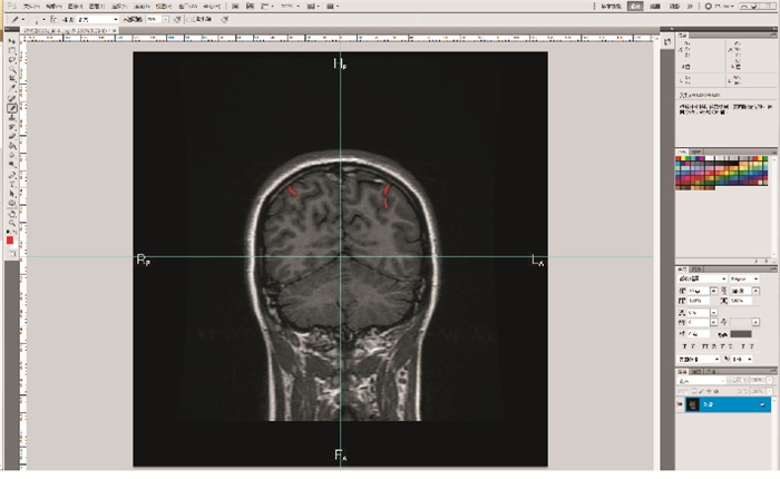
将30例头颅MRI T1WI图像以DICOM3.0的格式导入eFilm工作站,将冠状断层的图像导出保存为JPEG格式,然后将图像导入Adobe Photoshop CS系统,选择“图像”-“图像大小”,打开“图像大小”的对话框,将图像的高度和宽度均调整为240mm,分辨率调整为54.186像素/英寸,使其与图像实际参数相符,此时在Adobe Photoshop上测量的数值与eFilm的测得值一致,在顶内沟内侧缘测量其X值及Z值,Y值为所在图像距离AC-PC平面的层数乘以图像层厚的积,从而获得其三维坐标值,所有取样点的坐标组成顶内沟在三维坐标系中的立体定位数据集(见图 1)。将取样点的坐标数据集导入SPSS22.0软件中,求解出其在横断面、冠状面及矢状面上投影的回归方程。
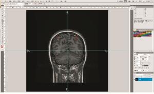
图 1 Photoshop软件中建立笛卡尔坐标系, 鼠标移到顶内沟感兴趣区,右上方显示该点的X、Z值
-
采用曲线拟合二次方程(Quadratic模型)回归分析。
-
30名健康成年人大脑T1WI MRI上,顶内沟所在的横断层MRI上共有795层,平均每例顶内沟13.22层,其中左侧大脑半球顶内沟419层(男性202层,女性217层),右侧376层(男性184层,男性192层)。男女性顶内沟各层面上的X、Z坐标值的均值及标准差(见表 1~2)。
Y/mm x±s d±sd 右侧 左侧 右侧 左侧 -20 -30.000 0±4.512 2 36.800 0±9.755 5 52.500 0±10.184 3 57.567 0±2.548 2 -22 -30.8375±3.295 4 35.200 0±7.412 4 46.357 1±9.264 8 52.583 0±11.373 6 -24 -31.333 3±4.734 2 34.671 4±6.024 6 44.437 5±6.933 3 48.686 0±11.478 6 -26 -29.700 0±3.740 6 32.680 0±7.448 0 45.350 0±7.239 9 50.330 0±10.276 0 -28 -29.923 1±4.236 4 31.300 0±7.533 6 47.646 2±7.460 6 48.477 0±8.060 6 -30 -29.820 0±5.097 7 30.133 3±5.753 0 49.433 3±7.765 3 49.253 0±8.662 3 -32 -29.806 7±4.886 2 29.773 3±5.371 7 47.066 7±7.699 9 48.227 0±8.742 5 -34 -29.086 7±4.931 5 28.980 0±4.750 8 48.586 7±7.805 0 48.287 0±8.598 3 -36 -29.533 3±5.560 7 28.860 0±4.329 3 50.573 3±6.952 1 48.233 0±8.022 6 -38 -28.586 7±5.363 5 28.233 3±5.420 3 50.593 3±7.402 2 49.673 3±7.507 9 -40 -28.420 0±5.525 8 28.020 0±7.084 8 52.833 3±9.064 0 50.253 3±7.974 2 -42 -28.133 3±6.113 1 27.060 0±6.650 7 53.576 9±7.854 1 50.740 0±8.800 7 -44 -28.010 0±6.949 9 26.014 3±7.607 0 53.827 3±8.349 0 52.357 1±8.083 8 -46 -26.544 4±2.958 1 27.135 7±7.931 3 54.790 0±7.950 1 52.535 7±8.352 5 -48 -25.037 5±3.837 4 25.515 4±6.740 8 53.587 5±7.194 9 54.453 8±7.609 4 -50 -26.300 0±2.653 3 22.725 0±5.891 3 50.325 0±4.217 7 52.162 5±6.339 4 -52 -24.800 0±2.545 6 21.916 7±4.643 0 48.550 0±3.747 7 51.333 3±3.537 6 合计 -28.949 0±5.042 3 28.546 0±6.925 5 46.487 0±7.975 0 50.095 0±7.975 0 表 1 男性顶内沟内侧缘各层取点三维坐标值(n=15)
Y/mm x±s d±sd 右侧 左侧 右侧 左侧 -16 -36.733 3±4.691 8 36.500 0±0.360 6 54.166 7±5.143 3 42.033 3±12.915 6 -18 -38.960 0±3.969 0 35.440 0±4.207 5 50.440 0±5.185 9 44.560 0±11.455 5 -20 -38.160 0±4.642 5 34.983 3±4.138 3 46.760 2±5.049 1 45.816 7±7.590 1 -22 -36.966 7±4.692 4 32.485 7±5.712 1 49.183 3±6.568 8 46.128 6±9.215 0 -24 -34.087 5±6.160 1 31.444 4±4.502 8 48.887 5±6.540 5 46.233 3±8.889 2 -26 -32.233 3±5.960 9 29.310 0±3.975 1 48.033 3±6.565 3 46.160 0±8.826 5 -28 -29.923 1±4.236 4 29.183 3±3.564 9 46.700 0±8.563 0 45.750 0±8.876 8 -30 -31.041 7±5.465 2 28.700 0±6.196 1 46.100 0±7.702 9 43.961 5±7.878 2 -32 -30.792 3±5.458 7 28.440 0±7.202 2 43.715 4±5.697 8 43.486 7±7.246 1 -34 -29.176 9±5.041 4 28.546 7±7.494 1 47.415 4±7.884 2 44.926 7±7.695 7 -36 -28.361 5±5.051 0 28.466 7±7.035 4 46.661 5±8.451 9 43.626 7±7.435 4 -38 -28.500 0±5.052 7 27.733 3±6.787 0 47.984 6±8.829 3 45.520 0±8.320 6 -40 -26.875 0±3.230 8 26.528 6±6.249 7 46.533 3±9.679 9 44.407 1±7.287 8 -42 -27.036 4±3.151 9 25.461 5±5.272 7 45.272 7±9.152 9 45.061 5±7.059 2 -44 -26.330 0±3.733 9 24.376 9±4.785 3 45.380 0±9.019 2 45.161 5±6.878 7 -46 -26.590 0±3.577 5 24.415 4±3.881 3 46.480 0±10.117 7 46.961 5±7.570 8 -48 -25.700 0±4.459 0 25.212 5±3.088 0 44.950 0±10.015 1 44.437 5±7.084 4 -50 -24.457 1±4.824 5 24.628 6±2.828 3 43.014 3±7.209 4 43.957 1±7.312 7 -52 -21.800 0±6.012 8 23.550 0±1.829 5 42.425 0±6.231 3 43.216 7±6.904 9 -54 -26.400 0±7.778 2 23.383 3±1.734 8 40.300 0±0.282 8 43.366 7±7.325 2 -56 -27.100 0±7.778 1 22.925 0±2.093 4 40.900 0±0.141 4 40.650 0±7.120 2 -58 -28.350 0±7.000 4 24.700 0±2.107 1 42.300 0±0.848 5 43.566 7±1.365 0 合计 -29.423 1±5.953 4 27.791 7±6.005 5 44.206 6±11.230 3 45.218 0±8.406 8 表 2 女性顶内沟内侧缘各层取点三维坐标值(n=15)
-
(1) 冠状面上Z值对X值回归分析,右侧:$ \hat Z = $60.646+1.114X+0.022X2(r=0.130,P < 0.05),左侧:$\hat Z = 73.409 - 1.939X - 0.032{X^2}$(r=0.155,P < 0.05)。(2)矢状面上Z值对Y值回归分析,右侧:$ \hat Z = $46.826-0.047Y-0.001Y2(r=0.032,P>0.05),左侧:$\hat Z = 36.822 - 0.480Y - 0.006{Y^2} $(r=0.077,P>0.05)。(3)横断面上Y值对X值回归分析,右侧:$\hat Z = - 53.739 - 0.752X - 0.{\rm{ }}003{X^2} $(r=0.130,P < 0.05),左侧:$\hat Z = - {\rm{ }}62.721 + 1.265X - 0.011{X^2} $(r=0.395,P < 0.05)。(4)各回归方程散点图(见图 2)。
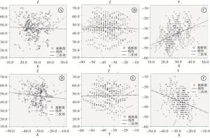
图 2 回归方程散点图
-
磁共振成像的高分辨率、高信噪比及获取任意方位断层影像等特点,促使立体定向手术更加完善和可靠[5-6]。顶内沟作为顶上小叶与顶下小叶的分界,顶下小叶又分为缘上回和角回,角回是视觉性语言区,这样的结构特征使得顶内沟位于视觉、体感和听觉联合皮层的交叉点[7],神经系统通过施加偏置信号自适应的调节正在进行的知觉、运动和认识功能来强化自上而下的控制,而顶内沟正是偏置信号的来源[8],侧脑室三角区的肿瘤选择经顶内沟入路,可以较好地保留脑皮质并不对脑的重要功能区造成损伤[9],这些都要求我们精准的识别和定位顶内沟。顶内沟位于大脑半球上外侧面,其在横断面上自前向后走形,平行于大脑纵裂,根据其形态学特征,可以分为连续型、两段型、3段型[10-11]。本研究通过对顶内沟立体定位数据集的构建,更好的对顶内沟进行识别和定位,为经顶内沟入路的手术提供解剖学基础,并且通过对顶内沟立体定位数据集的构建,我们发现左右两侧大脑顶内沟的坐标值存在非对称性,与张琪等[10]的研究结果大致相仿,在对猴脑的研究过程中[12],发现顶内沟可以分成5个部分且不同的部分对应着不同的功能,推测人脑顶内沟结构的不对称性可能也是导致功能不对称性的原因。同时,顶内沟内侧缘的立体定位数据集可以为未来机器人手术的策划提供一定的数据支持,在教育教学以及临床医生的培训等方面都有重要作用。
综上,本研究所构建的冠状面上大脑顶内沟在三维坐标系上的立体定位数据集不仅有助于大脑顶内沟的精准识别和定位,而且对顶内沟及其附近的病变的影像定位诊断及临床手术和治疗都有重要意义,对与顶内沟区域有关的疾病侧脑室三角区的手术入路和精准医学个性化治疗方案的制定方面,也具有重要的指导意义。
大脑顶内沟立体定位数据集的构建及投影回归分析
Construction of stereotactic localization data set and projection regression analysis of intraparietal sulcus
-
摘要:
目的构建大脑顶内沟基于联合间径(AC-PC)定位体系中的立体定位数据集及其平面投影回归方程。 方法将30名健康成年人颅脑横断层MRI数据经格式转化导入Photoshop CS软件包,经过严格的图像配准,建立以AC-PC中点为原点的三维立体坐标系,获得大脑冠状面图像上顶内沟的三维立体坐标值,以顶内沟最内侧点为起点,其中X值和Z值可直接在软件中读取,Y值为图像距离AC-PC平面的层数乘以图像层厚的积,所有取样点坐标值组成顶内沟在三维坐标系中的立体定位数据集。运用SPSS22.0对所获得的数据进行统计,求解其空间拟合平面回归方程。 结果成功构建大脑顶内沟在三维坐标系中的立体定位数据集及其在空间各平面上的投影回归方程。冠状面上Z值对X值回归方程右侧:$\hat{Z} $=60.646+1.114X+0.022X2,左侧:$\hat{Z} $=73.409-1.939X-0.032X2;矢状面上Z值对Y值回归方程右侧:$\hat{Z} $=46.826-0.047Y-0.001Y2, 左侧:$\hat{Z} $=36.822-0.480Y-0.006Y2;横断面上Y值对X值回归方程右侧:$\hat{Z} $=-53.739-0.752X-0.003X2,左侧:$\hat{Z} $=-62.721+1.265X-0.011X2。 结论构建的顶内沟立体定位数据集预期对该沟附近的皮质功能区定位、病灶定位及未来机器人精准手术的策划具有指导意义。 Abstract:ObjectiveTo construct the stereotatic localization data set and its plane projection regression equation of the intraparietal sulcus based on the AC-PC localization system. MethodsThe transverse tomography data of 30 healthy adults were imported into the Photoshop CS software package by format transformation.After strict image registration, a three dimensional coordinate system with AC-PC midpoint as the origin was established to obtain the three-dimensional coordinate values of intraparietal sulcus in the coronal plane images of brain.The X-value and Z-value of the sample point were recorded directly from the software, and the Y-value was the product of the number of layers between the image layer and AC-PC plane, and interlayer spacing.The stereotactic localization data set of the intraparietal sulcus were constructed using all the coordinate values of sample points in intraparietal sulcus.The SPSS 22.0 software was used to calculate the obtained data, and solve the spatial fitting plane regression equation. ResultsThe data set of three-dimensional localization in three-dimensional coordinate system and its regression equation of projection on every plane of space of intraparietal sulcus were successfully constructed.On the right side of the regression equation of Z-value to X-value on the coronal plane: $\hat{Z} $=60.646+1.114X+0.022X2, on the left side: $\hat{Z} $=73.409-1.939X-0.032X2.On the right side of the sagittal plane regression equation of Z-value to Y-value: $\hat{Z} $=46.826-0.047Y-0.001Y2, on the left side: $\hat{Z} $=36.822-0.480Y-0.006Y2.On the right side of the cross-sectional Y-value to X-value regression equation: $\hat{Z} $=-53.739-0.752X-0.003X2, on the left side: $\hat{Z} $=-62.721+1.265X-0.011X2. ConclusionsThe strereotatic localization data set of the intraparietal sulcus is expected to be of guiding significance for cortical functional area localization, lesion localization and robot precision surgery planning near the area of intraparietal sulcus. -
Key words:
- intraparietal sulcus /
- strereotatic localization /
- regression analysis
-
表 1 男性顶内沟内侧缘各层取点三维坐标值(n=15)
Y/mm x±s d±sd 右侧 左侧 右侧 左侧 -20 -30.000 0±4.512 2 36.800 0±9.755 5 52.500 0±10.184 3 57.567 0±2.548 2 -22 -30.8375±3.295 4 35.200 0±7.412 4 46.357 1±9.264 8 52.583 0±11.373 6 -24 -31.333 3±4.734 2 34.671 4±6.024 6 44.437 5±6.933 3 48.686 0±11.478 6 -26 -29.700 0±3.740 6 32.680 0±7.448 0 45.350 0±7.239 9 50.330 0±10.276 0 -28 -29.923 1±4.236 4 31.300 0±7.533 6 47.646 2±7.460 6 48.477 0±8.060 6 -30 -29.820 0±5.097 7 30.133 3±5.753 0 49.433 3±7.765 3 49.253 0±8.662 3 -32 -29.806 7±4.886 2 29.773 3±5.371 7 47.066 7±7.699 9 48.227 0±8.742 5 -34 -29.086 7±4.931 5 28.980 0±4.750 8 48.586 7±7.805 0 48.287 0±8.598 3 -36 -29.533 3±5.560 7 28.860 0±4.329 3 50.573 3±6.952 1 48.233 0±8.022 6 -38 -28.586 7±5.363 5 28.233 3±5.420 3 50.593 3±7.402 2 49.673 3±7.507 9 -40 -28.420 0±5.525 8 28.020 0±7.084 8 52.833 3±9.064 0 50.253 3±7.974 2 -42 -28.133 3±6.113 1 27.060 0±6.650 7 53.576 9±7.854 1 50.740 0±8.800 7 -44 -28.010 0±6.949 9 26.014 3±7.607 0 53.827 3±8.349 0 52.357 1±8.083 8 -46 -26.544 4±2.958 1 27.135 7±7.931 3 54.790 0±7.950 1 52.535 7±8.352 5 -48 -25.037 5±3.837 4 25.515 4±6.740 8 53.587 5±7.194 9 54.453 8±7.609 4 -50 -26.300 0±2.653 3 22.725 0±5.891 3 50.325 0±4.217 7 52.162 5±6.339 4 -52 -24.800 0±2.545 6 21.916 7±4.643 0 48.550 0±3.747 7 51.333 3±3.537 6 合计 -28.949 0±5.042 3 28.546 0±6.925 5 46.487 0±7.975 0 50.095 0±7.975 0  下载: 导出CSV
表 2 女性顶内沟内侧缘各层取点三维坐标值(n=15)
Y/mm x±s d±sd 右侧 左侧 右侧 左侧 -16 -36.733 3±4.691 8 36.500 0±0.360 6 54.166 7±5.143 3 42.033 3±12.915 6 -18 -38.960 0±3.969 0 35.440 0±4.207 5 50.440 0±5.185 9 44.560 0±11.455 5 -20 -38.160 0±4.642 5 34.983 3±4.138 3 46.760 2±5.049 1 45.816 7±7.590 1 -22 -36.966 7±4.692 4 32.485 7±5.712 1 49.183 3±6.568 8 46.128 6±9.215 0 -24 -34.087 5±6.160 1 31.444 4±4.502 8 48.887 5±6.540 5 46.233 3±8.889 2 -26 -32.233 3±5.960 9 29.310 0±3.975 1 48.033 3±6.565 3 46.160 0±8.826 5 -28 -29.923 1±4.236 4 29.183 3±3.564 9 46.700 0±8.563 0 45.750 0±8.876 8 -30 -31.041 7±5.465 2 28.700 0±6.196 1 46.100 0±7.702 9 43.961 5±7.878 2 -32 -30.792 3±5.458 7 28.440 0±7.202 2 43.715 4±5.697 8 43.486 7±7.246 1 -34 -29.176 9±5.041 4 28.546 7±7.494 1 47.415 4±7.884 2 44.926 7±7.695 7 -36 -28.361 5±5.051 0 28.466 7±7.035 4 46.661 5±8.451 9 43.626 7±7.435 4 -38 -28.500 0±5.052 7 27.733 3±6.787 0 47.984 6±8.829 3 45.520 0±8.320 6 -40 -26.875 0±3.230 8 26.528 6±6.249 7 46.533 3±9.679 9 44.407 1±7.287 8 -42 -27.036 4±3.151 9 25.461 5±5.272 7 45.272 7±9.152 9 45.061 5±7.059 2 -44 -26.330 0±3.733 9 24.376 9±4.785 3 45.380 0±9.019 2 45.161 5±6.878 7 -46 -26.590 0±3.577 5 24.415 4±3.881 3 46.480 0±10.117 7 46.961 5±7.570 8 -48 -25.700 0±4.459 0 25.212 5±3.088 0 44.950 0±10.015 1 44.437 5±7.084 4 -50 -24.457 1±4.824 5 24.628 6±2.828 3 43.014 3±7.209 4 43.957 1±7.312 7 -52 -21.800 0±6.012 8 23.550 0±1.829 5 42.425 0±6.231 3 43.216 7±6.904 9 -54 -26.400 0±7.778 2 23.383 3±1.734 8 40.300 0±0.282 8 43.366 7±7.325 2 -56 -27.100 0±7.778 1 22.925 0±2.093 4 40.900 0±0.141 4 40.650 0±7.120 2 -58 -28.350 0±7.000 4 24.700 0±2.107 1 42.300 0±0.848 5 43.566 7±1.365 0 合计 -29.423 1±5.953 4 27.791 7±6.005 5 44.206 6±11.230 3 45.218 0±8.406 8
下载: 导出CSV
-
[1] TURNER W.The convolutions of the human cerebrum topographically considered[J]. Edinb Med J, 1866, 11(12): 1105. [2] 魏柳青, 张学民. 多目标追踪的神经机制[J]. 心理科学进展, 2019, 27(12): 2007. [3] THIEL CM, ZILLES K, FINK GR.Cerebral correlates of alert-ing, orienting and reorienting of visuospatial attention: an event-related fMRI study[J]. Neuroimage, 2004, 21(1): 318 doi: 10.1016/j.neuroimage.2003.08.044 [4] BALDERSTON NL, HALE E, HSIUNG A, et al. Threat of shock increases excitability and connecyivity of the intraparietal sulcus[J]. Elife, 2017, 6: e23608. doi: 10.7554/eLife.23608 [5] 曲志峰. 立体定向神经外科技术的现状与进展[J]. 中国实用神经疾病杂志, 2015, 18(9): 125. [6] TANEI T, NAKAHARA N, TAKEBAYASHI S, et al. Endoscopic biopsy for lesins located in the parenchyma of the brain: preoperative planning based on stereotactic methods. Technical note[J]. Neurol Med Chir(Tokyo), 2012, 52(8): 617. doi: 10.2176/nmc.52.617 [7] ANDERSON JS, FERGUSON MA, LOPEZ-LARSON M, et al. Topographic maps of multisensory attention[J]. PNAS, 2010, 107(46): 20110. doi: 10.1073/pnas.1011616107 [8] HWANG K, SHINE JM, CELLIER D, et al. The human intraparietal sulcus modulates task-evoked functional connectivity[J]. Cerebral Cortex, 2020, 30(3): 875. doi: 10.1093/cercor/bhz133 [9] 张文彬, 郭志坤, 魏孟广, 等. 侧脑室三角区肿瘤顶内沟入路的解剖学定位研究[J]. 武警医学院学报, 2010, 19(2): 85. [10] 张琪, 孙博, 汤煜春, 等. 国人脑顶内沟的三维形态及其不对称性[J]. 解剖学报, 2019, 50(3): 334. [11] CUI ZT, CHEN ZX, HUANG XY, et al. The morphology of cerebral sulci of Chinese people[J]. Acta Anatomica Sinica, 1980, 11(2): 182. [12] GREFKES C, FINK GR.The functional organization of the intraparietal sulcus in humans and monkeys[J]. J Anat, 2005, 207(1): 3. doi: 10.1111/j.1469-7580.2005.00426.x -

 点击查看大图
图(2)表(2)
计量
- 文章访问数: 4456
- HTML全文浏览量: 2570
- PDF下载量: 18
- 被引次数: 0
