


-
随着我国步入老龄化社会,脑梗死发病率也在不断攀升,较高的死亡率和致残率给家庭和社会带来了沉重的负担。血栓形成的病因病理过程,对于脑梗死治疗方案和治疗药物选择均具有重要的临床意义。而现有的循证医学研究[1-2]表明,急性脑梗死发生时,给予病人及时足量的重组组织型纤溶酶原激活剂(recombinant tissue plasminogen activator,rt-PA)可改善病人临床预后,减轻临床症状,降低致残率和死亡率。但由于对血栓评估手段主要依靠于病理,使得这种方法无法在临床上广泛推广。随着多模态影像技术兴起,尤其是磁共振影像功能的开发,使得这一设想在临床上提供了技术可能。磁敏感加权成像(susceptiblity weighted imaging,SWI)通过对不同磁性物质在磁共振影像中的差异梯度和依赖于血管中血氧饱和度变化引起的血红细胞磁敏感变化效应,进而形成相应的影像[3]。本课题研究SWI上磁敏感血栓征(susceptiblity vessel sign,SVS)这一影像学现象,对于急性脑梗死病人的责任血管内血栓进行识别和定位,根据SVS提供的信息,对于指导临床治疗方案的选择以及病人预后评估可能具有重要的临床意义。现作报道。
-
入组的全部病人均为2018年5月至2019年5月入住我院神经内科并确诊为急性脑梗死的病人。入组标准:(1)使用中国缺血性脑卒中和短暂性脑缺血发作二级预防指南(2010版)作为急性脑梗死诊断标准[4],通过弥散加权成像(DWI)进一步证实有新出现的责任病灶。(2)全部病人脑梗死发生在5 d之内,既往脑梗死的后遗症等不会对本次神经功能缺损程度评分产生影响。(3)入组病人年龄均在18~90岁,男女不限,神经功能缺损程度评分4~22分。(4)头颅CT排除脑出血。(5)入组病人及其家属对本次研究均知情并签署相关知情同意书。排除标准:生命体征不平稳,有严重的呼吸循环系统功能不全,如心脏功能衰竭、呼吸功能衰竭等;妊娠妇女,体内植入永久性金属物品等,躁动,谵妄,未治疗或有效控制的精神疾病病人,及其他不能配合治疗和磁共振检查的病人。
-
采用美国国立卫生院神经功能缺损评分在病人入院时进行评分,评价病人神经功能缺损程度。对于SVS阳性和阴性急性脑梗死病人在发病90 d后进行电话随访,应用修订后的Rankin量表(mRS)对于病人临床预后进行评价,以0~2分为临床预后良好,以3~5或死亡为临床预后不良,以此判断SVS与病人临床预后是否有指示性意义。
-
Trio tim西门子3.0T超导全身磁共振扫描仪,标准头部线圈,平行于连接胼胝体膝部和压部的连线,作为扫描基线,扫描范围覆盖整个大脑。每位病人均接受MRI、磁共振血管成像(3D-TOF-MRA)和SWI检查。MRI检查包括以下序列(扫描参数):自旋回波(SE)序列(T1WI:TR 440 ms,TE 2.5 ms); FSE反转恢复(IR)序列T2FLAIR(TR 8 000 ms,TE 93 ms,TI 2 371.5 ms);DWI(TR 93 ms,TE 3 800 ms);快速自旋回波(FSE)序列T2WI(TR 3 000 ms,TE 93 ms);矢状,冠状和轴向扫描,层厚6 mm,间距1.2 mm,FOV 199 mm×220 mm,矩阵464×512, 高分辨率,3D梯度回波序列,TR=30 ms,TE=20 ms,翻转=15°,FOV=256 mm×256 mm,矩阵=256×512,层厚=2.0 mm,层数:56层,扫描时间为5分5秒。
-
采用配对χ2检验和两独立样本χ2检验。
-
本次符合入组标准的急性脑梗死病人共计25例,男13例(52%),女12例(48%);年龄(69.04±16.96)岁;患有高血压者15例(60%),糖尿病者5例(20%),心房颤动者5例(20%),高脂血症者9例(36%),有长期吸烟不良嗜好者8例(32%),长期饮酒者1例(4%),全部入组病人刚入院时NIHSS评分(7.52±5.27)分。
-
25例病人中,发现17例(68%)在SWI上有沿着病变血管走形的SVS,全部17例SVS阳性病人(100%)在对应的MRA上发现有相应血管的狭窄或者闭塞;8例SVS阴性病人在对应的MRA上发现有血管狭窄或闭塞(见图 1、2)。SVS阳性病人血管闭塞率高于阴性病人(P < 0.05)(见表 1)。
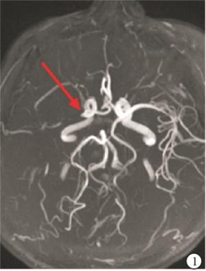
图 1 MRA示右侧大脑中动脉急性闭塞(红色箭头)
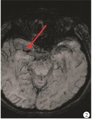
图 2 SWI示对应的右侧大脑中动脉有低信号的磁敏感血栓征(红色箭头)
SVS MRA 合计 血管狭窄 血管闭塞 阳性 4(16.0) 13(52.0) 17(68.0) 阴性 3(12.0) 5(20.0) 8(32.0) 合计 7(28.0) 18(72.0) 25(100.0) 表 1 SWI发现责任动脉内血栓[n;百分率(%)]
-
对25位脑梗死病人发病90 d后的随访,发现17例SVS阳性的病人中有10例(58.8%)病人临床预后不良,而在8例SVS阴性病人中仅有1例(12.5%)病人临床预后不良;SWI上的SVS阳性病人较阴性病人临床预后不良率高(χ2=4.74,P < 0.05)。
-
以往的研究[5-6]认为,SWI可以根据血栓成分,尤其是血氧饱和度变化,在影像上显示血栓形态,其可以定位颅内大血管内血栓,如颈内动脉、大脑中动脉M1段等,且灵敏性和特异性在相关研究中均得到了证实,此外对SWI进一步分析研究,其还可以定位颅内动脉远端的血栓,以及颅内静脉血管内血栓形成。由于SWI具有的磁共振特点,使得其在临床应用前景更为广阔,具有重要的临床应用价值。
目前国内外学者认为SVS在影像学发生是由于当急性脑梗死发生时,颅内责任动脉急性闭塞,血流动力学改变使得血流减慢,而血管内残存的血液,由于供血区域组织细胞代谢,而使得区域内血红细胞,氧和血红蛋白和去氧血红蛋白比例明显下调,使得梗死区域组织间的磁敏感属性发生了显著变化,而SWI对不同物质磁敏感性甚至微弱的变化仍很敏感,故可以对责任动脉内血栓,尤其是富含红细胞的血栓磁敏感属性形成磁敏感信息,具有充当造影剂的功能,结合计算机软件进行分析,最终形成功能影像,得以显示出血管内血栓。HUANG等[7-8]研究通过对44例急性脑梗死病人的SWI影像学回顾性分析,发现SWI中的SVS具有定位颅内动脉血栓的影像学意义,通过对比MRA等影像学工具,最终认为SWI可以显示颅内动脉血栓,甚至颅内动脉远端以往不易识别的血栓。此外有学者[6]在后续的研究中指出SWI对于动脉血栓定位的敏感性高达83%,准确性为100%,且存在SVS责任血管在后续DSA等检查中均得到了证实,无假阳性,进一步证实了SWI对于识别颅内责任动脉血栓的意义,本研究结论与之基本一致。不过在本研究中也存在8例急性脑梗死病人未发现SVS征,我们根据SWI属性和SVS形成机制认为:(1)由于急性脑梗死病人责任动脉形成的血栓,如为富含血小板的白色血栓或者是长期陈旧性的附壁血栓脱落,可能由于缺乏氧合指数变化,无法引起空间磁敏感性变化,而不能在SWI上进行显影;(2)目前对于SWI的国内外研究主要是对颅内前循环血管分析研究,主要是由于后颅窝等骨质结构与富含气体的窦腔可以影响SVS的显示。
目前对急性脑梗死的治疗,rt-PA溶栓治疗得到国际公认和治疗指南的推崇,而这得益于长期以来对于梗死血栓的病理病因研究。rt-PA主要是针对富含纤维蛋白的血栓、纤维蛋白网内具有大量的红细胞,这一不稳定的血栓特性,使得rt-PA容易进入血栓内,并溶解纤维蛋白网,从而分解和破坏血栓,达到溶解血栓、促进血管再通的目的。但是如果是富含血小板的白色血栓、或者是时间较久的陈旧血栓,则由于其结构更为致密,甚至有的血栓发生了机化和钙化,使得rt-PA不能渗透进去,或者是渗透困难,无法溶解血栓内的纤维蛋白网架结构,自然难以达到溶栓的目的。VIDMAR等[9]通过对不同成分血栓进行分析研究,就指出富含红细胞的红色血栓更易于溶解,而富含血小板的白色血栓则结构更为致密,更为难以发生溶解。在对不同成分的血栓进行的rt-PA药物溶栓实验中,就发现由于白色血栓的致密结构,从而导致血液内的rt-PA无法有效渗透到血栓内部,由于其无法充分接触到纤维蛋白网等结构,而使得其溶解血栓的作用大大下降[10-11],这也就是一些病人rt-PA溶栓效果差的原因,提示这样的病人更需要采用的是取栓治疗,而非溶栓治疗,因此SWI的SVS可能对于急性脑梗死治疗方法选择也具有重要的提示意义,我们将在后续的研究中进一步深入探讨。根据SWI的SVS形成机制,可能对判断血栓成分具有一定帮助。以往的研究认为SVS对急性脑梗死病人临床预后无指示性[12-13],本研究中我们发现SVS可能指示病人临床预后较差。与以往研究不一致,我们考虑可能与样本量较小,一些SVS阴性病人是由于血栓过小未被发现和识别等因素有关。我们将在以后的研究中加大样本量等进一步研究。
综上,我们认为SWI可以在血栓定位方面具有重要的临床意义,此外对病人临床预后也具有一定指示意义。因此我们认为SWI应该作为一个常规的影像学工具应用于急性脑梗死诊断和预后评估方面有重要的临床意义,值得推广。
磁敏感血栓征在急性缺血性脑卒中病人诊治中的意义
Clinical application value of susceptibility vessel sign in patients with acute ischemic stroke
-
摘要:
目的探讨磁敏感加权成像上磁敏感血栓征(SVS)在急性脑梗死临床诊断和治疗中的意义。 方法对25例急性脑梗死病人入院5 d内完善常规MRI、DWI、SWI和MRA等相关检查。对SWI对颅内责任血管血栓定位进行统计分析,并对全部脑梗死病人入院时进行神经缺损程度评分,发病90 d后进行残障评分,判断磁敏感血栓征对病人临床预后是否具有指示意义。 结果25例病人中,发现17例病人(68%)在SWI上有沿着病变血管走形的SVS,且SVS阳性病人血管闭塞率高于阴性病人(P < 0.05);对25位脑梗死病人发病90 d后随访发现,17例SVS阳性的病人有10例病人临床预后不良,而8例SVS阴性病人,仅有1例病人临床预后不良,SWI上的SVS阳性病人较阴性病人临床预后不良率高(P < 0.05)。 结论脑梗死急性期,SWI的SVS征可以提示颅内责任动脉内血栓,并能提示病人临床预后不良。 Abstract:ObjectiveTo investigate the significance of magnetic sensitive weighted imaging(SVS)in the diagnosis and treatment of acute cerebral infarction. MethodsThe routine detection of MRI, DWI, SWI and MRA in 25 patients with acute cerebral infarction were implemented.The location of intracranial responsible blood vessel thrombosis was analyzed using SWI, the nerve defect degree score on admission and disability score after 90 days of onset were performed in all cases, and the significance of magnetically sensitive thrombus sign in judging the prognosis of patients was analyzed. ResultsAmong 25 patients with acute cerebral infarction, the SVS along lesion vessel in 17 cases(68%)were found in SWI, and the vascular occlusion rate in SVS-positive patients was higher than that in SVS-negative patients(P < 0.05).During the following-up for 90 days of 25 patients with cerebral infarction, the clinical prognosis in 10 cases with positive SVS, 8 cases with negative SVS and 1 case were poor, and the poor clinical prognosis rate of positive SVS patients in SWI was higher compared with negative SVS patients(P < 0.05). ConclusionsDuring the acute phase of cerebral infarction, the SVS sign in SWI can indicate the intracranial responsible intra-arterial thrombosis, and poor clinical prognosis. -
表 1 SWI发现责任动脉内血栓[n;百分率(%)]
SVS MRA 合计 血管狭窄 血管闭塞 阳性 4(16.0) 13(52.0) 17(68.0) 阴性 3(12.0) 5(20.0) 8(32.0) 合计 7(28.0) 18(72.0) 25(100.0)  下载: 导出CSV
-
[1] JAUCH EC, SAVER JL, ADAMS HP, et al. Guidelines for the early management of patients with acute ischemic stroke: a guideline for healthcare professionals from the american heart association/american stroke association[J]. Stroke, 2013, 44(3): 870. doi: 10.1161/STR.0b013e318284056a [2] 孙世光. 中国急性缺血性脑卒中诊治指南2010[J]. 中华神经科杂志, 2010, 43(2): 146. doi: 10.3760/cma.j.issn.1006-7876.2010.02.022 [3] HAACKE EM, MITTAL S, WU Z, et al. Susceptibility-weighted imaging: technical aspects and clinical applications, part1[J]. AJNR, 2009, 30(1): 19 doi: 10.3174/ajnr.A1400 [4] 中华医学会神经病学分会脑血管病学组急性缺血性脑卒中诊治指南撰写组. 中国急性缺血性脑卒中诊治指南2010[J]. 中华神经科杂志, 2010, 43: 146. doi: 10.3760/cma.j.issn.1006-7876.2010.02.022 [5] MITTAL S, WU Z, NEELAVALLI J, et al. Susceptibility-weighted imaging: technical aspects and clinical applications, part2[J]. AJNR, 2009, 30(2): 232. doi: 10.3174/ajnr.A1461 [6] ROVIRA A, ORELLANA P, ALVAREZ-SABIN J, et al. Hyperacute ischemic stroke: middle cerebral artery susceptibility sign at echo-planar gradient-echo MR imaging[J]. Radiology, 2004, 232(2): 466. doi: 10.1148/radiol.2322030273 [7] HUANG P, CHEN CH, LIN WC, et al. Clinical applications of susceptibility weighted imaging in patients with major stroke[J]. J Neurol, 2012, 259(7): 1426. doi: 10.1007/s00415-011-6369-2 [8] LUO S, YANG L, WANG L.Comparison of susceptibility-weighted and perfusion-weighted magnetic resonance imaging in the detection of penumbra in acute ischemic stroke[J]. J Neuroradiol, 2015, 42: 255. doi: 10.1016/j.neurad.2014.07.002 [9] VIDMAR J, BLINC A, KRALJ E, et al. An MRI study of the differences in the rate of thrombolysis between red blood cell-rich and platelet-rich components of venous thrombi ex vivo[J]. J Magn Reson Imaging, 2011, 34(5): 1184. doi: 10.1002/jmri.22731 [10] SABOVIC M, LIJNEN HR, KEBER D, et al. Effect of retraction on the lysis of human clots with fibrin specific and non-fibrin specific plasminogen activators[J]. Thromb Haemost, 1989, 62(4): 1083. doi: 10.1055/s-0038-1647122 [11] SABOVIC M, BLINC A.Biochemical and biophysical conditions for blood clot lysis[J]. Pflugers Arch, 2000, 440(5 Suppl): 134. [12] 佘德君. 磁敏感加权成像诊断脑梗死颅内动脉血栓的临床应用价值[J]. 临床放射学杂志, 2013, 32(11): 1546. [13] SCHELLINGER PD, CHALELA JA, KANG DW, et al. Diagnostic and prognostic value of early MR Imaging vessel signs in hyperacute stroke patients imaged < 3 hours and treated with recombinant tissue plasminogen activator[J]. AJNR, 2005, 26(3): 618. -

 点击查看大图

图(2)表(1)
计量
- 文章访问数: 4391
- HTML全文浏览量: 2018
- PDF下载量: 18
- 被引次数: 0
