


-
腰椎终板改变即Modic改变,是指MRI上可见的邻近椎体终板退变引起的骨髓信号改变[1]。1987年DeRoos第一次提出腰椎退行性病变的病人中MRI影像靠近终板椎体信号有异常改变,随后Modic在1988年提出了Modic的定义。Modic变化共分为Ⅰ型、Ⅱ型和Ⅲ型变化,以说明在MRI上观察到的椎体区终板的炎症期。在骨组织活检中PERILLI等[2]发现Modic Ⅰ型改变的病理变化为骨组织代谢旺盛,Modic Ⅱ型改变表现为成骨减少,而Modic Ⅲ型改变则表现为广泛地骨组织硬化状态。XIAO等[3]在模拟骨质疏松的动物模型中发现去除卵巢组的大鼠出现了椎体骨质疏松和软骨终板骨软骨重建,促进了骨-软骨表面血管生成和孔隙率增加,影响了基质代谢,从而对椎间盘产生不利影响,但在临床上骨质疏松和Modic变化的研究涉及不多。本研究通过分析骨质疏松和Modic变化的相关性,旨在探究椎体骨密度在Modic改变发生中的意义。现作报道。
-
选择2018年9月至2020年9月在蚌埠医学院第一附属医院脊柱外科行椎体骨密度测定及腰椎MRI检查的152例腰痛病人,年龄50~90岁,行椎体骨密度测定及腰椎MRI检查,测定椎体骨量并观察影像学资料。排除以往有腰椎手术史、年龄不足、合并腰椎骨折、肿瘤、感染或腰椎先天性异常、移行椎、严重的脊柱侧弯。
-
采用双能X线骨密度仪(Hologic公司,010-0575型)进行腰椎椎体骨密度,使病人取仰卧位,测量L1、L2、L3、L4椎体和腰椎总骨密度。腰椎MRI检查采用GE Signa Excite型1.5 T MRI扫描仪,行腰椎矢状位T1WI、T2WI及横断位T2WI检查,扫描参数:T1WI,TR 450 ms,TE 13.6 ms;T2WI,TR 2 000 ms,TE 102 ms,采集次数2次,矢状位视野(FOV)32 cm×32 cm,横断位FOV 20 cm×20 cm。
-
根据2018年骨质疏松诊断标准:使用T值表示骨密度,T值≥-1.0为骨量正常,-2.5 < T值< -1.0为骨量减少,T值≤-2.5为骨质疏松,T值≤-2.5伴脆性骨折史为严重骨质疏松症;所有腰痛病人采用疼痛视觉模拟法(VAS)进行评分,0分表示无痛,10分表示最痛。
-
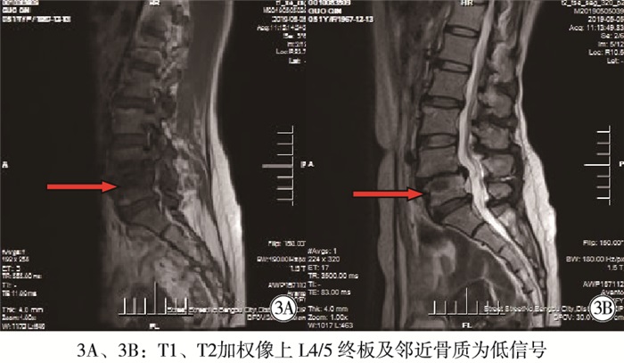
由一位经验丰富的放射核医学科主治医师和一名脊柱外科医师分别独立观察分析和记录病人MRI资料。腰椎椎体Modic分型评价标准: Ⅰ型, T1加权像上呈低信号,T2加权像上呈高信号;Ⅱ型, T1加权像上呈高信号,T2加权像上呈等或者轻度高信号;Ⅲ型, T1、T2加权像上均呈低信号。
-
采用Kappa检验、χ2检验、方差分析、q检验和Spearman等级相关检验。
-
一位经验丰富的放射核医学科主治医师和一名脊柱外科医师对腰椎椎体Modic改变的评估一致性良好(Kappa=0.80)。
-
152例腰痛病人中,骨量正常89例,其中Modic改变29例,发生率32.6%;骨量减少43例,其中Modic改变23例,发生率53.5%;骨质疏松20例,其中Modic改变17例,发生率85.0%。3组Modic改变发生率差异有统计学意义(P < 0.05)。骨质疏松Modic改变率与骨量正常组比较差异有统计学意义(P < 0.05)(见表 1)。Modic Ⅰ型VAS评分最高,无Modic改变组VAS评分最低,Modic Ⅰ型组和Ⅱ、Ⅲ、无改变组腰痛程度比较差异有统计学意义(P < 0.05~P < 0.01);Modic Ⅱ型组和Ⅲ型组之间比较差异无统计学意义(P>0.05)(见表 2)。Spearman等级相关检验证明骨质疏松和Modic改变呈正相关关系(r=0.396,P < 0.01)。3型Modic改变图示见图 1~3。
分组 n 正常 Ⅰ型 Ⅱ型 Ⅲ型 发生情况 骨质正常组 89 60 12 15 2 29(32.58) 骨质减少组 43 20 10 12 1 23 (53.49) 骨质疏松组 20 3 5 8 4 17 (85.00)* χ2 — — — — — 6.57 P — — — — — < 0.05 与骨质正常组比较*P < 0.05 表 1 骨密度评价与Modic改变比较[n; 百分率(%)]
Modic改变 n VAS评分/分 无改变组 83 3.52±1.54 Ⅰ组 27 7.00±1.17** Ⅱ组 35 5.00±1.28**## Ⅲ组 7 5.57±0.97**# F — 44.76 P — < 0.01 MS组内 — 1.969 与无改变组比较**P < 0.01;与Ⅰ组比较#P < 0.05,##P < 0.01 表 2 Modic改变三种类型下腰痛VAS评分情况(x±s)

图 1 女,60岁,T值为-2.0,L4/L5Ⅰ型Modic改变,腰椎退行性变

图 2 男,57岁,T值为0.4,L5/S1 Ⅱ型Modic改变,腰椎退行性变

图 3 女,60岁,T值为-1.9,L4/5 Ⅲ型Modic改变,腰椎退行性变
-
骨质疏松症是骨量降低、骨组织微细结构平衡被打破导致骨脆性增加易发生骨折的全身性骨病。在正常的骨质代谢中依靠成骨细胞介导的骨形成和破骨细胞调节的骨吸收维持平衡[4]。当骨形成降低、骨吸收增加时,骨代谢失衡引起骨量丢失[5]。老年性骨质疏松症主要与激素、营养及年龄有关,与遗传具有一定相关性。发生机制: (1)老年人性激素水平下降,降钙素等激素分泌紊乱,加上胃肠吸收功能减弱,导致营养不足,激素对机体的作用减弱,骨形成减少,骨吸收增加,骨平衡被打破,最终导致骨代谢紊乱[6]; (2)老年人活动减少,导致骨骼应力作用降低,骨形成下降,同时活动减少也促使骨吸收。
-
Modic改变的机制:(1)终板出现损伤后,相邻的椎体及椎间盘会发生不同程度的退行性病变。因为终板损伤使椎间盘局部应力发生改变,影响组织的新陈代谢导致Modic改变的发生[2, 7]。(2)低毒性细菌感染学说认为椎间盘内的厌氧环境、易损伤性及低修复特性为入侵椎间盘的细菌造成Modic改变提供一个良好环境。DUDLI等[8-9]在动物模型中证明痤疮丙酸杆菌导致Modic改变,CHEN等[10]利用从Modic改变病人体内提取低毒性细菌转注入兔椎间盘的方法模拟出了骨髓的信号变化。HAN等[11]通过提取兔自体髓核包埋在椎体软骨下骨的方法同样观察到了椎体的信号变化。(3)自身免疫反应学说认为髓核具有抗原封闭特性,在椎间盘被各种因素损伤后,髓核暴露出来导致机体产生免疫应答反应,进而发生Modic改变。(4)近年有研究表明Modic的发生与基因遗传学密切相关, PERERA等[12]采用单核苷酸变异分析聚蛋白多糖在MCs中的代谢途径,发现白细胞介素-1基因簇、聚蛋白多糖酶S4基因及聚蛋白多糖酶基因在Modic过程中起重要作用。
-
贺宪等[13]研究表明下腰段同上腰段相比更易发生Modic信号变化,腰椎间盘退变严重程度和Modic改变的发生率成正相关。其原因可能与脊柱所受负荷及椎体骨质疏松等因素有关系。HILTON等[14]研究表明,长期反复的压力作用在脊柱功能单位时,最初会导致终板的微骨折但椎间盘完整性并没有损伤,当17%的椎间盘发生改变时,58%的终板会发生破裂,证明终板在脊柱功能单位中是比较脆弱的部位。当腰椎间盘退变后,脊柱功能单位所受负荷将重新分布,椎间盘所承受的压力增加,导致终板发生显微骨折;腰椎间盘内髓核物质通过显微骨折裂隙进入终板引起局部的炎症反应,终板长期的显微骨折可能是Modic形成的重要原因[14-15]。
本研究结果显示骨量正常组、减少组及疏松组之间Modic改变发生率有显著差异,表明椎体骨密度与Modic改变之间具有相关性。骨量正常组与骨质疏松组及减少组Modic改变发生率差异均有统计学意义,表明随着腰椎椎体骨密度水平的降低,Modic改变的发生率升高。骨量减少组与骨质疏松组之间Modic改变发生率也有统计学差异,在本研究中,在无Modic改变组以及各类型Modic变组的腰痛病人腰痛程度比较中, Modic Ⅰ型改变组VAS腰痛评分高于其他各组(P < 0.05~P < 0.01), 可见腰椎Modic Ⅰ型改变与腰痛的发生有密切关系; Spearman等级相关检验证明骨质疏松和Modic改变呈弱正相关(r=0.396,P < 0.01),表明椎体骨密度与Modic改变之间的关联性较弱,提示除骨密度外,Modic改变形成可能还受其他因素的作用。研究[13]表明, 老年人腰椎椎体骨密度与Modic改变之间具有相关性,随着椎体骨密度的降低,腰椎Modic改变的发生率逐渐升高,但是这种相关性较弱,说明Modic改变的发生与发展还受其他因素影响。老年人骨质疏松症的预防上可以在一定程度上能够降低Modic改变的发生率。本研究不足在于获得的老年人病人中骨质疏松样本量太小,并且对于男性病人的年龄选择存在问题,可能对研究结果有一定影响,有待收集大量样本进一步研究。
骨质疏松和Modic改变的相关性分析
Correlation analysis of osteoporosis and Modic changes
-
摘要:
目的研究50岁以上的腰痛病人椎体骨密度与腰椎MRI上Modic改变的相关性,探讨椎体骨密度在Modic改变中的意义。 方法收集脊柱外科行椎体骨密度测定及腰椎MRI检查的152例腰痛病人,测定椎体骨量并观察影像学资料。将所有病人根据其骨量水平分为骨量正常组(89例)、骨量减少组(43例)及骨质疏松组(20例)。比较3组Modic改变的发生率,分析椎体骨密度与Modic改变的关系,并用疼痛视觉模拟法(VAS)进行腰痛程度评分,分析Modic改变和骨质疏松及腰痛的关系。 结果152例腰痛病人中,骨量正常89例,其中Modic改变29例,发生率32.6%;骨量减少43例,其中Modic改变23例,发生率为53.5%;骨质疏松20例,其中Modic改变17例,发生率为85.0%。3组Modic改变发生率差异有统计学意义(P < 0.05)。骨质疏松Modic改变率与骨量正常组比较差异有统计学意义(P < 0.05);ModicⅠ型VAS评分最高,无Modic改变组VAS评分最低,ModicⅠ型组和Ⅱ、Ⅲ、无改变组腰痛程度比较差异有统计学意义(P < 0.05~P < 0.01);ModicⅡ型组和Ⅲ型组之间比较差异无统计学意义(P>0.05),Ⅲ组和Ⅱ组均高于无改变组(P < 0.01)。Spearman等级相关检验证明,骨质疏松和Modic改变呈正相关关系(r=0.396,P < 0.01)。 结论50岁以上的老年人椎体骨密度与Modic改变有相关性,且Modic改变的发生率随着椎体骨密度的减低而升高,ModicⅠ型改变和腰痛关系密切。 Abstract:ObjectiveTo study the correlation between vertebral bone mineral density and Modic changes of lumbar, and explore the significance of vertebral bone mineral density in Modic changes. MethodsThe vertebral bone mineral density(BMD) and lumbar MRI examination in 152 patients with low back pain were measured to detect the vertebral bone mass, and the imaging data were observed.According to the level of bone mass, the patients were divided into the normal bone mass group(n=89), osteopenia group(n=43) and osteoporosis group(n=20).Among three groups, the incidence rates of Modic changes were compared, and the relationship between vertebral bone mineral density and Modic changes were analyzed.The degree of low back pain was scored using pain visual simulation(VAS), and the relationship between Modic changes and osteoporosis, low back pain were analyzed. ResultsAmong the 152patients, there were 89 cases of normal bone mass(including 29 cases of Modic changes with incidence rate of 32.6%), 43 cases of osteopenia(includimg 23 cases of Modic changes with incidence rate of 53.5%), and 20 cases of osteoporosis(including 17 cases of Modic changes with incidence rate of 85.0%).The differences of the incidence rate of Modic changes among three groups were statistically significant(P < 0.05).The differences of the incidence rate of Modic changes between normal bone mass group and osteoporosis group were statistically significant(P < 0.05).The VAS score in Modic type Ⅰ and nochange Modic group were the highest and lowest, repsectively.The differences of lumbago degree between Modic type Ⅰ group and Ⅱ, Ⅲ type, nochange group were statistically significant(P < 0.05 to P < 0.01).The differences of lumbago degree between Modic type Ⅱand Ⅲ was not statistically significant(P>0.05), which in type Ⅱ and Ⅲ groups were higher than that in no change group(P < 0.01).The results of Spearman rank correlation test proved that there was a weak positive correlation between osteoporosis and Modic changes(r=0.396, P < 0.01). ConclusionsIn the elderly over 50 years old, there is a correlation between vertebral bone mineral density and Modic changes, and the incidence rate of Modic changes increases with the decreasing of vertebral bone mineral density.Modic type Ⅰ changes is closely related to low back pain. -
Key words:
- osteoporosis /
- back pain /
- bone mineral density /
- magnetic resonance imaging /
- Modic change
-
表 1 骨密度评价与Modic改变比较[n; 百分率(%)]
分组 n 正常 Ⅰ型 Ⅱ型 Ⅲ型 发生情况 骨质正常组 89 60 12 15 2 29(32.58) 骨质减少组 43 20 10 12 1 23 (53.49) 骨质疏松组 20 3 5 8 4 17 (85.00)* χ2 — — — — — 6.57 P — — — — — < 0.05 与骨质正常组比较*P < 0.05  下载: 导出CSV
表 2 Modic改变三种类型下腰痛VAS评分情况(x±s)
Modic改变 n VAS评分/分 无改变组 83 3.52±1.54 Ⅰ组 27 7.00±1.17** Ⅱ组 35 5.00±1.28**## Ⅲ组 7 5.57±0.97**# F — 44.76 P — < 0.01 MS组内 — 1.969 与无改变组比较**P < 0.01;与Ⅰ组比较#P < 0.05,##P < 0.01
下载: 导出CSV
-
[1] 吴凯, 封志云, 胡小坚. 经皮脊柱内镜手术治疗伴Modic Ⅱ型改变的腰椎间盘突出症的疗效观察[J]. 浙江医学, 2019, 41(15): 1657. doi: 10.12056/j.issn.1006-2785.2019.41.15.2019-1242 [2] PERILLI E, PARKINSON IH, TRUONG LH, et al. . Modic (endplate) changes in the lumbar spine: bone micro-architecture and remodelling[J]. Eur Spine J, 2015, 24(9): 1926. doi: 10.1007/s00586-014-3455-z [3] XIAO ZF, HE JB, SU GY, et al. Osteoporosis of the vertebra and osteochondral remodeling of the endplate causes intervertebral disc degeneration in ovariectomized mice[J]. Arthritis Res Ther, 2018, 20(1): 207. doi: 10.1186/s13075-018-1701-1 [4] 董冰子, 孙晓方. 骨质疏松症治疗新进展: 从分子机制到药物靶点[J]. 中华骨质疏松和骨矿盐疾病杂志, 2018, 11(6): 620. doi: 10.3969/j.issn.1674-2591.2018.06.016 [5] 刘丹丹. 1, 25(OH) 2D3对类风湿关节炎破骨细胞形成影响可能机制的研究[D]. 太原: 山西医科大学, 2016: 1. [6] 才林, 艾光禹, 孙强. 老年骨质疏松机制及股骨转子间骨折治疗的研究进展[J/CD]. 中华损伤与修复杂志(电子版), 2016, 11(6): 469. [7] DUDLI S, HASCHTMANN D, FERGUSON SJ. Persistent degenerative changes in the intervertebral disc after burst fracture in an in vitro model mimicking physiological post-traumatic conditions[J]. Eur Spine J, 2015, 24(9): 1901. doi: 10.1007/s00586-014-3301-3 [8] DUDLI S, LIEBENBERG E, MAGNITSKY S, et al. Propionibacterium acnes infected intervertebral discs cause vertebral bone marrow lesions consistent with Modic changes[J]. J Orthop Res, 2016, 34(8): 1447. doi: 10.1002/jor.23265 [9] DUDLI S, FIELDS AJ, SAMARTZIS D, et al. Pathobiology of Modic changes[J]. Eur Spine J, 2016, 25(11): 3723. doi: 10.1007/s00586-016-4459-7 [10] CHEN Z, ZHENG Y, YUAN Y, et al. Modic changes and disc degeneration caused by inoculation of propionibacterium acnes inside intervertebral discs of rabbits: a pilot study[J]. Biomed Res In, 2016, 2016: 9612437. doi: 10.1155/2016/9612437 [11] HAN C, WANG T, JIANG HQ, et al. An animal model of Modic changes by embedding autogenous nucleus pulposus inside subchondral bone of lumbar vertebrae[J]. Sci Rep, 2016, 6: 35102. doi: 10.1038/srep35102 [12] PERERA RS, DISSANAYAKE PH, SENARATH U, et al. Single nucleotide variants of candidate genes in aggrecan metabolic pathway are associated with lumbar disc degeneration and Modic changes[J]. PLoS One, 2017, 12(1): e0169835. doi: 10.1371/journal.pone.0169835 [13] 贺宪, 黄东生, 孔畅, 等. 下腰痛病人中腰椎终板Modic改变的分布情况及与腰椎间盘退变的关系[J/CD]. 中华临床医师杂志(电子版), 2015, 9(8): 1283. [14] HILTON RC, BALL J. Vertebral rim lesions in the dorsolumbar spine[J]. Ann Rheum Dis, 1984, 43(2): 302. doi: 10.1136/ard.43.2.302 [15] LIU J, HAO L, SUYOU L, et al. Biomechanical properties of lumbar endplates and their correlation with MRI findings of lumbar degeneration[J]. J Biomech, 2016, 49(4): 586. -

 点击查看大图

图(3)表(2)
计量
- 文章访问数: 4085
- HTML全文浏览量: 2331
- PDF下载量: 19
- 被引次数: 0
