

-
甲状腺肿瘤是临床常见疾病,伴随高频超声的广泛应用,高达30%~67%的正常人可以检测出甲状腺结节[1]。因此,甲状腺癌的发病率逐年增加,临床检出的结节中约5%~15%为恶性[2]。肿瘤的产生依赖于血管的形成,良恶性肿瘤的生长与其血供特点密不可分[3]。随着医学影像技术的日益发展,超微血管三维立体成像(smart three-dimensional superb microvascular imaging,Smart 3D SMI)出现并日益发展[4], 有文献报道称[5]SMI能够检测乳腺肿瘤内部的穿支血管,有助于辨别肿瘤的良恶性。但Smart 3D SMI在甲状腺领域的研究甚少。本研究通过分析甲状腺结节的血流灌注特性,探讨Smart 3D SMI技术评估结节微血管灌注的可行性。
-
纳入2018年3月至2019年12月在我院接受诊疗的甲状腺病人107例。男21例,女86例,年龄11~76岁。纳入标准:(1)实性部分>50%的结节;(2)所有结节手术前均接受二维灰阶超声和超微血管三维立体成像检查,检查数据完整。排除标准:未经手术治疗,无明确手术及病理结果。本组每个病例只纳入1个结节,对于多发病灶,只纳入符合纳入标准的一个病灶。所有病人均有完整的手术和病理资料。
-
使用配有Smart 3D SMI软件的阿波罗500彩超仪器,配用SLI5~4线阵探头。病人平卧,头后伸以完全露出颈部。首先通过常规超声检测甲状腺结节的大小,形状,位置,边界和内部回声,然后使用仪器自带的彩色多普勒,超微血管二维和三维成像软件检测结节的血流状况。同时,存储静态和动态图像以获得甲状腺结节完整的超声数据,包括结节中血管的数量,血流空间分布等特征。
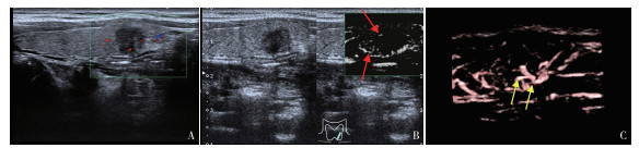
本研究基于Adler的半定量分类标准[6],血流分布和穿支血管,分析病灶的血液灌注特性。(1)半定量分级:O级:结节内及边缘无血流;Ⅰ级,1~2个点状或短棒血流;Ⅱ级,3~4个星点状或一条状血流,其长度接近或超过病灶半径;Ⅲ级,5个点状或2条以上较长血流。(2)根据结节血流分布的形态特征分为:无血流型、穿入型、周缘型、中心型和混合型。穿入型定义为任意切面检测结节的外部血管向内部伸展或由结节内部向外周延伸,且血管长度超过结节最大径的1/3。混合型指存在两种或两种以上的血流分布模式。
-
采用秩和检验和χ2检验。
-
本研究共选入甲状腺结节107枚,所有结节均通过二维超声和三维超微血管成像检查。44枚良性结节:包括16枚结节性甲状腺肿,28枚腺瘤。63枚恶性结节:包含62枚乳头状癌,1枚髓样癌。
-
彩色多普勒(CDFI)、2D SMI和Smart 3D SMI三种方法对同一结节的血流评估差异有统计学意义(P<0.05~P<0.01)。Smart 3D SMI评估结节的血流分级优于CDFI(P<0.05),且良、恶性结节的Smart 3D SMI血流分级存在差异性(P<0.01)(见表 1、图 1)。
方法 结节数 0 Ⅰ级 Ⅱ级 Ⅲ级 Hc P 良性结节 44 CDFI 18(40.9) 19(43.2) 5(11.4) 2(4.5) 2D SMI 14(31.8) 17(38.7) 10(22.7) 3(6.8) 8.91 <0.05 Smart 3D SMI 9(20.5) * 15(34.1)* 14(31.8)* 6(13.6)* 恶性结节 63 CDFI 7(11.1) 15(23.8) 27(42.9) 14(22.2) 2D SMI 5(7.9) 10(15.9) 25(39.7) 23(36.5) 17.46 <0.01 Smart 3D SMI 2(3.2)*## 5(7.9)*## 21(33.3)*## 35(55.6)*## 与CDFI比较*P<0.05;恶性结节Smart 3D SMI与良性结节Smart 3D SMI比较##P<0.01 表 1 三种方式评估甲状腺良、恶性结节的血流分级[个; 百分率(%)]
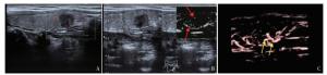
图 1 良、恶性结节的Smart 3D SMI血流分级
-
良性结节的血流分布模式多为周围型和混合型,恶性结节多为穿入型和中心型。在良性组中,5枚结节经2D SMI检查为无血流型,但经Smart 3D SMI检查其中3枚为周围型,2枚为混合型。在恶性组中,2枚经2D SMI检查为无血流型,其中1枚Smart 3D SMI示为穿入型,另1枚示为中心型(见表 2)。三种扫查方式对良性结节血管分布的评估差异无统计学意义(P>0.05),但对恶性结节的评估有明显差异(P<0.01)。
方法 结节数 无血流型 穿入型 周围型 中心型 混合型 Hc P 良性结节 44 CDFI 19(43.2) 1(2.3) 13(29.5) 2(4.5) 9(20.5) 2D SMI 13(29.5) 3(6.8) 16(36.4) 4(9.1) 8(18.2) 8.09 >0.05 Smart 3D SMI 8(18.2)** 3(6.8) ** 20(45.5)** 3(6.8)** 10(22.7)** 恶性结节 63 CDFI 8(12.7) 9(14.3) 11(17.4) 19(30.2) 16(25.4) 2D SMI 4(6.3) 16(25.4) 7(11.1) 25(39.7) 11(17.5) 20.27 <0.01 Smart 3D SMI 2(3.2)** 24(38.1)** 4(6.3)** 27(42.9)** 6(9.5)** 与CDFI组、2D SMI组比较**P<0.01 表 2 三种方法评价良、恶性结节的血流分布模式[n; 百分率(%)]
-
随着甲状腺癌发病率的快速增长,它已成为30岁之前女性常见的恶性肿瘤之一[7]。肿瘤的形成与繁殖、良恶性肿瘤的分化与肿瘤血管的形成和其血流特性息息相关[3]。常规超声可通过检测血流信息评估肿瘤的良恶性,但无法判断血流变化较小的肿瘤,会出现漏诊、误诊的情况[8]。随着医学影像技术的发展,三维超声在临床上的应用日益广泛,包括肝脏、心脏、妇科及产科等[9-12]。Smart 3D SMI是一种更加直观的、立体的微血管成像技术,它在二维超声基础上通过血管重建对肿瘤血管进行三维立体成像。因此,Smart 3D SMI不仅具有2D SMI的优点能够检测到细小血管和低速血流信号[13],同时还能够突显血管轮廓,改善病变的血流显示程度。本研究结果表明,根据Adler半定量血流分类,Smart 3D SMI显示甲状腺恶性结节的血流以Ⅲ型为主,占55.6%,而采用CDFI和2D SMI评估甲状腺结节以Ⅱ级血流为主,Ⅲ级血流分别占22.6%和36.5%,由此表明Smart 3D SMI技术对甲状腺恶性结节新生血管的血流显示程度更高,这与李伟伟等[4]研究结果一致。
据国内外相关文献报道[14-15]甲状腺结节中的血管生成是肿瘤增殖和发展的基本条件,因此检测甲状腺结节的血管非常重要。与CDFI不同,SMI可以识别由血流和组织运动产生的噪声,并使用自适应计算方法显示真实的血液流动信息,可以显示非常低速的血流和穿支血管。本研究结果表明,恶性结节的血流分布多为穿入型和中心型,良性结节多为周围型和混合型。与2D SMI相比,Smart 3D SMI能够更直观地显示穿支血管,二者在评估良恶性结节穿支血管方面差异具有统计学意义。分析原因可能是2D SMI基于二维超声成像原理,仅能通过某一个切面显示病灶的血流灌注情况,而3D SMI基于三维立体成像原理,病变的血管构造和血流空间分布方式等可以在多个角度和方向显示,并且可以一次获得病变的所有血流信息,与2D SMI比较,Smart 3D SMI可增加病变新生血管的显示程度。本组有5枚良性结节性经2D SMI检查为无血流型,但由Smart 3D SMI检查其中3枚结节的血流分型为外周型,另2枚为混合型。在恶性组中,2枚结节经2D SMI检查为无血流型,再由Smart 3D SMI检测其中1枚为穿入型,另1枚为中心型。其原因可能是由于结节较小,2D SMI仅在某一切面未检测到血流信号,而Smart 3D SMI可全方位立体获取小结节的全部血流信息。由此可见,Smart 3D SMI不仅能较敏感的检测出结节内极低速的穿支血管,3D SMI还可对2D SMI检测的血流灌注情况进行校正,它可以更真实地反映各种结节的血流特征,例如血管构造、血流分布方式等,进一步丰富了超声医生的检查信息,有助于良恶性结节的鉴别诊断。
综上所述,与CDFI和2D SMI相比,Smart 3D SMI技术可以更灵敏地检测甲状腺结节的微血管构造和血流分布方式,使结节的血流特征更加直观化、可视化,值得临床推广应用。
超微血管三维立体成像评估甲状腺结节血流的临床应用价值
Clinical application value of smart three-dimensional superb microvascular imaging in the evaluation of thyroid nodule blood flow
-
摘要:
目的通过观察甲状腺结节的超微血管三维立体成像(Smart 3D SMI)声像图特征,探讨Smart 3D SMI评估甲状腺结节血流的应用价值。 方法对2018年3月至2019年12月107例病人采用彩色多普勒CDFI、超微血管二维(2D SMI)和三维成像(Smart3D SMI)技术,观察结节的血管走行、血流空间分布等特征,通过Alder半定量分级、穿支血管及血管分布形态评估结节的血管灌注特征。 结果Smart 3D SMI对甲状腺结节的血流评估优于CDFI,且良、恶性结节的血流分型差异有统计学意义(P<0.01)。三种方法对恶性结节穿入血管的评估:CDFI检测率为14.3%、2D SMI 25.4%、Smart 3D SMI 38.1%,Smart 3D SMI对穿支血管的检测率最高。根据血管分布评估,5枚良性结节:2D SMI检查为无血流类型,其中3枚Smart 3D SMI显示为外周型,2枚为混合型。2枚恶性结节:2D SMI检查均为无血流类型,其中1枚Smart 3D SMI示为穿入型,另1枚为中心型。 结论Smart 3D SMI能够有效的甲状腺结节的微血管灌注特性,值得临床进一步推广和实践。 -
关键词:
- 甲状腺结节 /
- 超微血管三维立体成像 /
- 超声
Abstract:ObjectiveTo observe the ultrasonic characteristics of smart three-dimensional superb microvascular imaging (Smart 3D SMI) in thyroid nodule, and explore the application value of Smart 3D SMI in the evaluation of thyroid nodule blood flow. MethodsFrom March 2018 to December 2019, 107 patients(107 nodules) were detected using the color Doppler Flow Imaging(CDFI), 2D SMI and Smart 3D SMI to observe the blood vessels line and blood flow spatial distribution of the nodules.The vascular perfusion characteristics of nodules were evaluated using Alder semi-quantitative classification, perforator vessels and vascular distribution morphology. ResultsThe blood flow evaluation of Smart 3D SMI for thyroid nodule was better than that of CDFI, and the difference of blood flow classification between benign and malignant nodule was statistically significant(P<0.01).The results of three methods in the evaluation of malignant nodules penetrating into blood vessels showed that the detection rates of CDFI, 2D SMI and Smart 3D SMI were 14.3%, 25.4%, 38.1%, respectively, and the detection rates of Smart 3D SMI was the highest.According to the vascular distribution assessment, there were 5 benign nodules.The results of 2D SMI showed that 5 benign nodules were blood flow type, and the results of Smart 3D SMI showed that 3 benign nodules were peripheral type, and 2 benign nodules were mixed type.Among 2 malignant nodules, the results of 2D SMI showed that 2 malignant nodules were no blood flow type, and the results of Smart 3D SMI showed that 1 malignant nodule was penetrating type, and 1 malignant nodule was central type. ConclusionsSmart 3D SMI has high clinical application value in the evaluation of the microvascular perfusion characteristics of thyroid nodule, which is worthy of further promotion and practice in clinic. -
表 1 三种方式评估甲状腺良、恶性结节的血流分级[个; 百分率(%)]
方法 结节数 0 Ⅰ级 Ⅱ级 Ⅲ级 Hc P 良性结节 44 CDFI 18(40.9) 19(43.2) 5(11.4) 2(4.5) 2D SMI 14(31.8) 17(38.7) 10(22.7) 3(6.8) 8.91 <0.05 Smart 3D SMI 9(20.5) * 15(34.1)* 14(31.8)* 6(13.6)* 恶性结节 63 CDFI 7(11.1) 15(23.8) 27(42.9) 14(22.2) 2D SMI 5(7.9) 10(15.9) 25(39.7) 23(36.5) 17.46 <0.01 Smart 3D SMI 2(3.2)*## 5(7.9)*## 21(33.3)*## 35(55.6)*## 与CDFI比较*P<0.05;恶性结节Smart 3D SMI与良性结节Smart 3D SMI比较##P<0.01  下载: 导出CSV
表 2 三种方法评价良、恶性结节的血流分布模式[n; 百分率(%)]
方法 结节数 无血流型 穿入型 周围型 中心型 混合型 Hc P 良性结节 44 CDFI 19(43.2) 1(2.3) 13(29.5) 2(4.5) 9(20.5) 2D SMI 13(29.5) 3(6.8) 16(36.4) 4(9.1) 8(18.2) 8.09 >0.05 Smart 3D SMI 8(18.2)** 3(6.8) ** 20(45.5)** 3(6.8)** 10(22.7)** 恶性结节 63 CDFI 8(12.7) 9(14.3) 11(17.4) 19(30.2) 16(25.4) 2D SMI 4(6.3) 16(25.4) 7(11.1) 25(39.7) 11(17.5) 20.27 <0.01 Smart 3D SMI 2(3.2)** 24(38.1)** 4(6.3)** 27(42.9)** 6(9.5)** 与CDFI组、2D SMI组比较**P<0.01
下载: 导出CSV
-
[1] HAUGEN BR, ALEXANDER EK, BIBLE KC, et al. 2015 American thyroid association management guidelines for adult patients with thyroid nodules and differentiated thyroid cancer: the american thyroid association guidelines task force on thyroid nodules and differentiated thyroid cancer[J]. Thyroid, 2016, 26(1): 1. [2] SHIN JH, BAEK JH, CHUNG J, et al. Ultrasonography diagnosis and imaging-based management of thyroid nodules: revised korean society of thyroid radiology consensus statement and recommendations[J]. Korean J Radiol, 2016, 17(3): 370. doi: 10.3348/kjr.2016.17.3.370 [3] WANG M, FNG HL, LIU YQ, et al. Angiogenesis research in mouse mammary cancer based on contrast-enhanced ultrasonography: Exploratory study[J]. Acad Radiol, 2018, 25(7): 889. doi: 10.1016/j.acra.2017.12.004 [4] 李伟伟, 周庆华, 吴迎, 等. 超微血管三维立体成像技术对乳腺肿块血流的评估价值[J]. 中国超声医学杂志, 2019, 35(7): 587. doi: 10.3969/j.issn.1002-0101.2019.07.005 [5] 翟栋材, 张涛, 张申杰, 等. 乳腺癌穿支血管及超声生长方式与预后的关系[J]. 中国医药导刊, 2012, 14(z2): 536. doi: 10.3969/j.issn.1009-0959.2012.z2.078 [6] ADLER DD, CARSON PL, RUBIN JM, et al. Doppler ultrasound color flow imaging in the study of breast cancer: preliminary findings[J]. Ultrasound Med Biol, 1990, 16(6): 553. doi: 10.1016/0301-5629(90)90020-D [7] CHEN W, ZHENG R, BAADE PD, et al. Cancer statistics in China, 2015[J]. CA Cancer J Clin, 2016, 66(2): 115. doi: 10.3322/caac.21338 [8] 方超, 吴婷婷, 周莹颖, 等. 超声联合诊断甲状腺乳头状癌颈淋巴结转移的应用价值[J]. 中国全科医学, 2019, 21(24): 3000. doi: 10.12114/j.issn.1007-9572.2018.00.428 [9] OPOLSKIENE G, SLADKEVICIUS P, JOKUBKIENE L, et al. Three-dimensional ultrasound imaging for discrimination between benign and malignant endometrium in women with postmenopausal bleeding and sonographic endometrial thickness of at least 4.5mm[J]. Ultrasound Obstet Gynecol, 2010, 35(1): 94. [10] LEVINE EM, FERNANDEZ CM, MILLER D, et al. Clinical value of 3-dimensional ultrasound in gynecology[J]. J Ultrasound Med, 2018, 37(10): 2445. doi: 10.1002/jum.14587 [11] VELASCO O, BECKETT MQ, JAMES AW, et al. Real-time three-dimensional echocardiography: characterization of cardiac anatomy and function-current clinical applications and literature review update[J]. Biores Open Access, 2017, 6(1): 15. doi: 10.1089/biores.2016.0033 [12] HOTTA N, AYADA M, OKUMURA A, et al. Usefulness of live 3-D echocardiography during radiofrequency ablation in a case of hepatocellular case of hepatocellular carcinoma[J]. Clin Imaging, 2007, 31(4): 283. doi: 10.1016/j.clinimag.2007.01.009 [13] PARK AY, SEO BK, WOO OH, et al. The utility of ultrasound superb microvascular imaging for evaluation of breast tumour vascularity: comparison with colour and power Doppler imaging regarding diagnostic performance[J]. Clin Radiol, 2018, 73(3): 304. doi: 10.1016/j.crad.2017.10.006 [14] LU R, MENG Y, YAN Z, et al. Superb microvascular imaging(SMI) compared with conventional ultrasound for evaluating thyroid nodules[J]. Bmc Med Imaging, 2017, 17(1): 65. doi: 10.1186/s12880-017-0241-5 [15] 张丽波, 张波, 曹京, 等. 超微血管成像技术在TI-RADS 4类甲状腺结节检测中的应用价值[J]. 中华超声影像学杂志, 2017, 26(12): 1029. doi: 10.3760/cma.j.issn.1004-4477.2017.12.004 -

 点击查看大图
图(1)表(2)
计量
- 文章访问数: 3703
- HTML全文浏览量: 1864
- PDF下载量: 6
- 被引次数: 0
