

-
对肿块的诊疗首要问题是区分良恶性,超声对此有其独特之处[1]。美国放射学会(ACR)据声像图信息提出TI-RADS分层可提高诊断敏感性,但假阳性率增加[2]。超微血管成像(SMI)可以测得低速细微血管[3-4]。剪切波弹性成像(SWE)主要通过传输声音辐射脉冲在人体中发出横波,目前报道的研究尚少。本研究通过综合结节的硬度和血流信息,调整TI-RADS风险分层,评估TI-RADS、SMI和SWE三者联合诊断的价值。现作报道。
-
选取2018年3月至2019年12月期间行超声甲状腺结节检查病人。入选标准:(1)实性成分>50%的结节;(2)所有病人超声检查资料完整;(3)所有结节均有手术及病理确认。排除标准:(1)有放射线暴露和甲状腺手术史;(2)囊性成分>50%,伴有粗钙化者(钙化>2 mm);(3)结节长径超过20 mm;(4)伴有严重甲状腺机能亢进等。最终共纳入病人61例,其中男15例,女46例;年龄23~70岁,平均(48.53±11.46)岁;结节长径3~19 mm,平均(9.67±3.96)mm。
-
采用阿波罗500及法国声科Supper Sonic Aix Plar型机器,配用高频探头。本组采用了浅表组织器官检查的事先设置条件。
-
(1) 灰阶超声检查:病人平卧位,颈部完全显示。获取图像信息。(2)在二维确定病变部位后,选择血流最丰富的部位,让病人在图像最清晰时安静地屏住呼吸,按照SMI检查的运行和参数,使用SMI技术检测甲状腺结节,并观察其血管的分布情况。(3)先通过二维成像获取肿瘤最佳切面,再切换到SWE模式,获得相应的参数(Emax和Emean)。
-
(1) TI-RADS量化评分:收集所有结节参数,基于ACR TI-RADS分层标准[5],参考纪欢等[6]提出的TI-RADS量化评分:1类,评0分;2类,评1分;3类,评2分;4a类,评3分;4b类,评4分;4c类,评5分;5类,评6分。(2)SMI血流等级:主要观察结节的血流灌注状况,包括血管形态、血管走行和血流分类,参考ADLER血流分级[7],对病灶的血流信号、形态进行分级。(3)SWE图像:使用SWE技术观察甲状腺结节的硬度状况,在得到的弹性图像中红色渲染部分表示较硬的组织,而蓝色渲染示为较软的组织,选择感兴趣的区域后,通过采样框中的弹性模量参数(Emax和Emean)来了解采样框内甲状腺结节的硬度情况。(4)绘制ROC曲线,获取曲线下面积(AUC)及弹性临界值:若结果 < 临界值,评为良性结节;结果≥临界值,评为恶性结节。
-
(1) 比较2组风险分层和定量评分;(2)比较恶性和良性组的SMI血流分类;(3)比较弹性参数(Emax和Emean);(4)基于病理,评价风险分层、SMI和SWE单独及联合诊断价值。
-
采用χ2检验、秩和检验及t检验。
-
本组恶性结节50枚,其中乳头状癌48枚,髓样癌1枚,腺癌1枚;良性结节35枚,其中腺瘤3枚,结甲32枚;2组TI-RADS均分为2~5类,危险分级差异有统计学意义(P<0.01)(见表 1)。恶性组量化评分(4.34±0.49)分高于良性组(4.12±0.45)分,差异有统计学意义(t=2.11,P<0.01)。
分组 n 2类 3类 4a类 4b类 4c类 5类 uc P 良性组 35 1(2.86) 27(77.14) 7(20.00) 0(0.00) 0(0.00) 0(0.00) 7.98 <0.01 恶性组 50 0(0.00) 0(0.00) 4(8.00) 23(46.00) 22(44.00) 1(2.00) 合计 85 1(1.18) 27(31.76) 11(12.84) 23(27.06) 22(25.88) 1(1.18) 表 1 良性组和恶性组TI-RADS风险分层比较[n; 百分率(%)]
-
2组SMI血流分级中,良性组0级15例、Ⅰ级12例、Ⅱ级6例、Ⅲ级2例,恶性组0级9例、Ⅰ级11例、Ⅱ级17例、Ⅲ级13例,2组差异有统计学意义(uc=3.47,P<0.01)。
-
恶性组弹性最大值和弹性平均值均高于良性组(P<0.01)(见表 2)。
分组 n 弹性最大值 弹性平均值 良性组 35 104.41±10.26 93.49±9.51 恶性组 50 125.47±13.63 112.67±14.37 t — 7.73 6.90 P — < 0.01 < 0.01 表 2 良、恶性组SWE测值对比(x±s;kPa)
-
联合诊断恶性结节的敏感性、特异性均强于单独诊断(见表 3~4)。
诊断方法 类型 病理结果 合计 恶性 良性 ACR TI-RADS危险分级 恶性 36 3 39 良性 14 32 46 SMI 恶性 37 6 43 良性 13 29 42 SWE 恶性 34 5 39 良性 16 30 46 三者联合诊断 恶性 46 2 48 良性 4 33 37 表 3 联合诊断甲状腺结节的结果(n)
诊断方法 灵敏度/% 特异度/% 约登指数 AUC(95%CI) ACR TI-RADS危险分级 72.00 91.43 0.628 6 0.814(0.769~0.860) SMI 74.00 82.86 0.557 2 0.779(0.742~0.841) SWE 68.00 85.71 0.553 5 0.777(0.710~0.840) 三者联合诊断 92.00 94.29 0.853 6 0.927(0.892~0.967) 表 4 ACR TI-RADS危险分级结合SMI、SWE诊断甲状腺恶性的效能比较
-
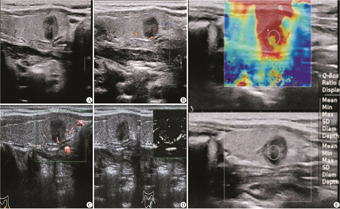
病人女,左侧甲状腺低回声结节,病理:甲状腺乳头状癌。灰阶超声显示,外形欠规整,轮廓欠佳(见图 1A);彩超示其边缘有少许血流(见图 1B);cSMI示血流由周边向内部走行(见图 1C);mSMI成像显示,结节的血流由周边向内部延伸,呈“纠集征”,Adler血流分级为Ⅲ级(见图 1D);SWE成像,弹性平均值Mean=100.7 kPa(见图 1E)。

图 1 典型病例
-
甲状腺肿瘤血管的形成尤为重要,但在100 μm以下的血管中检测血液的流动信号时敏感性较差,因此,采用彩超评估甲状腺肿物的血管存在局限性。SMI是一种新型滤波手段,主要用于消除杂波,显示极低流速的血流信号,同时能够区分伪像[8]。如YOO等[9]报道,SMI可通过甲状腺结节诊断更清楚地显示结节的微小血流信号;SWE技术能较好地反映硬度信息,适用范畴越来越广泛[10]。
YOON等[11]报道3、4类结节的恶性率参考范围为1.7%和3.3%~72.4%。本研究结果显示,3类、4a类、4b类和4c类结节的恶性率分别0%、8.00%、46.00%、44.00%,这与国外研究报道相近,可见甲状腺恶性结节主要在4类和5类中常见;恶性组的风险分层和量化评分优于良性组,表明甲状腺恶性结节的危险分级更高,可作为筛查甲状腺结节的首选方法。然而,此法仅适用于灰阶图像,未包含血流及硬度情况,诊断时仅应用二维特征来进行鉴别是不够的,因而区分4类结节的亚型尤显紧要。
甲状腺病变的生长与血管的生成和分布紧密相连。特别是细微血管,通常彩色多普勒仅呈现高流速、大管径的血流信息。本研究结果显示,良、恶性组的SMI血流等级差异有统计学意义,恶性组血流分级主要为Ⅱ、Ⅲ级为,良性组主要为0、Ⅰ级。SMI能更好地显示低速细微血管。SWE具有操作独立性、可重复性和通过跟踪组织内横波传播的组织定量等优点,可作为一种评估甲状腺结节的新技术[12]。本研究中SWE中,恶性组弹性最大值及弹性平均值均高于良性组,说明SWE技术能较好地反映甲状腺结节的硬度,从而为临床诊断提供依据。据报道[6]SMI与SWE联合应用对良恶性结节的敏感性和特异性为91.3%和92.5%。
本研究ROC曲线分析灵敏度、特异性为92.00%、94.29%,均胜于单独采取TI-RADS分级诊断。另外,联合检查时,ROC曲线下的面积最大为0.937。因此,联合诊断的效能最佳。彩色多普勒对低速血流灵敏度差,加之甲状腺结节复杂多样,常常存在误诊的可能,因此灰阶超声检查、TI-SADS的单独诊断受限[13]。SMI对检测微血流信号更敏感,能够较真实反映结节内部的血流情况,但SMI的诊断特异性无明显提高。本组采用SMI诊断恶性结节的特异性仅为82.86%,可能是由于良、恶性病变的血流分布模式存在重叠,不易辨识微小钙化和细微血管。若结节中存在微钙化,如果结节的血流信号被错误识别,结节分类就会上调一级,所以实际工作中首先要仔细观察声像图,明确是否有微钙化[14]。在本研究中,SWE检测4个结节出现假阳性结果,分析原因可能是因良性结节合并钙化后组织变硬,病灶受力形变小,弹性参数相对于不合并钙化的甲状腺良性结节增高,这会导致假阳性,另3枚病变假阴性,考虑与其体积、位置相关。因此,基于病灶的硬度和血液流动的特征,调整新的分类标准,并评价鉴定甲状腺结节良性的临床价值[15]。
综上所述,TI-RADS分类、SMI和SWE三种诊断技术均具有一定的价值,特别是将三者联合诊断时效能显著提高。
ACR TI-RADS危险分级结合多模态超声影像技术对甲状腺良恶性病变的诊断价值
Diagnostic value of ACR TI-RADS risk grading combined with multimodal ultrasonography in benign and malignant thyroid lesions
-
摘要:
目的探讨美国放射学会(ACR)甲状腺TI-RADS风险分层与多模态超声对甲状腺病变的诊断价值。 方法对61例病人进行超微血管(SMI)及剪切波弹性成像扫查(SWE)。最终根据手术获取的病理报告将结节分成良性和恶性组,比较TI-RADS风险分层及定量评分,并对2组的SMI血流分类和SWE弹性参数进行检测并分析。 结果2组TI-RADS均分为2~5类,危险分级差异有统计学意义(P<0.01);恶性组量化评分高于良性组(P<0.01)。2组SMI血流分级、SWE弹性参数比较差异均有统计学意义(P<0.01)。联合诊断恶性结节的敏感性、特异性均强于单独诊断。 结论ACR TI-RADS风险分层、SMI和SWE单独诊断甲状腺病变均有价值,联合诊断的效能显著提高。 -
关键词:
- 甲状腺肿瘤 /
- ACR TI-RADS危险分级 /
- 超微血管成像 /
- 剪切波
Abstract:ObjectiveTo analyze diagnostic value of American College of radiology thyroid imaging reporting and data system(ACR TI-RADS) risk grading combined with multimodal ultrasonography in benign and malignant thyroid lesions. MethodsSixty-one patients were detected using superb microvascular imaging(SMI) and shear wave elastography(SWE), and divided into benign group and malignant group according to the pathological results.The ACR TI-RADS risk grading and quantitative scoring in two groups were implemented, and the SMI blood flow grading and SWE elastic parameters in two groups were detected and analyzed. ResultsThe ACR TI-RADS risk grading in two groups were divided into 2-5 classes, and the difference of risk grading was statistically significant(P < 0.01).The quantitative scores in malignant group were higher than those in benign group(P < 0.01).The differences of the SMI blood flow grading and SWE elastic parameters between two groups were statistically significant(P < 0.01).The sensitivity and specificity of combined diagnosis of malignant nodules were better than that of single diagnosis. ConclusionsThe risk stratification of ACR TI-RADS, SMI and SWE are all valuable in the diagnosis of thyroid lesions alone, and the efficacy of combined diagnosis can be significantly improved. -
表 1 良性组和恶性组TI-RADS风险分层比较[n; 百分率(%)]
分组 n 2类 3类 4a类 4b类 4c类 5类 uc P 良性组 35 1(2.86) 27(77.14) 7(20.00) 0(0.00) 0(0.00) 0(0.00) 7.98 <0.01 恶性组 50 0(0.00) 0(0.00) 4(8.00) 23(46.00) 22(44.00) 1(2.00) 合计 85 1(1.18) 27(31.76) 11(12.84) 23(27.06) 22(25.88) 1(1.18)  下载: 导出CSV
表 2 良、恶性组SWE测值对比(x±s;kPa)
分组 n 弹性最大值 弹性平均值 良性组 35 104.41±10.26 93.49±9.51 恶性组 50 125.47±13.63 112.67±14.37 t — 7.73 6.90 P — < 0.01 < 0.01
下载: 导出CSV
表 3 联合诊断甲状腺结节的结果(n)
诊断方法 类型 病理结果 合计 恶性 良性 ACR TI-RADS危险分级 恶性 36 3 39 良性 14 32 46 SMI 恶性 37 6 43 良性 13 29 42 SWE 恶性 34 5 39 良性 16 30 46 三者联合诊断 恶性 46 2 48 良性 4 33 37
下载: 导出CSV
表 4 ACR TI-RADS危险分级结合SMI、SWE诊断甲状腺恶性的效能比较
诊断方法 灵敏度/% 特异度/% 约登指数 AUC(95%CI) ACR TI-RADS危险分级 72.00 91.43 0.628 6 0.814(0.769~0.860) SMI 74.00 82.86 0.557 2 0.779(0.742~0.841) SWE 68.00 85.71 0.553 5 0.777(0.710~0.840) 三者联合诊断 92.00 94.29 0.853 6 0.927(0.892~0.967)
下载: 导出CSV
-
[1] ZHAO YF, ZHOU P, PENG H, et al. Superb microvascular imaging comparedwith contrast-enhanced ultrasound to assess microvessels in thyroid nodules[J]. J Med Ultrason, 2020, 47(2): 287. doi: 10.1007/s10396-020-01011-z [2] 姚建锋, 张煜华, 王全江, 等. ACR TI-RADS与Kwak TI-RADS对比在甲状腺结节定性诊断中的效能[J]. 中国临床医学影像杂志, 2019, 30(8): 537. [3] ERCAN A, AHMET A, ĪBRAHIMĪ, et al. Evaluation of ovarian vascularity in children by using the "Superb Microvascular Imaging" ultrasound technique in comparison with conventional Doppler ultrasound techniques[J]. J Ultrasound Med, 2019, 38(10): 2751. doi: 10.1002/jum.14983 [4] 陶玲玲, 詹维伟, 樊金芳, 等. 超微血管成像结合TI-RADS鉴别诊断甲状腺良恶性结节[J]. 中国医学影像技术, 2020, 36(5): 671. [5] TESSLER FN, MIDDLETON WD, GRANT EG, et al. ACR Thyroid Imaging, Reporting and Data System (TI-RADS): White Paper of the ACR TI-RADS Committee[J]. J Am Coll Radiol, 2017, 14(5): 587. doi: 10.1016/j.jacr.2017.01.046 [6] 纪欢, 张蕾, 李守强, 等. 剪切波弹性成像联合超微血管显像在甲状腺肿块良恶性鉴别诊断中的应用[J]. 中华超声影像学杂志, 2018, 27(2): 143. [7] ADLER DD, CARSON PL, RUBIN JM, et al. Doppler ultrasound color flow imaging in the study of breast cancer: preliminary findings[J]. Ultrasound Med Biol, 1990, 16(6): 553. doi: 10.1016/0301-5629(90)90020-D [8] 张剑, 陈卉, 徐斌, 等. 超微血管成像、高级动态血流显像、彩色多普勒血流显像对乳腺微小癌的诊断价值及其与病理肿瘤微血管密度的相关性研究[J]. 中华超声影像学杂志, 2019, 28(9): 787. [9] YOO J, JE BK, JI YC. Ultrasonographic demonstration of the tissue microvasculature in children: microvascular ultrasonography versus conventional color Doppler ultrasonography[J]. Korean J Radiol, 2020, 21(2): 146. [10] 阮吟, 石彦, 宁艳, 等. 常规超声BI-RADS分类结合实时剪切波弹性成像对三阴性乳腺癌的诊断价值[J]. 蚌埠医学院学报, 2020, 45(5): 630. [11] YOON JH, LEE HS, KIM EK, et al. Malignancy risk stratification of thyroid nodules comparison between the thyroid imaging reporting and data system and the 2014 American thyroid association management guidelines[J]. Radiology, 2016, 278(3): 917. [12] 施国荣, 方丽丽, 沈红英, 等. 乳腺影响报告和数据系统分级联合剪切波弹性成像对鉴别甲状腺结节良恶性的诊断价值[J]. 实用临床医药杂志, 2019, 23(22): 8. [13] 李小娟, 温德惠, 张利英, 等. TI-RADS联合SMI技术对甲状腺良恶性结节的鉴别诊断价值[J]. 山西医科大学学报, 2018, 49(6): 670. [14] 赵永锋, 周平, 彭洪, 等. 超微血管成像及超声造影在甲状腺结节鉴别诊断中的应用[J]. 中南大学学报(医学版), 2019, 44(6): 649. [15] 李永红. TI-RADS、SWE与SMI联合应用对甲状腺良恶性结节的鉴别诊断价值[J]. 中国实用医刊, 2019, 46(1): 47. -

 点击查看大图
图(1)表(4)
计量
- 文章访问数: 3539
- HTML全文浏览量: 2032
- PDF下载量: 5
- 被引次数: 0
