


-
目前,乳腺癌已经成为世界上严重威胁女性生命健康的恶性肿瘤之一。乳腺癌发病原因并不明确,一般与病人的内分泌水平及遗传因素存在相关性,高发人群为40~60岁围绝经妇女,且于近年来呈现年轻化之趋[1-2]。临床循证研究[3]表明, 早诊断、早治疗是提高乳腺癌病人生存期的关键。目前,乳腺彩色多普勒超声和X线钼靶是乳腺癌早期普查和临床检测手段运用最多的方法,两种方法的检查结果在一定程度上对临床治疗及手术方式的选择起到了至关重要的影响。但由于乳腺癌病理分型具有复杂性及多样性,尤其针对早期乳腺癌,声像表现不典型,给诊断工作带来了困难[4-5]。本研究通过对太和县中医院拟行乳腺癌保乳术的病人进行了超声弹性成像技术联合钼靶X线检查方法诊断,克服了临床上单一超声和钼靶X线检查准率低的弊端。现作报道。
-
收集2017年2月至2019年12月期间因乳腺癌在太和县中医院外科拟行保乳术的84例病人。纳入标准:(1)以往有生育史;(2)术前经乳腺肿物穿刺活检病理学确诊;(3)对本研究所行保乳手术治疗方案均知情且已经签订知情同意书等。排除标准:(1)术前经乳腺肿物穿刺活检病理学无法确诊确诊;(2)合并严重心、脑、肝、肾等疾病无法耐受手术的病人;(3)病人拒绝行保乳手术。84例病人年龄30~45岁;病程2~8个月;浸润性导管癌39例,浸润性小叶癌25例,导管内原位癌20例。
-
采用ClearVue 8彩色多普勒超声诊断仪(东芝公司生产)进行检查。病人取仰卧位,上举双臂显露双侧乳腺以观察乳腺是否存在凹陷、突出和皮肤颜色等改变。用探头接触乳腺,频率设定为7.5~12 MHz,以乳头为中心顺时针对乳房肿物的位置、形态、大小、边界、回声、纵横比、是否钙化、有无衰减、血流信号、频谱和腋窝淋巴结等进行观察,然后将取样框的面积扩大到病灶大小的2倍,将探头垂直于体表,并施加外力,并对比病灶区域和周围组织硬度,对乳腺肿物弹性图像进行评分。在行超声操作时可采用多种扫描方式相结合,如扇形扫查、纵扫和横扫三者联合。探及到乳腺肿物时要综合、多角度观察病灶与周围腺体、血管和乳腺导管的关系,着重收集病灶内部及边缘血流信号、血流数量、血流形态以及血流阻力指数等情况。
-
乳腺钼靶检查采取SenoClaire型号钼靶X线(CE公司生产)进行检查。病人常规取头尾位和内外斜位,并根据病人病情需要加摄侧位和外内斜位等。记录病人双侧乳房的轮廓、乳房肿物的位置、形态、大小、边界、内部回声、密度及是否钙化等。
-
超声弹性成像评分:1分,病灶为整体的绿色,无蓝色呈现;2分,病灶中央范围为蓝色,周边组织呈现绿色;3分,蓝色区域与绿色区域的面积比近似为1∶1;4分,病灶大部分为蓝色,仅有周围少量绿色区域;5分,病灶以及周边组织全部呈现为蓝色。弹性评分≤3分系良性病变,弹性评分≥4分系恶性病变。
-
统计比较乳腺超声弹性成像、钼靶X线及2种方法联合检查对乳腺病灶和腋窝淋巴结的检出率,并与乳腺病灶病理结果对照;3种检查方法对病人是否可行保乳术的准确度比较,准确度=(阳性例数+阴性例数)/总例数×100%。
-
采用χ2检验。
-
结果显示,乳腺超声弹性成像联合钼靶X线对乳腺病灶的检出率和病理学结果浸润性导管癌检出率均高于单一乳腺超声弹性成像及单一乳腺钼靶X线的检出率,差异均有统计学意义(P < 0.05~P < 0.01);本次研究纳入的乳腺癌病人保乳术后腋窝淋巴结病理结果提示有41例转移,在腋窝淋巴结转移检出率方面,乳腺超声弹性成像联合钼靶X线检查在病灶检出率为70.73%,乳腺超声弹性成像检出率为60.98%,乳腺钼靶X线检出率为53.31%。联合检查的检出率高于单一乳腺超声弹性成像和单一钼靶X线的检出率,但差异无统计学意义(P>0.05)(见表 1)。
检测方法 病灶检出率/% 淋巴结检出率/% 病理结果/% 浸润性导管癌 浸润性小叶癌 导管内原位癌 乳腺超声弹性成像 83.33(70/84) 60.98(25/41) 82.05(32/39) 84.00(21/25) 85.00(17/20) 钼靶X线 78.57(66/84) 53.31(22/41) 76.92(30/39) 80.00(20/25) 80.00(16/20) 乳腺超声弹性成像联合钼靶X线 95.24(80/84)*△△ 70.73(29/41) 97.44(38/39)*△ 92.00(23/25) 95.00(19/20) χ2 10.11 2.55 7.16 1.59 2.26 P < 0.01 >0.05 < 0.05 >0.05 >0.05 注:与乳腺超声弹性成像组比较,*P < 0.05;与钼靶X线组比较比较,△P < 0.05, △△P < 0.01 表 1 3种检查方法的病灶、腋窝淋巴结检出率及病理结果比较
-
术前行乳腺超声弹性成像联合钼靶X线检查,对是否可行保乳术的准确度高于超声弹性成像组(χ2=3.86,P < 0.05)和乳腺钼靶X线组(χ2=6.93, P < 0.01)(见表 2)。
分组 病理结果 可保乳 不可保乳 乳腺超声弹性成像 可保乳 51 14 不可保乳 7 12 钼靶X线 可保乳 41 15 不可保乳 10 18 乳腺超声弹性成像联合钼靶X线 可保乳 65 9 不可保乳 2 8 表 2 3种检查方法对病人是否可行保乳术的评估比较(n)
-
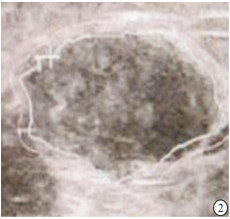
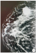
病人,女,43岁,行右侧乳腺癌保乳术,术后病理为浸润性导管癌,Ⅲ级。术前头足位钼靶,肿块边界尚清,BI-RADS 4a(见图 1); 术前二维超声图,显示一边界清晰的低回声肿块,内部回声不均匀(见图 2); 术前超声弹性图,显示肿块弹性成像特点为红绿混合,以绿色为主的表现,评分3分(见图 3)。
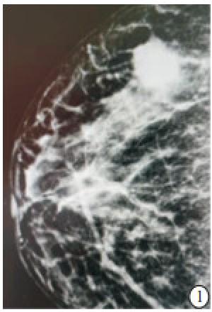
图 1 术前头足位钼靶
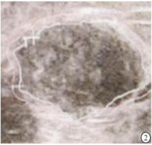
图 2 术前二维超声图
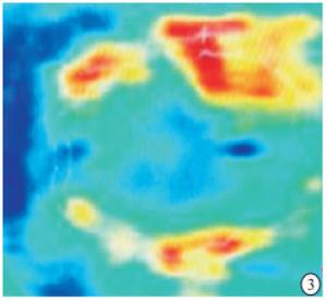
图 3 术前超声弹性图
-
随着乳腺癌的发病率逐年升高,对乳腺治疗方式的研究越来越深入,研究[6-7]发现,针对乳腺癌疾病而言,早期浸润性乳腺癌较之非浸润性乳腺癌的预后相对更差。值得注意的是,由于此类疾病的早期症状隐匿,不会出现显著临床表现,且其术后生存率和疾病分期存在紧密关联。因此早期诊断、早期治疗已经成为提高乳腺癌生存期的关键。目前认为保乳手术也是乳腺癌有效治疗手段之一,不仅可以达到同样的生存期,还可以尽量减少病人心理的负担和打击,极大的提高了病人的生存质量[8-9]。
目前临床上乳腺癌的检测手段主要是乳腺彩色多普勒超声和乳腺X线钼靶。这两种方法均可以为乳腺癌手术方案提供较为准确的依据[10-14]。但是近些年来研究发现乳腺癌病灶内部及周围组织明显的间质反应导致纤维组织增生,硬度值升高,从而使乳腺常规超声和钼靶检查的特异性降低[15]。同时乳腺超声检查在检查乳腺病灶时存在假阴性以及对成簇状微小钙化的检出率极低、敏感性不如X线等缺点[16]。因此,近些年出现的弹性成像技术通过获得乳腺组织的硬度信息,弥补了传统超声在诊断乳腺癌时特异度低、假阳性率高的不足,对提高乳腺癌诊断的准确度具有重大意义。世界医学生物学超声联合会于2015年指出超声弹性成像技术是二维超声的补充。LEE等[17]研究表明在二维超声的基础上运用弹性成像技术对乳腺进行联合检查未出现乳腺癌漏诊情况,避免了67.7%的活检手术,发现乳腺病灶的特异度提高到了近3倍,阳性预测值也提高到两倍多。PU等[15]研究表明乳腺弹性成像可以对二维超声和钼靶X线检查的乳腺肿块进行再评估,从而明确诊治方法和发现隐匿型病变。同时乳腺弹性成像在评估乳腺肿物和乳腺病理类型、预测评估乳腺癌新辅助化疗效果、判断乳腺癌是否合并腋窝淋巴结转移和病灶边界的评估等方面也显现出了明显的优势和敏感性[18-21]。
但是目前关于乳腺弹性成像技术单独或者联合运用于乳腺癌保乳手术的评估方面的研究相对较少,从而限制了该技术在乳腺癌保乳手术的运用,基于此本研究通过搜集临床上乳腺癌病人,术前评估均运用乳腺超声弹性成像技术和乳腺钼靶X线单独和联合检查,并与病理结果做比较。本研究表明对于乳腺二维超声和钼靶X线检测时乳腺病灶中的钙化病变显示的不全,后通过乳腺弹性成像检测得到证实和确诊,有效的弥补了乳腺二维超声和钼靶X线的不足,因乳腺超声弹性成像技术有利于区分病灶边界,更加全面地观察早期乳腺肿物内部回声和形态,提高早期乳腺肿物的检出率,增加诊断的灵敏度和特异度,为早期乳腺癌保乳手术提供参考。
本研究结果显示乳腺超声弹性成像检查联合乳腺钼靶的乳腺病灶的检出率提高96.12%,明显更高于单一乳腺超声检查和单一的乳腺钼靶检测;在腋窝淋巴结转移检出率方面,乳腺超声弹性成像联合钼靶X线检查在病灶检出率为70.73%,乳腺超声弹性成像检出率为60.98%,乳腺钼靶X线腋检出率为53.31%.联合检查的检出率高于单一钼靶X线的检出率, 但差异无统计学意义,本研究组考虑系本研究临床病例数量较少,需待后续的研究中增加样本量,进一步明确。
本研究中,通过超声弹性成像联合乳腺钼靶检测结果进行乳腺癌的保乳手术评估,术前行乳腺弹性成像联合钼靶X线检查,判断保乳术的准确度高于单一乳腺超声弹性成像检测和单一钼靶X线线检查,从而在早期诊疗和治疗时机的把握上具有明显优势,两者互有侧重,充分的弥补了二者独立应用的不足。合理地用全数字化乳腺摄影联合超声成像检查进行成像技术上的优势互补,可以最大程度提供完整的影像学资料,对于评估肿瘤的分级和预后,正确分析保乳手术的可行性,制定规范的手术细节以及术后化疗药物的疗效检测,术后随访等工作有着极为重要的意义。两者联合应用取长补短,相得益彰,可对乳腺癌保乳手术的广泛开展提供十分重要的价值。总之,乳腺超声弹性成像和钼靶X线在乳腺癌病人早期诊断中各具优势,两者联用可以更有助于提高疾病的诊断率,具有更高的临床应用价值。
超声弹性成像联合钼靶X线在乳腺癌手术中的应用研究
Study on the application value of ultrasound elastography combined with mammography in the breast cancer surgery
-
摘要:
目的探讨超声弹性成像技术联合钼靶X线在乳腺癌保乳手术中的临床应用效果。 方法搜集因乳腺癌拟行保乳手术病人84例,术前评估均运用乳腺超声弹性成像技术和乳腺钼靶X线单独及联合检查。并在术后与病理结果作比较,明确乳腺超声弹性成像技术联合钼靶X线与单一超声和钼靶X线诊断对乳腺癌拟行保乳术前的影像学表现和准确度。 结果乳腺超声弹性成像联合钼靶X线的病灶检出率高于单一的乳腺超声弹性成像检测或单一的钼靶X线检查的病灶检出率(P < 0.05和P < 0.01);在腋窝淋巴结转移检出率方面,乳腺超声弹性成像联合钼靶X线检查在病灶检出率为70.73%,乳腺超声弹性成像检出率为60.98%,乳腺钼靶X线检出率为53.31%,但差异无统计学意义(P>0.05);在可行保乳术的准确度方面联合检查均高于单一的乳腺超声弹性成像和钼靶X线(P < 0.05)。 结论超声弹性成像和乳腺钼靶联合检查运用于乳腺癌保乳术更有助于提高疾病的准确率,为临床提供更多的参考价值。 Abstract:ObjectiveTo investigate the clinical application value of ultrasound elastography combined with molybdenum target in the breast conserving surgery of breast cancer. MethodsEighty-four breast cancer patients scheduled by breast conserving surgery were detected using ultrasound elastography and mammography alone and in combination before operation.The imaging findings and accuracy of breast ultrasound elastography combined with mammography and mammography alone were determined by comparing with the postoperative pathological results. ResultsThe detection rate of breast lesions detected by ultrasound elastography combined with molybdenum target X-ray was significantly higher than that detected by ultrasound elastography alone and mammography alone(P < 0.05 and P < 0.01).The detection rates of axillary lymph node metastasis of ultrasound elastography, mammography and ultrasound elastography combined with mammography were 60.98%, 53.31% and 70.73%, respectively, but the difference of which was not statistically significant(P>0.05).The ultrasound elastography combined with mammography in evaluating the feasibility of breast conserving surgery was more accurate than the other two methods(P < 0.05). ConclusionsThe application of ultrasound elastography combined with mammography in breast conserving surgery is more helpful to improve the accuracy of the disease, and provide more reference value for clinical practice. -
Key words:
- breast neoplasms /
- breast conserving therapy /
- ultrasound elastography /
- mammography
-
表 1 3种检查方法的病灶、腋窝淋巴结检出率及病理结果比较
检测方法 病灶检出率/% 淋巴结检出率/% 病理结果/% 浸润性导管癌 浸润性小叶癌 导管内原位癌 乳腺超声弹性成像 83.33(70/84) 60.98(25/41) 82.05(32/39) 84.00(21/25) 85.00(17/20) 钼靶X线 78.57(66/84) 53.31(22/41) 76.92(30/39) 80.00(20/25) 80.00(16/20) 乳腺超声弹性成像联合钼靶X线 95.24(80/84)*△△ 70.73(29/41) 97.44(38/39)*△ 92.00(23/25) 95.00(19/20) χ2 10.11 2.55 7.16 1.59 2.26 P < 0.01 >0.05 < 0.05 >0.05 >0.05 注:与乳腺超声弹性成像组比较,*P < 0.05;与钼靶X线组比较比较,△P < 0.05, △△P < 0.01  下载: 导出CSV
表 2 3种检查方法对病人是否可行保乳术的评估比较(n)
分组 病理结果 可保乳 不可保乳 乳腺超声弹性成像 可保乳 51 14 不可保乳 7 12 钼靶X线 可保乳 41 15 不可保乳 10 18 乳腺超声弹性成像联合钼靶X线 可保乳 65 9 不可保乳 2 8
下载: 导出CSV
-
[1] ENGEL JM, STANKOWSKI-DRENGLER TJ, STANKOWSKI RV, et al. All-cause mortality is decreased in women undergoing annual mammography before breast cancer diagnosis[J]. Ajr Am J Roentgenol, 2015, 204(4): 898. doi: 10.2214/AJR.14.12666 [2] 郭聪敏. 超声血流参数对乳腺癌的诊断价值分析[J]. 医学理论与实践, 2020, 33(3): 463. [3] SIEGEL RL, MILLER KD, JENMAL A. Cancer statistics. 2018[J]. CA Cancer J Clin, 2018, 68(1): 7. doi: 10.3322/caac.21442 [4] 陆梦鸥, 陈方红, 杨伟斌. X线钼靶联合彩色多普勒超声在乳腺癌筛查中的应用价值[J]. 全科医学临床与教育, 2020, 18(1): 32. [5] 叶芬, 王丽萍. 超声显像特征与乳腺癌组织中HER-2表达的关系研究[J]. 实用癌症杂志, 2020, 35(1): 50. doi: 10.3969/j.issn.1001-5930.2020.01.014 [6] 曹文, 张尹. MR增强扫描和X线钼靶应用于早期乳腺癌诊断价值对比研究[J]. 四川医学, 2016, 37(6): 674. [7] 阮吟, 石彦, 宁艳, 等. 常规超声BI-RADS分类结合实时剪切波弹性成像对三阴性乳腺癌的诊断价值[J]. 蚌埠医学院学报, 2020, 45(5): 630. [8] DOWN SK, JHA PK, BURGE A, et al. Oncological advantages of oncoplastic breast-conserving surgery in treatment of early breast cancer[J]. Breast J, 2013, 19(1): 56. doi: 10.1111/tbj.12047 [9] DIRIX LY, TAKACS I, JERUSALEM G, et al. Avelumab, an anti-PD-L1 antibody, in patients with locally advanced or metastatic breast cancer: a phase 1b JAVELIN solid tumor study[J]. Breast Cancer Res Treat, 2018, 167(3): 671. doi: 10.1007/s10549-017-4537-5 [10] 吕坤, 徐丽艳. 乳腺钼靶X线片簇状钙化灶对乳腺癌的诊断价值[J]. 安徽医学, 2017, 38(1): 42. doi: 10.3969/j.issn.1000-0399.2017.01.011 [11] 吕彦利, 康秀梅, 岳胜南, 等. 乳腺癌的彩色多普勒血流特征与ER、PR、HER-2、P53和KI-67表达的相关性研究[J]. 医学影像学杂志, 2019, 29(11): 1919. [12] BARR RG, DESTOUNIS S, LACKEY LB, et al. Evaluation of breast lesions using sorographic elasticity imaging: a multicenter trial[J]. J Ultrasound Med, 2012, 31(2): 281. doi: 10.7863/jum.2012.31.2.281 [13] 刘芳欣, 王洲, 李健, 等. 不同声触诊组织成像定量技术在乳腺良恶性结节鉴别诊断中的应用价值[J]. 蚌埠医学院学报, 2020, 45(2): 238. [14] 陈光玉, 金永红, 项金凤. 超声对乳腺结节BI-RADS分类的声像图表现与病理结果的对比分析[J]. 蚌埠医学院学报, 2019, 44(8): 1097. [15] PU H, ZHANG XL, XIANG LH, et al. The efficacy of added shear wave elastography(SWE) in breast screening for women with inconsistent mammography and conventional ultrasounds(US)[J]. Clin Hemorheol Microcirc, 2019, 71(1): 83. doi: 10.3233/CH-180398 [16] 石玲萍, 刘艳, 蒋顺和, 等. 乳腺癌患者保乳术前超声联合钼靶X射线评估的效果[J]. 中国肿瘤临床与康复, 2019, 26(6): 701. [17] LEE SH, CHUNG J, CHOI HY, et al. Evaluation of screening US-detected breast masses by combined use of elastography and color Doppler US with B-mode US in women with dense breasts: a multicenter prospective study[J]. Radiology, 2017, 285(2): 660. doi: 10.1148/radiol.2017162424 [18] ZHU YC, ZHANG Y, DEN SH, et al. Correlation between histopathological grading and shear-wave elastography in evaluating invasive carcinoma of no special type[J]. Exp Ther Med, 2018, 16(6): 4700. [19] EVANS A, SIM YT, POURREYRON C, et al. Pre-operative stromal stiffness measured by shear wave elastography is independently associated with breast cancer-specific survival[J]. Breast Cancer Res Treat, 2018, 171(2): 383. doi: 10.1007/s10549-018-4836-5 [20] YEO SH, KIM GR, LEE SH, et al. Comparison of ultrasound elastography and color Doppler ultrasonography for distinguishing small triple -negative breast cancer from fibroadenoma[J]. J Ultrasound Med, 2018, 37(9): 2135. doi: 10.1002/jum.14564 [21] WANG D, ZHU K, TIAN J, et al. Clinicopathological and ultrasonic features of triple-negative breast cancers: a comparison with hormone receptor-positive/human epidermal growth factor receptor-2-negative breast cancers[J]. Ultrasound Med Biol, 2018, 44(5): 1124. doi: 10.1016/j.ultrasmedbio.2018.01.013 -

 点击查看大图
图(3)表(2)
计量
- 文章访问数: 3111
- HTML全文浏览量: 1478
- PDF下载量: 6
- 被引次数: 0
