

-
近年我国乳腺癌的发病率、发现率呈不断上升趋势[1],引发社会各界的广泛关注。乳腺癌有多种检测方法,但超声由于其安全、可靠、便捷的优势,仍然是乳腺癌筛查的首选[2]。普通二维彩色超声可综合应用形态、边界、内部回声、钙化等进行全面的分类评价,临床上有较高参考价值[3]。此外在彩色多普勒技术的发展带动下,有研究者[4]认为在常规超声中引入血管征象有利于提高诊断准确率,不过在实际应用过程中,由于彩超对低速血流的敏感性低,且难以进行量化分析,因而其应用价值和效果并未明确[5-6]。超微血管成像技术(SMI) 可较好地弥补这种缺陷,其具有彩色模式超微血管成像和灰阶模式超微血管成像,对血流信号可进行更加敏感的提取[7]。其优势为微血管自发显影,能够直观、整体呈现病变微血管特征,更加充分地明确乳腺病变血管特征。本研究分析SMI应用于乳腺良恶性病变鉴别诊断的效果。现作报道。
-
采集2020年9月至2022年1月在我院进行进行诊治的乳腺肿瘤病人75例,其中确诊为乳腺良性肿瘤病人45例,乳腺恶性肿瘤病人30例,共获得有效的病灶结节数量为94个,均为女性,年龄24~78岁,肿瘤长径0.5~4.6 cm。经病理活检证实,其中良性结节45个(良性组),恶性结节49个(恶性组)。2组年龄、肿瘤长径均具有可比性。
-
采用Aplio 500A型彩超仪(日本东芝公司),配置线阵高频5L12探头。告知病人保持仰卧位,举起双臂,充分暴露乳腺及腋窝,扫描采集二维灰阶图像,对肿瘤尺寸、边界、回声特征、纵横比相关情况进行观察分析;肿物血管丰富程度、血管分布特征及走行方向等通过SMI进行分析。对取样框尺寸和多普勒入射角度(0°~60°)等参数进行适当调节后,根据彩色多普勒检查要求进行详细检测,记录所得结果。
-
(1) 乳腺病变血管丰富与否:以Adler血管分级为依据,若Adler血管分级0级(未探及明显血管)或1级(探及很少量血管,1~2个点状或细条状血管),则属于血管信号不丰富型;若为2级(中量血管,内部探及到3~4个点状或1条重要血管(长度接近或超过肿物1/2)或3级(血管丰富,至少出现2条重要血管或探及5个以上点状血管),则属于血管信号丰富型[8]。(2)穿支血管:有无从结节外部贯穿入结节内部血管。(3)主干血管与分支血管夹角:根据主干血管的同在结节内走行的分支血管的夹角,分为直角、非直角两类。(4)血管走向:分为中心型和无明显中心型。
-
以2组病人各血管特征差异是否具有统计学意义(P < 0.05)为赋分依据,同时结合诊断乳腺恶性病变的阳性预测值(PPV),若血管特征在良恶性间差异P>0.05,则赋值0;若P < 0.05,则基于PPV赋值如下:PPV≥80%计3分,PPV>20%~ < 80%计2分,PPV≤20%计1分。
-
采用χ2检验和ROC曲线分析。
-
SMI显示,2组病人乳腺病变血管特征中,血管走向、主干与分支夹角、穿支血管差异均有统计学意义(P < 0.01),而2组血管丰富与否差异无统计学意义(P>0.05)(见表 1)。
分组 n 血管数量 血管走向 穿支血管 主干与分支夹角 不丰富 丰富 无中心 有中心 无 有 非直角 直角 良性组 45 12(66.7) 33(43.4) 39(63.9) 6(18.2) 29(61.7) 16(34.0) 39(79.6) 6(13.3) 恶性组 49 6(33.3) 43(56.6) 22(36.1) 27(81.8) 18(38.3) 31(66.0) 10(20.4) 39(86.7) χ2 — 3.15 17.96 7.21 41.27 P — >0.05 < 0.01 < 0.01 < 0.01 表 1 2组病人乳腺病变血管特征比较[n;百分率(%)]
-
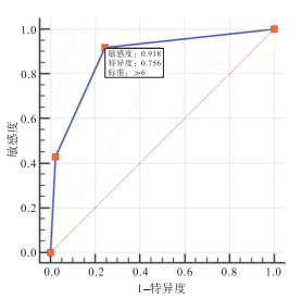
乳腺良性、恶性的敏感血管特征分别为乳腺恶性病变诊断时PPV≤20%、PPV≥80%,各血管分型的乳腺恶性病变诊断PPV与赋分见表 2。基于血管特征赋分绘制ROC曲线,将乳腺恶性病变诊断临界值确定为血管特征分值>6,ROC曲线下面积(AUC)为0.879,敏感性、特异性、准确性、PPV、阴性预测值分别为91.84%、75.56%、67.39%、80.4%、89.5%(见图 1~3)。
血管特征指标 PPV 赋分 血管数量 不丰富 33.30% 0 丰富 56.60% 0 血管走向 无中心 36.10% 2 有中心 81.80% 3 穿支血管 无 38.30% 2 有 66.00% 2 主干与分支夹角 非直角 20.40% 2 直角 86.70% 3 表 2 SMI诊断乳腺恶性病变PPV及赋分

图 1 血管特征诊断乳腺病变的ROC曲线

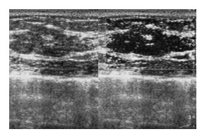
图 2 浸润性导管癌, 管径粗细不均匀, 走行迁曲, 可见杂乱细小分支

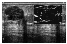
图 3 纤维腺瘤, 弥散分布狭细血管, 管径粗细较一致, 走行较平直
-
对于肿瘤的生长与转移,新生血管在支撑营养方面发挥着关键作用,故在肿瘤检查方面,明确血管特征可以极大地帮助分析其生物学特性,在预后评估方面提供关键指导[9]。乳腺肿物内部及其周边均可见明显的血管形成,通常良性肿瘤血管具有较均匀的管径,且呈平直走行。恶性肿瘤存在明显的浸润性表现,使得血管分支明显异常,呈较为迂曲走行,血管走行角度与乳腺良性肿物存在较大差异,并且血管管径的变化也较为复杂,故能够借助彩色多普勒方法对肿物血管形态特征开展定性分析评估[10]。本研究中,借助SMI获知病灶血管数量,借鉴Adler血管分级,发现就血管数量而言,乳腺良恶性病变间不存在明显区别,而其他3项血管特征(血管走向、穿支血管、血管主干与分支夹角)在乳腺良、恶性病变间均存在明显差异,可见通过SMI检查观察乳腺肿物血管特征,在乳腺肿物良恶性鉴别中存在较大临床意义。
上述三项有意义的血管特征,在鉴别乳腺肿物良恶性过程中,还存在着一定区别,通过本次研究可以发现,乳腺良性结节中比较有价值血管特征是血管主干与分支夹角非直角型,而在乳腺恶性结节中比较有价值的血管特征为中心型血管走向。为了在SMI模式视角下,更为全面地评价血管特点在乳腺良恶性结节中临床诊断价值,基于诊断乳腺恶性病结节PPV和各血管特征的统计学差异,对不同血管特征进行赋值,基于赋分的诊断模型,完成ROC曲线的绘制。基于AUC愈趋于1,诊断准确度愈高,本研究发现,若截断值为>6,AUC为0.879,敏感性、特异性、准确性、PPV、阴性预测值分别为91.84%、75.56%、67.39%、80.4%、89.5%。提示在乳腺病变方面,血管特征具有较高的临床诊断效能,对良恶性的鉴别诊断有重要意义。由于本次研究并未对血管特征中的血管参数进行分析,且研究样本量有所限制,今后仍需通过更具规模的大样本研究和在此基础上增加血管参数来进行进一步研究。
超微血管成像技术对乳腺良恶性结节血管特征诊断价值
Diagnostic value of superb microvascular imaging in the vascular characteristics of benign and malignant breast nodules
-
摘要:
目的通过超微血管成像技术(superb microvascular imaging, SMI)分析乳腺良恶性结节中的血管特征表现,判断此项技术的运用前景与价值。 方法选取乳腺肿瘤病人75例,其中确诊为乳腺良性肿瘤病人45例,乳腺恶性肿瘤病人30例,共获得有效病灶结节94个,其中良性结节45个(良性组),恶性结节49个(恶性组)。通过SMI观察血管走向、血管主干与分支夹角、穿支血管和血管数量4项病变血管特征,以病理结果作为金标准,根据乳腺良恶性结节内血管特征的不同表现,采用ROC曲线分析SMI技术鉴别乳腺恶性结节内部血管特征的临界值,并进一步分析得出其敏感性与特异性指标。 结果在SMI技术的视角下,2组血管走行方向、血管主干与分支夹角、穿支血管差异均有统计学意义(P < 0.01),2组血管丰富情况差异无统计学意义(P>0.05)。SMI诊断乳腺恶性结节的ROC曲线下面积为0.879,敏感性91.84%、特异性75.56%、准确性67.39%、阳性预测值80.4%、阴性预测值89.5%。 结论SMI可以很好地反映乳腺良恶性结节内部血管特征区别,对于乳腺恶性病变有较好的诊断价值。 Abstract:ObjectiveTo analyze the vascular characteristics in benign and malignant breast nodules using superb microvascular imaging (SMI), and judge the application prospect and value of this technology. MethodsA total of 75 patients with breast cancer were selected, among them, 45 patients were diagnosed as benign breast tumors, 30 patients with malignant breast cancer.A total of 94 effective lesions and nodules were obtained, there were 45 nodules in benign group, 49 nodules in malignant group.Four pathological vascular characteristics were observed through SMI, including the direction of blood vessels, the angle between main vessels and branches, perforator vessel and vessel number.Taking pathological results as the gold standard, according to the different manifestations of vascular characteristics in benign and malignant breast nodules, ROC curve was used to analyze the cut-off value of SMI technology to identify the internal vascular characteristics of malignant breast nodules, and its sensitivity and specificity were further analyzed. ResultsFrom the perspective of SMI technology, there was significant difference in direction of blood vessels, angle between main blood stream and branch, and perforator vessel number(P < 0.01), however, there was no significant difference in the vascular abundance between the two groups(P>0.05).The area under the ROC curve of SMI in the diagnosis of breast malignant nodules was 0.879, the related indicators were also obtained, including sensitivity (91.84%), specificity(75.56%), accuracy(67.39%), positive predictive value (80.4%), and negative predictive value (89.5%). ConclusionsSMI can well reflect the difference of internal vascular characteristics between benign and malignant breast nodules, and has a good diagnostic value for malignant breast lesions. -
表 1 2组病人乳腺病变血管特征比较[n;百分率(%)]
分组 n 血管数量 血管走向 穿支血管 主干与分支夹角 不丰富 丰富 无中心 有中心 无 有 非直角 直角 良性组 45 12(66.7) 33(43.4) 39(63.9) 6(18.2) 29(61.7) 16(34.0) 39(79.6) 6(13.3) 恶性组 49 6(33.3) 43(56.6) 22(36.1) 27(81.8) 18(38.3) 31(66.0) 10(20.4) 39(86.7) χ2 — 3.15 17.96 7.21 41.27 P — >0.05 < 0.01 < 0.01 < 0.01  下载: 导出CSV
表 2 SMI诊断乳腺恶性病变PPV及赋分
血管特征指标 PPV 赋分 血管数量 不丰富 33.30% 0 丰富 56.60% 0 血管走向 无中心 36.10% 2 有中心 81.80% 3 穿支血管 无 38.30% 2 有 66.00% 2 主干与分支夹角 非直角 20.40% 2 直角 86.70% 3
下载: 导出CSV
-
[1] LEE EJ, CHANG YW, OH ES, et al. Reproducibility and diagnostic performance of the vascular index of superb microvascular imaging in real-time breast ultrasonography for evaluating breast masses[J]. Ultrasonography(Seoul, Korea), 2021, 40(3): 398. [2] UYSAL E, ÖZTVRK M, KILINÇER A, et al. Comparison of the effectiveness of shear wave elastography and superb microvascular imaging in the evaluation of breast masses[J]. Ultrasound Q, 2021, 37(2): 191. doi: 10.1097/RUQ.0000000000000562 [3] CHAE EY, YOON GY, CHA JH, et al. Added value of the vascular index on superb microvascular imaging for the evaluation of breast masses: comparison with grayscale ultrasound[J]. J Ultrasound Med, 2021, 40(4): 715. doi: 10.1002/jum.15441 [4] 车丹丹, 李玉宏, 于晓溪, 等. 超微血管成像技术对乳腺肿物的诊断价值[J]. 暨南大学学报(自然科学与医学版), 2019, 40(4): 352. [5] 蔡思曼, 王红燕, 张晓燕, 等. 智能三维超微血管成像评估乳腺病变血管的观察者间一致性分析[J]. 中华超声影像学杂志, 2020, 29(7): 613. doi: 10.3760/cma.j.cn131148-20200219-00089 [6] PARK AY, SEO BK, WOO OH, et al. The utility of ultrasound superb microvascular imaging for evaluation of breast tumour vascularity: comparison with colour and power Doppler imaging regarding diagnostic performance[J]. Clin Radiol, 2018, 73(3): 304. doi: 10.1016/j.crad.2017.10.006 [7] ZHANG XY, CAI SM, ZHANG L, et al. Association between vascular index measured via superb microvascular imaging and molecular subtype of breast cancer[J]. Front Oncol, 2022, 12(16): 861151. [8] 薛雯, 杨柳茵, 范丽, 等. 超微血管成像技术鉴别乳腺良恶性病变[J]. 中国医学影像技术, 2019, 35(1): 77. [9] 肖露,褚雯,王华. 超微血管成像技术对乳腺肿瘤血管形态分布特征及其诊断效能的初步分析[J]. 中华超声影像学杂志,2018,27(11):973. doi: 10.3760/cma.j.issn.1004-4477.2018.11.012 [10] 李响,康姝,王学梅,等. 超微血管成像与彩色多普勒血流成像在乳腺肿瘤诊断中的应用[J]. 中国医学影像技术,2015,31(5):663. -

 点击查看大图
图(3)表(2)
计量
- 文章访问数: 1956
- HTML全文浏览量: 860
- PDF下载量: 7
- 被引次数: 0
