


-
根尖周炎是一种常见的口腔炎症性疾病,特征是牙髓中的微生物感染引起根尖周组织的炎症反应和骨破坏[1-2]。有研究[2-3]表明,在根尖周炎的病损组织中可发现调节性T细胞、白细胞介素(interleukin, IL)、肿瘤坏死因子等各种免疫细胞及细胞因子。根尖周炎对病人的口腔健康及生活质量影响严重,为了解该疾病复杂的免疫反应,促进该疾病的治疗,免疫细胞因子在根尖周炎中的作用近年来成为研究的热点。IL-35是2007年COLLISON等国外学者新命名的一种在机体免疫系统中发挥重要作用的新型细胞因子, 其主要由调节性T细胞(regulatory T cell, Treg细胞)分泌产生, 与Treg有互相正向调节的作用[4-5]。叉头样转录因子3(foxhead box p3, Foxp3)在Treg发育分化和功能中是不可或缺的, 是其特异性标志物,检测Foxp3的表达量能够直接反映Treg的表达水平[6]。目前关于IL-35在根尖周炎中的研究较少,课题组前期研究证实了IL-35在人根尖周肉芽肿中有表达, 较正常牙龈组织显著增高[7],但其在根尖周炎中发挥什么作用,以及是否与Treg有关尚不清楚。因此本课题拟建立大鼠根尖周炎模型,通过实时荧光定量PCR检测在根尖周炎发展过程中IL-35、Foxp3 mRNA的表达情况,进而探讨IL-35在该疾病进展中的作用及其可能的机制。
-
SPF级SD大鼠40只(常州卡文斯动物实验公司);逆转录试剂盒(Fermentas,加拿大);TRIZOL(Invitrogen,美国);牙科手机MIO-35M(NSK,日本);实时荧光定量PCR仪(ABI,美国)等。
-
经蚌埠医学院伦理委员会批准(伦动科批字[2022]第371号),将40只SPF级SD大鼠随机分为5组,每组8只,禁食禁水12 h后,10%水合氯醛(4.5 mL/100 g) 腹腔麻醉,用牙科手机在下颌第一磨牙牙合面近中窝处开髓,髓腔打开后直接暴露于口腔环境中。大鼠苏醒后常规标准饲养, 分别于术后第0、7、14、21、28天各随机脱颈处死一组,取下颌第一磨牙根尖周组织和分离下颌骨。
-
将术后每组大鼠分离出的下颌骨置于4%多聚甲醛溶液中固定24 h后取出进行X线拍摄,观察下颌第一磨牙根尖暗影变化。
-
固定后的下颌骨进行10%EDTA脱钙, 不同浓度乙醇逐级脱水后石蜡包埋,置于切片机上制作近远中向4 μm厚的连续切片,按HE染色步骤对切片进行染色,后中性树胶封片,镜检观察下颌第一磨牙根尖周组织病理变化。
-
取大鼠根尖周组织加入Trizol,进行RNA的提取。后逆转录进行实时荧光定量PCR反应检测每组大鼠根尖周组织Foxp3、IL-35 mRNA相对表达水平。实时荧光定量PCR引物见表 1。
引物名称 上游引物 下游引物 Foxp3 5′GTA TCA CTT GCC ACT AAC C 3′ 5′GCT CTC TGC TTA TCT GT TC 3′ IL-35 5′CCT CCT AAA CCA CCT CAC TTC 3′ 5′GTC GGT GTG AAC GGA TTT G 3′ Gapdh 5′AGC CAG GCA ACT CTC ATT C 3′ 5′TCC CAT TCT CAG CCT TGA C 3′ 表 1 实时荧光定量PCR引物
-
采用单因素方差分析与LSD检验或Tanhane′s T2检验和Pearson相关分析。
-
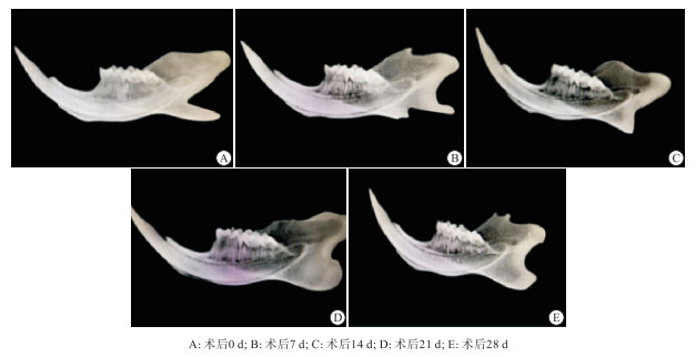
术后0 d: 第一磨牙根尖周影像未见明显异常;术后7 d: 第一磨牙根尖牙周膜间隙稍增宽,根尖骨组织密度降低;术后14 d: 第一磨牙根尖周牙周膜间隙增宽明显,根尖周见低密度病变区;术后21 d: 第一磨牙牙周膜间隙持续加宽,根尖可见稍变大的低密度阴影;术后28 d: 根尖低密度阴影较术后21 d组未见较明显改变(见图 1)

图 1 大鼠下颌第一磨牙根尖周炎术后X线片
-
术后0 d: 可见具有正常特征的根尖周区域,包括牙周韧带和牙骨质区域,未见明显的炎性细胞浸润;术后7 d: 在牙槽骨和牙周韧带区域可见明显炎性细胞聚集;术后14 d: 牙槽骨与牙周韧带区域除炎性细胞聚集外还可见朗罕氏巨细胞;术后21 d: 炎症浸润区域进一步扩大,可见多核巨细胞与破骨细胞,伴有局部骨溶解;术后28 d: 炎症浸润较21 d有所减轻,可见破骨细胞及骨溶解(见图 2)。

图 2 大鼠下颌第一磨牙根尖周炎组织HE染色(蓝色箭头示炎性细胞; 黑色箭头示郎罕巨细胞; 黄色箭头示多核巨细胞; 橙色箭头示破骨细胞; 绿色方框示骨溶解)
-
RT-qPCR结果显示,术后IL-35、Foxp3 mRNA相对表达量在不同时期中呈波动性升高趋势(P < 0.01): 术后0~7 d迅速上升,7~21 d表达量呈下降趋势(P < 0.05),21~28 d又呈上升趋势(P < 0.05) (见表 2)。
分组 n IL-35 mRNA Foxp3 mRNA 术后0 d 8 0.020 9±0.0020 0.021 5±0.002 3 术后7 d 8 0.028 1±0.003 7* 0.028 0±0.002 5* 术后14 d 8 0.024 4±0.002 6*△ 0.025 0±0.001 8*△ 术后21 d 8 0.024 1±0.002 9*△ 0.024 7±0.001 5*△ 术后28 d 8 0.027 7±0.003 0*#▲ 0.026 9±0.001 0*▲ F — 8.46 13.64 P — < 0.01 < 0.01 MS组内 — 0.000 0.000 LSD-t检验: 与术后0 d比较*P < 0.05;与术后7 d比较△P < 0.05;与术后14 d比较#P < 0.05;与术后21 d比较▲P < 0.05 表 2 RT-qPCR检测各种细胞因子在术后不同时期的表达情况(x±s)
-
Pearson相关分析显示,在大鼠根尖周炎中IL-35 mRNA与Foxp3 mRNA呈正相关关系(r=0.609, P < 0.01)。
-
根尖周炎是发生在牙齿根尖周组织的一种常见的临床口腔炎性疾病,其发病机制源于牙髓坏死后细菌及其产物进入根尖周组织刺激宿主引起的多种免疫细胞因子参与的复杂免疫反应[8-10]。有文献[11-12]报道采用直接开髓暴露牙髓的方法进行根尖周炎造模,在开髓7 d后根尖区即出现低密度阴影,其炎症范围随着时间的推迟而延长。本次实验建立大鼠根尖周炎模型,X线及HE染色结果显示,随着7~21 d牙髓暴露时间的延长, 根尖牙周膜持续加宽,根尖低密度阴影范围逐渐增大, 根尖部炎性浸润区域增大, 证实了大鼠根尖周炎造模成功。
IL-35是一种新型细胞因子, 参与种植体周围炎、类风湿关节炎、慢性牙周炎等慢性炎症性疾病的发生发展。有学者[13]通过酶联免疫吸附方法检测种植体周围炎龈沟液中IL-35的表达,炎症组治疗后较治疗前升高。郭燕妳等[14]检测类风湿关节炎病人血清中IL-35的水平,发现IL-35的水平随着类风湿关节炎病人病情严重程度的加重而降低。CAFFERATA等[15]构建小鼠牙周炎模型,向其体内注射IL-35, 同未注射IL-35的患有牙周炎的小鼠比较其牙周组织破坏情况, 发现注射IL-35的小鼠较对照组破坏较轻。基于以上文献的调研,我们推测IL-35在根尖周炎中可能也发挥重要的作用。本实验RT-qPCR结果显示,IL-35 mRNA在根尖周炎不同时期的表达具有差异性, 其表达量与根尖周炎炎症程度成反比, 这提示我们IL-35参与根尖周炎的进展, 且可能发挥抑制炎症进展的作用。
学者们在研究IL-35时发现: IL-35由Treg细胞分泌, 可以抑制辅助性T细胞17(T helper cell 17, Th17)分化和IL-17释放发挥免疫抑制作用, 亦可以正反馈调节Treg, 促进其增殖分化,两者相互促进,共同限制炎症的进展[16-17]。IL-35能够扩增Treg细胞, 是Treg增殖和IL-10生成的关键介质[18]。学者在研究小鼠胶原诱导型关节炎时发现IL-35可以促进Treg的增殖及其细胞因子IL-10的分泌从而阻止小鼠胶原诱导型关节炎的进一步发展[19]。在本实验中通过相关性分析证明在根尖周炎中IL-35表达水平与Treg呈正相关,说明在根尖周炎中IL-35的表达可能也与Treg的表达有关。而Treg是大家公认的免疫抑制细胞,其在根尖周炎中控制根尖周组织炎症的进展[20]。本次研究也显示Treg表达量与根尖周炎炎症程度成反比,与前人研究[21-22]结果一致。因此我们推测IL-35在根尖周炎中发挥抑制炎症的作用且可能与Treg有关。
综上所述,IL-35在根尖周炎中可能通过调控Treg的表达发挥抑炎作用参与根尖周炎的发生发展。通过本实验对IL-35与根尖周炎的探讨,为更进一步了解根尖周炎复杂的免疫反应提供可能的理论依据,也给我们新的启示,IL-35是否代表了一种新的潜在治疗根尖周炎的细胞因子,这需要后期进行更多的研究证实。
白细胞介素-35和叉头样转录因子3在大鼠根尖周炎中的表达
Expressions of interleukin-35 and foxhead box P3 in periapical periodontitis
-
摘要:
目的观察大鼠实验性根尖周炎中白细胞介素-35(interleukin-35, IL-35)与叉头样转录因子3(foxhead box p3, Foxp3)的动态表达情况, 探讨IL-35在根尖周炎中的作用。 方法选取40只SPF级SD大鼠, 随机分为5组, 单侧下颌第一磨牙开髓进行根尖周炎造模, 分别于术后0、7、14、21、28 d各随机处死一组, 下颌骨拍摄X线片, 观察根尖周变化情况。取下颌第一磨牙根尖周组织进行苏木精-伊红染色(HE)观察其组织病理变化。实时荧光定量PCR反应(real-time qPCR, RT-qPCR)动态检测根尖周病损组织中IL-35和Foxp3 mRNA的表达情况。 结果X线和HE染色显示随着牙髓暴露时间的延长, 根尖部炎症程度逐渐加重, 待髓腔开放21~28 d时炎症则趋于稳定, 从临床表现和组织病理学两方面证实了大鼠根尖周炎造模成功。RT-qPCR结果显示: IL-35和Foxp3 mRNA相对表达量在术后不同时期均呈波动性升高趋势(P < 0.01), 术后0~7 d迅速上升, 7~21 d表达量呈下降趋势(P < 0.05), 21~28 d又呈上升趋势(P < 0.05); Pearson相关分析显示: IL-35mRNA与Foxp3mRNA相对表达量呈正相关关系(r=0.609, P < 0.05)。 结论IL-35在大鼠根尖周炎中的表达量与炎症程度成反比, 可能通过调控Treg的表达参与根尖周炎的发生发展。 Abstract:ObjectiveTo observe the dynamic expressions of interleukin-35 (IL-35) and foxhead box p3 (Foxp3) in experimental rats with periapical periodontitis, and to explore the role of IL-35 in periapical periodontitis. MethodsForty SPF-grade SD rats were selected and randomly divided into 5 groups.The model of periapical periodontitis was established by pulpotomy of the unilateral mandibular first molar.One group was sacrificed at random on the 0, 7, 14, 21 and 28 d after operation, respectively.X-ray films were taken in the mandible to observe periapical changes.The periapical tissues of the mandibular first molars were collected and stained with hematoxylin-eosin(HE) to observe their histopathological changes.The expressions of IL-35 and Foxp3 mRNA in the periapical lesions were dynamically detected using real-time quantitative PCR (RT-qPCR). ResultsX-ray and HE staining showed that with the extension of pulp exposure time, the degree of periapical periodontitis increased gradually, and the inflammation tended to be stable after the pulp cavity was opened for 21 to 28 days.The clinical manifestations and histopathology confirmed the successful establishment of the periapical periodontitis.RT-qPCR results showed that the mRNA relative expression levels of IL-35 and Foxp3 increased in a fluctuating trend at different periods of operation (P < 0.01), increased rapidly at 0-7 days after pulp opening, decreased at 7-21 days (P < 0.05), and increased at 21-28 days (P < 0.05).Pearson correlation analysis showed that there was a positive correlation between IL-35 mRNA and Foxp3 mRNA expression (r=0.609, P < 0.05). ConclusionsThe expression level of IL-35 in periapical periodontitis is inversely proportional to the degree of inflammation, which may be involved in the occurrence and development of periapical periodontitis by regulating the expression of Treg. -
Key words:
- periapical periodontitis /
- interleukin-35 /
- foxhead box p3 /
- rat
-
表 1 实时荧光定量PCR引物
引物名称 上游引物 下游引物 Foxp3 5′GTA TCA CTT GCC ACT AAC C 3′ 5′GCT CTC TGC TTA TCT GT TC 3′ IL-35 5′CCT CCT AAA CCA CCT CAC TTC 3′ 5′GTC GGT GTG AAC GGA TTT G 3′ Gapdh 5′AGC CAG GCA ACT CTC ATT C 3′ 5′TCC CAT TCT CAG CCT TGA C 3′  下载: 导出CSV
表 2 RT-qPCR检测各种细胞因子在术后不同时期的表达情况(x±s)
分组 n IL-35 mRNA Foxp3 mRNA 术后0 d 8 0.020 9±0.0020 0.021 5±0.002 3 术后7 d 8 0.028 1±0.003 7* 0.028 0±0.002 5* 术后14 d 8 0.024 4±0.002 6*△ 0.025 0±0.001 8*△ 术后21 d 8 0.024 1±0.002 9*△ 0.024 7±0.001 5*△ 术后28 d 8 0.027 7±0.003 0*#▲ 0.026 9±0.001 0*▲ F — 8.46 13.64 P — < 0.01 < 0.01 MS组内 — 0.000 0.000 LSD-t检验: 与术后0 d比较*P < 0.05;与术后7 d比较△P < 0.05;与术后14 d比较#P < 0.05;与术后21 d比较▲P < 0.05
下载: 导出CSV
-
[1] CAVALLA F, LETRA A, SILVA RM, et al. Determinants of periodontal/periapical lesion stability and progression[J]. J Dent Res, 2021, 100(1): 29. doi: 10.1177/0022034520952341 [2] BRAZ-SILVA PH, BERGAMINI ML, MARDEGAN AP, et al. Inflammatory profile of chronic apical periodontitis: a literature review[J]. Acta Odontol Scand, 2019, 77(3): 173. doi: 10.1080/00016357.2018.1521005 [3] GEGEN T, ZHU Y, SUN Q, et al. Role of interleukin-33 in the clinical pathogenesis of chronic apical periodontitis[J]. J Int Med Res, 2019, 47(7): 3332. doi: 10.1177/0300060519854630 [4] COLLISON LW, WORKMAN CJ, KUO TT, et al. The inhibitory cytokine IL-35 contributes to regulatory T-cell function[J]. Nature, 2007, 450(7169): 566. doi: 10.1038/nature06306 [5] 刘芳, 蔡扬. 白细胞介素-35及其与口腔疾病的关系[J]. 国际口腔医学杂志, 2018, 45(6): 716. [6] HORI S, NOMURA T, SAKAGUCHI S. Control of regulatory T cell development by the transcription factor Foxp3[J]. Science, 2003, 299(5609): 1057. doi: 10.1126/science.1079490 [7] 陈直, 宋伟, 朱永娜, 等. 白细胞介素-35(IL-35)在人慢性根尖肉芽肿中的表达情况及临床意义[J]. 吉林医学, 2020, 41(2): 261. doi: 10.3969/j.issn.1004-0412.2020.02.001 [8] DONG M, JIN H, ZUO M, et al. The potential effect of Bruton's tyrosine kinase in refractory periapical periodontitis[J]. Biomed Pharmacother, 2019, 112: 108710. doi: 10.1016/j.biopha.2019.108710 [9] GOLDMAN E, REICH E, ROSHIHOTZKI, et al. A mouse model for studying the development of apical periodontitis with age[J]. Cells, 2021, 10(3): 671. doi: 10.3390/cells10030671 [10] DESSAUNE NETO N, PORPINO MTM, ANTUNES HDS, et al. Pro-inflammatory and anti-inflammatory cytokine expression in post-treatment apical periodontitis[J]. J Appl Oral Sci, 2018, 26: e20170455. [11] 张竹砚, 格根塔娜. 慢性根尖周炎动物模型的研究进展[J]. 口腔材料器械杂志, 2020, 29(2): 47. [12] 布尔兰·杰恩斯. 慢性根尖周炎中PD-1/PD-L1与RANKL/OPG相关性分析[D]. 乌鲁木齐: 新疆医科大学, 2021. [13] 徐乙娜, 杨朝晖, 孔祥波, 等. 种植体周龈沟液IL-17A和IL-35水平检测及意义[J]. 口腔医学, 2016, 36(2): 135. doi: 10.3877/cma.j.issn.1674-1366.2016.02.010 [14] 郭燕妳, 刘小君, 许海涛. 类风湿性关节炎患者血清中IL-35、VitD、IL-33及其受体ST2表达水平及临床意义[J]. 中外医学研究, 2020, 18(21): 4. [15] CAFFERATA EA, TERRAZA-AGUIRRE C, BARRERA R, et al. Interleukin-35 inhibits alveolar bone resorption by modulating the Th17/Treg imbalance during periodontitis[J]. J Clin Periodontol, 2020, 47(6): 676. doi: 10.1111/jcpe.13282 [16] WANG CJ, ZHANG M, WU H, et al. IL-35 interferes with splenic T cells in a clinical and experimental model of acute respiratory distress syndrome[J]. Int Immunopharmacol, 2019, 67: 386. doi: 10.1016/j.intimp.2018.12.024 [17] 徐家新, 李娟, 王磊, 等. Th17/Treg失衡与毛细支气管炎发病关系研究[J]. 蚌埠医学院学报, 2021, 46(6): 726. [18] CASTELLANI ML, ANOGEIANAKI A, FELACO, et al. IL-35, an anti-inflammatory cytokine which expands CD4+CD25+ Treg Cells[J]. J Biol Regul Homeost Agents, 2010, 24(2): 131. [19] KOCHETKOVA I, GOLDEN S, HOLDERNESS K, et al. IL-35 stimulation of CD39+ regulatory T cells confers protection against collagen II-induced arthritis via the production of IL-10[J]. J Immunol, 2010, 184(12): 7144. doi: 10.4049/jimmunol.0902739 [20] WEI L, XU M, XIONG H. An update of knowledge on the regulatory role of Treg cells in apical periodontitis[J]. Oral Dis, 2021, 27(6): 1356. [21] 吕广应, 刘乙臻, 张秀梅, 等. 叉头样转录因子3和白细胞介素10在慢性根尖周炎组织中的表达研究[J]. 实用口腔医学杂志, 2019, 35(5): 653. [22] 杜萱. PD-1/PD-L1与Treg/Th17在大鼠慢性根尖周炎中的表达变化及其相关性分析[D]. 乌鲁木齐: 新疆医科大学, 2021. -

 点击查看大图
图(2)表(2)
计量
- 文章访问数: 2183
- HTML全文浏览量: 930
- PDF下载量: 6
- 被引次数: 0
