• 中国科技论文统计源期刊
  • 中国科技核心期刊
  • 中国高校优秀期刊
  • 安徽省优秀科技期刊
Volume 45 Issue 10
Nov.  2020
Article Contents
Turn off MathJax

Citation:

Comparsion of the clinical effects between intrapascial and interfascial nerve-sparing in laparoscopic radical prostatectomy for prostate cancer

  • Received Date: 2020-02-09
    Accepted Date: 2020-05-16
  • ObjectiveTo compare the clinical effects between intrafascial and interfascial nerve-sparing in laparoscopic radical prostatectomy.MethodsThe clinical data of 41 prostate cancer patients treated with laparoscopic radical prostatectomy were retrospectively analyzed.Twenty cases were treated with intrafascial nerve-sparing technique(intrafascial group), and 21 cases were treated with interfascial nerve-sparing technique(interfascial group).The perioperative indicators, erectile function and early urinary control were compared between two groups.ResultsThe number of using urinal pads in intrafascial group was significantly better than that in interfascial group after 3 and 6 months of operation(P < 0.05).In the intrafascial group, the recovery of erectile function was significantly improved after 6 months of operation(66.7% vs 33.3%, P < 0.05).ConclusionsCompared with the interfascial nerve-sparing technique, the intrafascial nerve-sparing technique can provide similar surgical results, and obstain better early urinary control and restoration of erectile function.
  • 加载中
  • [1] 王超, 郑美霞, 贾国, 等.经腹膜外途径腹腔镜下根治性前列腺切除术治疗局限性前列腺癌(附27例报告)[J].微创泌尿外科杂志, 2019, 8(3):178.
    [2] MITA K, KOBATAKE K, KOHADA Y, et al.Laparoscopic radical prostatectomy of 926 patients at the hiroshima endourological association[J].Nippon Hinyokika Gakkai Zasshi, 2019, 110(1):1.
    [3] MOTTET N, BELLMUNT J, BOLLA M, et al.EAU-ESTRO-SIOG Guidelines on Prostate Cancer.Part 1:Screening, Diagnosis, and Local Treatment with Curative Intent[J].Eur Urol, 2017, 71(4):618.
    [4] MASTERSON TA, CHENG L, BORIS RS, et al.Open vs.robotic-assisted radical prostatectomy:a single surgeon and pathologist comparison of pathologic and oncologic outcomes[J].Urol Oncol, 2013, 31(7):1043. doi: 10.1016/j.urolonc.2011.12.002
    [5] WALSH PC, DONKER PJ.Impotence following radical prostatectomy:insight into etiology and prevention[J].J Urol, 1982, 128(3):492. doi: 10.1016/S0022-5347(17)53012-8
    [6] SCHUESSLER WW, SCHULAM PG, CLAYMAN RV, et al.Laparoscopic radical prostatectomy:initial short-term experience[J].Urology, 1997, 50(6):854. doi: 10.1016/S0090-4295(97)00543-8
    [7] RABOY A, FERZLI G, ALBERT P, et al.Initial experience with extraperitoneal endoscopic radical retropubic prostatectomy[J].Urology, 1997, 50(6):849. doi: 10.1016/S0090-4295(97)00485-8
    [8] RAPOPORT L, YOSSEPOWITCH O, SHPOT E, et al.Radical prostatectomy performed via robotic, transperitoneal and extraperitoneoscopic approaches:functional and early oncological outcomes[J].Cent European J Urol, 2018, 71(4):378.
    [9] BOUKHEIR G, AOUN F, ALBISINNI S et al.Update on laparoscopic electrosurgical devices and their use in complex urologic procedures[J].Prog Urol, 2017, 27(5):267. doi: 10.1016/j.purol.2017.02.002
    [10] SOARES R, DI BENEDETTO A, DOVEY Z, et al.Minimum 5-year follow-up of 1 138 consecutive laparoscopic radical prostatectomies[J].BJU Int, 2015, 115(4):546. doi: 10.1111/bju.12887
    [11] RASSWEILER J, WAGNER AA, MOAZIN M, et al.Anatomic nerve-sparing laparoscopic radical prostatectomy:comparison of retrograde and antegrade techniques[J].Urology, 2006, 68(3):587.
    [12] GANZER R, BLANA A, GAUMANN A, et al.Topographical anatomy of periprostatic and capsular nerves:quantification and computerised planimetry[J].Eur Urol, 2008, 54(2):353.
    [13] WALZ J, BURNETT AL, COSTELLO AJ, et al.A critical analysis of the current knowledge of surgical anatomy related to optimization of cancer control and preservation of continence and erection in candidates for radical prostatectomy[J].Eur Urol, 2010, 57(2):179.
    [14] COSTELLO AJ, BROOKS M, COLE OJ.Anatomical studies of the neurovascular bundle and cavernosal nerves[J].BJU Int, 2004, 94(7):1071. doi: 10.1111/j.1464-410X.2004.05106.x
    [15] STOLZENBURG JU, RABENALT R, TANNAPFEL A, et al.Intrafascial nerve-sparing endoscopic extraperitoneal radical prostatectomy[J].Urology, 2006, 67(1):17.
    [16] WALSH PC.Anatomic radical prostatectomy:evolution of the surgical technique[J].J Urol, 1998, 160(6 Pt 2):2418.
    [17] STOLZENBURG JU, RABENALT R, DO M, et al.Intrafascial nerve-sparing endoscopic extraperitoneal radical prostatectomy[J].Eur Urol, 2008, 53(5):931. doi: 10.1016/j.eururo.2007.11.047
    [18] POTDEVIN L, ERCOLANI M, JEONG J, et al.Functional and oncologic outcomes comparing interfascial and intrafascial nerve sparing in robot-assisted laparoscopic radical prostatectomies[J].J Endourol, 2009, 23(9):1479. doi: 10.1089/end.2009.0369
    [19] NEILL MG, LOUIE-JOHNSUN M, CHABERT C.Does intrafascial dissection during nerve-sparing laparoscopic radical prostatectomy compromise cancer control?[J].BJU Int, 2009, 11(11):1730.
    [20] WANG X, WU Y, GUO J, et al.Intrafascial nerve-sparing radical prostatectomy improves patients' postoperative continence recovery and erectile function:a pooled analysis based on available literatures[J].Medicine (Baltimore), 2018, 97:e11297. doi: 10.1097/MD.0000000000011297
    [21] STOLZENBURG JU, SCHWALENBERG T, HORN LC, et al.Anatomical landmarks of radical prostatectomy[J].Eur Urol, 2007, 51(3):629. doi: 10.1016/j.eururo.2006.11.012
    [22] MONDAINI N, CAI T, SARTI E, et al.A case series of patients who underwent laparoscopic extraperitoneal radical prostatectomy with the simultaneous implant of a penile prosthesis:focus on penile length preservation[J].World J Mens Health, 2018, 36(2):132. doi: 10.5534/wjmh.17043
    [23] LUCIANI LG, MATTEVI D, MANTOVANI W, et al.Retropubic, laparoscopic, and robot-assisted radical prostatectomy:a comparative analysis of the surgical outcomes in a single regional center[J].Curr Urol, 2017, 11(1):36.
    [24] NIELSEN ME, SCHAEFFER EM, MARSCHKE P, et al.High anterior release of the levator fascia improves sexual function following open radical retropubic prostatectomy[J].J Urol, 2008, 180(6):2557. doi: 10.1016/j.juro.2008.08.047
    [25] ALEMOZAFFAR M, DUCLOS A, HEVELONE ND, et al.Technical refinement and learning curve for attenuating neurapraxia during robotic-assisted radical prostatectomy to improve sexual function[J].Eur Urol, 2012, 61(6):1222. doi: 10.1016/j.eururo.2012.02.053
    [26] MENON M, SHRIVASTAVA A, KAUL S, et al.Vattikuti Institute prostatectomy:contemporary technique and analysis of results[J].Eur Urol, 2007, 51(3):648. doi: 10.1016/j.eururo.2006.10.055
    [27] GRAEFEN M, WALZ J, HULAND H.Open retropubic nerve-sparing radical prostatectomy[J].Eur Urol, 2006, 49(1):38.
    [28] STOLZENBURG JU, KALLIDONIS P, DO M, et al.A comparison of outcomes for interfascial and intrafascial nerve-sparing radical prostatectomy[J].Urology, 2010, 76(3):743. doi: 10.1016/j.urology.2010.03.089
    [29] SAVERA AT, KAUL S, BADANI K, et al.Robotic radical prostatectomy with the "Veil of Aphrodite" technique:histologic evidence of enhanced nerve sparing[J].Eur Urol, 2006, 49(6):1065. doi: 10.1016/j.eururo.2006.02.050
    [30] SIEVERT KD, HENNENLOTTER J, LAIBLE I, et al.The periprostatic autonomic nerves-bundle or layer[J].Eur Urol, 2008, 54(5):1109. doi: 10.1016/j.eururo.2008.06.007
    [31] MITIDIERI E, CIRINO G, D'EMMANUELE DI VILLA BIANCA R, et al.Pharmacology and perspectives in erectile dysfunction in man[J].Pharmacol Ther, 2020, 208:107493. doi: 10.1016/j.pharmthera.2020.107493
  • 加载中
通讯作者: 陈斌, bchen63@163.com
  • 1. 

    沈阳化工大学材料科学与工程学院 沈阳 110142

  1. 本站搜索
  2. 百度学术搜索
  3. 万方数据库搜索
  4. CNKI搜索

Figures(1) / Tables(3)

Article views(5224) PDF downloads(10) Cited by()

Related
Proportional views

Comparsion of the clinical effects between intrapascial and interfascial nerve-sparing in laparoscopic radical prostatectomy for prostate cancer

  • Department of Urinary Surgery, The First Affiliated Hospital of Bengbu Medical College, Bengbu Anhui 233004, China

Abstract: ObjectiveTo compare the clinical effects between intrafascial and interfascial nerve-sparing in laparoscopic radical prostatectomy.MethodsThe clinical data of 41 prostate cancer patients treated with laparoscopic radical prostatectomy were retrospectively analyzed.Twenty cases were treated with intrafascial nerve-sparing technique(intrafascial group), and 21 cases were treated with interfascial nerve-sparing technique(interfascial group).The perioperative indicators, erectile function and early urinary control were compared between two groups.ResultsThe number of using urinal pads in intrafascial group was significantly better than that in interfascial group after 3 and 6 months of operation(P < 0.05).In the intrafascial group, the recovery of erectile function was significantly improved after 6 months of operation(66.7% vs 33.3%, P < 0.05).ConclusionsCompared with the interfascial nerve-sparing technique, the intrafascial nerve-sparing technique can provide similar surgical results, and obstain better early urinary control and restoration of erectile function.

  • 根治性前列腺切除术被认为是对局限性前列腺癌病人最有效治疗的方法之一[1-3]。该治疗方案的目的是肿瘤学的控制、勃起功能以及排尿功能的恢复[4]。为此,国内外学者开展了许多研究来改善根治性前列腺切除术中保留尿控和勃起功能的技术,WALSH等[5]最先描述了前列腺周围神经血管束的背外侧解剖位置及其对病人术后生活质量改善的重要性,并于1982年首次提出了保留血管神经束的根治性前列腺切除术,以保护病人术后的勃起和排尿功能。1997年,SCHUESSLER等[6-7]首次报道了腹腔镜下实施前列腺根治性切除术。近年来,随着手术中对前列腺精细解剖的不断进展,针对局限性前列腺癌的病人,保留神经的前列腺根治性切除技术已被广泛开展,用于改善病人术后的勃起功能和早期尿控恢复[8-11]。并且,许多解剖学专家重新阐明了对海绵状神经血管束的理解,提出前列腺周围神经分散在前列腺的腹外侧和背侧表面,而不是局限在单一的背外侧束中的新观点[12],前列腺周围神经纤维的弥散范围可达前列腺外侧的2点和10点位置[13]。因此,保留这些纤维不仅对保留病人的术后勃起功能有显著作用,并且对早期尿控的恢复也有积极作用[14-15]。随后,许多研究团队也已研究报道了通过腹腔镜下的精细操作来最大限度地保留前列腺周围血管神经束的新进展[16]。同时,STOLZENBURG等[15, 17]也提出了,采用筋膜内保留神经技术的前列腺根治性切除术能够最大限度地在完整切除前列腺的同时,尽可能降低对前列腺表面筋膜与神经血管束的损伤。

    我科从2014年开始采用筋膜间保留神经技术实施腹腔镜下前列腺根治性切除术,2016年尝试通过筋膜内保留神经技术开展腹腔镜前列腺根治性切除术。本研究回顾性分析2018年1月至2019年8月41例行腹腔镜下前列腺根治性切除的前列腺癌病人病例资料,其中20例采用筋膜内保留神经技术为筋膜内组,21例采用筋膜间保留神经技术为筋膜间组。比较2组病人间的围手术期、术后控尿及勃起功能等临床指标,进一步探究2种技术手段的治疗效果差异。

1.   资料与方法
  • 行筋膜间腹腔镜前列腺癌根治术病人的纳入条件:术前勃起功能正常,临床分期T1~T2期,Gleason评分≤8且血清前列腺特异性抗原(PSA)较高的病人;行筋膜间腹腔镜前列腺癌根治术病人的纳入条件:术前勃起功能正常,临床分期T1~T2期, Gleason评分≤7且血清PSA<20 μg/L的病人。所有病人均通过前列腺增强磁共振检查以及经直肠超声引导下前列腺穿刺活检,明确病理为局限性前列腺癌,并且核素全身动态骨显像提示为阴性。所有病人均未接受手术前放疗或激素治疗,无前列腺手术史,所有病人均同意手术。

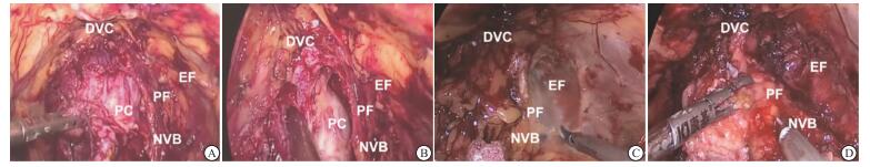
  • 病人气管插管全麻后取平卧位,所有病人均使用3个Torcar进行手术。脐下缘做一2 cm切口,钝性分离腹直肌后间隙,气囊扩张间隙。由该切口置入10 mm Trocar,气腹为15 mmHg。腹腔镜直视下建立双侧腹直肌外缘通道,于脐下2~3 cm置入12 mm Trocar。建立通道后使用超声刀清理耻骨后及膀胱附近脂肪,暴露髂血管、闭孔、盆内筋膜反折等重要解剖结构。2组病人手术到此步骤时均相同,术中病人手术图像见图 1

    筋膜间神经保留技术:首先打开盆内筋膜,暴露耻骨前列腺膀胱韧带,完全分离肛提肌与前列腺之间的纤维组织,离断耻骨前列腺韧带,缝扎阴茎背深静脉复合体(dorsal vascular complex, DVC)。利用拉动导尿管气囊识别膀胱颈,然后在膀胱颈和前列腺之间进行解剖分离。解剖膀胱后颈后,暴露离断双侧输精管后分离离断精囊。水平切开狄氏(Denonvilliers)筋膜的后层水平打开。在前列腺筋膜和盆腔内筋膜之间锐性分离直至前列腺尖部,此操作过程中尽量避免使用电凝,防止过多损伤神经血管束。离断DVC,剪刀离断前列腺与尿道,尽可能多的保留功能尿道。将前列腺组织装入标本袋,吻合尿道膀胱。检查无明显漏尿后,留置盆腔引流管1根,脐下切口完整取出标本袋。

    筋膜内神经保留技术:行筋膜内前列腺癌根治术时与筋膜间技术不同的地方在于不切开盆腔内筋膜、不离断耻骨前列腺韧带、不结扎背静脉复合体。从盆腔内筋膜,膀胱颈和前列腺的表面轻轻分离脂肪和疏松结缔组织后,切开膀胱颈。解剖膀胱后颈部,然后暴露并切除双侧输精管和精囊。并且不切开狄氏筋膜,在前列腺包膜和筋膜之间的间隙钝性分离至前列腺尖部,将神经血管束从前列腺完全游离,余留置引流管及取标本步骤同前。

  • 所有病人术前、术后均采用男性尿控及性健康问卷进行评估,出院后病人在门诊收集随访资料。控尿正常定义为不需要尿垫或每天仅预防性使用1块尿垫;轻度尿失禁指正常活动(行走)每天2~3块尿垫;尿失禁指每天3块以上尿垫;勃起功能正常定义为问卷总得分≥22分。本研究中所有病人术前男性性健康问卷总得分都≥22分。

  • 采用t检验、χ2检验、秩和检验。

2.   结果
  • 纳入本次研究的所有病人手术均成功完成,2组病人均无输血、中转开放手术或行二次手术。2组病人一般资料比较,差异均无统计学意义(P>0.05)(见表 1),围手术期各指标比较,差异均无统计学意义(P>0.05)(见表 2)。筋膜内组切缘阳性率15.00%,筋膜间组19.00%,差异亦无统计学意义(χ2=0.01,P>0.05)。纳入研究的所有病人均随访6个月以上,无失访病例。

    分组 n 年龄/岁 前列腺体积/mL BMI/ (kg/m2) 术前PSA (ng/mL) 活检Gleason评分/分
    筋膜内组 20 67.20±6.46 48.17±22.16 24.76±2.21 13.74±5.70 6.80±0.41
    筋膜间组 21 67.95±5.42 42.09±12.53 24.19±2.49 13.70±7.58 6.71±0.46
    t 0.40 1.07* 0.77 0.02 0.66
    P > 0.05 > 0.05 > 0.05 > 0.05 > 0.05
      *示t′值
    分组 n 手术时间/min 吻合时间/min 术中出血量/mL 引流管留置时间/d 尿管拔除时间/d 术后进食时间/d 术后住院时间/d
    筋膜内组 20 108.50±20.17 19.65±3.11 132.85±30.44 2.87±0.36 7.02±0.68 1.93±0.37 11.25±1.20
    筋膜间组 21 105.52±15.87 19.71±3.00 160.71±66.95 3.09±0.49 6.92±0.48 1.83±0.33 11.19±1.32
    t 0.53 0.06 1.73 1.63 0.50 0.91 0.15
    P > 0.05 > 0.05 > 0.05 > 0.05 > 0.05 > 0.05 > 0.05

    所有病人在手术前均为尿控正常(0垫/天),勃起功能正常,病人术后3个月及6个月统计结果见表 3。统计病人术后尿控情况后发现,在6个月的随访期间,2组病人的尿控能力随术后恢复时间的延长而得到改善。筋膜内组3个月及6个月时的尿垫数量级别使用明显优于筋膜间组(P < 0.05)。行筋膜内腹腔镜前列腺癌根治术病人在3个月时有55%的病例报告为控尿基本正常(0至1个垫/天),而25%的病人报告了压力性尿失禁(2个垫/天)和20%的病人需要大于2个垫/天。3个月时筋膜间组分别为19%、33.3%和47.6%。行筋膜内腹腔镜前列腺癌根治术病人术后6个月的尿控正常率(每天0~1个护垫)更高(85% vs 47.6%)。筋膜内组在6个月时的勃起功能恢复显著提高(66.7%vs 33.3%,P < 0.05)(见表 3)。

    分组 n 3个月尿垫数量 6个月尿垫数量 6个月勃起功能恢复
    0~1块 2~3块 >3块 0~1块 2~3块 >3块
    筋膜内组 20 11(55.0) 5(25.0) 4(20.0) 17(85.0) 2(10.0) 1(5.0) 12(66.7)
    筋膜间组 21 4(19.0) 7(33.3) 10(47.6) 10(47.6) 5(23.8) 6(28.6) 6(33.3)
    uc 2.40 2.54 4.11*
    P < 0.05 < 0.05 < 0.05
      *示χ2
3.   讨论
  • 我科自2013年开始开展筋膜间保留神经的腹膜外腹腔镜前列腺癌根治术,2016年3月开始开展筋膜内保留神经的腹膜外腹腔镜前列腺癌根治术。目前筋膜内保留神经手术已经是一种非常成熟的技术[18-20]。采用筋膜内技术保留神经的前列腺根治性切除术中,需将前列腺包膜和筋膜的间隙钝性结合锐性进行分离,由于分离更加的靠近腺体使得前列腺包膜表面几乎无组织覆盖,这使得术后的病理切缘阳性会增加。有学者就临床分期T3a-b的前列腺癌病人进行了回顾分析,发现采用筋膜内保留神经技术的病人比采用筋膜间的切缘阳性率更高[18]。但是,国际著名泌尿外科专家NEILL等[19]随后公布了新的研究成果,发现临床分期在T1~T2期的病人,采用筋膜内保留神经技术不会导致切缘阳性率的增高。在我们本次的回顾性分析中,2组病人的术中出血量、术后拔管时间、切缘阳性率以及引流管留置时间等围手术期指标差异均无统计学意义,我们认为筋膜内手术能够达到与筋膜间手术相等的术后肿瘤控制效果。

    自从对男性骨盆筋膜和神经血管束进行解剖以来,专家学者们在根治性前列腺切除术中对血管神经束的保留技术进行了不断改进,以达到更好的术后尿控及勃起功能的恢复[21]。从解剖学上分析,盆丛神经发出的海绵体神经走行于前列腺后外侧的基底部,和前列腺血管及尿道伴行,形成了支配及营养阴茎的神经血管束,筋膜内前列腺癌根治术正是通过保留神经血管术从而达到保留性功能的术后效果[22-23],有专家报道[18, 20],采用筋膜内技术保留神经的病人的术后勃起功能及早期尿控对的恢复率显著提高。同时,NEILL等[19]也发现采用筋膜内技术保留神经的根治性切除术,术后病人早期尿控的获得时间更短。由于筋膜内神经保留技术对神经血管束的牵拉作用较小,所以能够提高对神经血管束保护的准确性[24-25]。我们的研究也表明,利用筋膜内技术保留神经的病人术后的勃起功能恢复在6个月时出现明显的改善,这与其他学者报道的研究结果相一致[24, 26-27]

    在早期尿控恢复的观点上,有研究发现术后3个月及6个月,行筋膜内前列腺癌根治术的病人的勃起功能恢复率更高[27-28]。本研究结果表明,2组病人在术后在3个月和6个月两个时间点上,筋膜内早期尿控恢复率明显优于筋膜间。这与采用筋膜内技术术中未切开盆内筋膜及未离断耻骨前列腺韧带密不可分,此方法减少了术中对尿道周围括约肌的损伤和分离[19]。SAVERA等[29]提供的组织学证据表明,与筋膜间技术相比,筋膜内技术可有效的保护前列腺前外侧区域的神经纤维束。尽管神经血管束在尿控或勃起功能中的作用尚不明确,但是在不影响肿瘤学完整控制的前提下,尽可能多地保留神经纤维束可以有效地改善前列腺癌根治术后的功能结果[28, 30]。此外在本研究中,不论进行哪种方式的前列腺癌根治术,均存在少部分随访一年以上仍未恢复性功能的病人,因影响性功能因素较多[31],如年龄、心理因素和个体差异等,且病例数较少,本研究暂不进行展开讨论。

    通过以上比较分析我们得出结论,采用筋膜内保留神经技术的腹腔镜前列腺根治性切除术的病人在早期尿控和勃起功能恢复方面要明显优于筋膜间手术组,并且2组手术对肿瘤的完整控制效果相当,对于术前勃起和排尿功能正常的病人(病理分期T1~T2期)推荐采用筋膜内保留神经技术的腹腔镜前列腺根治性切除术。

Reference (31)

Catalog

    /

    DownLoad:  Full-Size Img  PowerPoint
    Return
    Return