• 中国科技论文统计源期刊
  • 中国科技核心期刊
  • 中国高校优秀期刊
  • 安徽省优秀科技期刊
Volume 44 Issue 4
Apr.  2019
Article Contents
Turn off MathJax

Citation:

Expression characteristics of HERG protein in gastric cancer tissue and its correlation with pathological features

  • Received Date: 2018-05-25
    Accepted Date: 2018-11-28
  • ObjectiveTo investigate the expression characteristics of human eag-related gene (HERG) protein in gastric cancer tissue, and its correlation with pathological features.MethodsThe expression levels of HERG protein in 78 gastric cancer tissue specimens and normal tissue adjacent to the cancer (from the margin of cancer >5 cm) from February 2014 to March 2017.were detected using immunohistochemistryResultsThe positive rate of HERG protein expression in gastric cancer was 76.92%, which was significantly higher than that in adjacent tissue(0.00%) (P < 0.01).The positive rates of HERG protein expression in patients with low-middle differentiation and lymph node metastasis were 86.67% and 92.86%, which were significantly higher than those in patients with high differentiation and no lymph node metastasis (63.64% and 58.33%), respectively(P < 0.05 and P < 0.01).The positive expression rates of HERG protein were not significantly correlated with age, sex, tumor infiltration, TNM staging and tumor size in gastric cancer patients(P>0.05).The median survival time and progression-free survival time in positive HERG patients were 23 months and 20 months, respectively, the median survival time and progression-free survival time in patients with negative HERG were 30 months and 24 months, respectively, and the difference of which was not statistically significant(P>0.05).ConclusionsThe expression of HERG protein is related to the degree of differentiation and lymph node metastasis of gastric cancer, but it is not clear with the prognosis of the patients.
  • 加载中
  • [1] 季加孚, 陕飞, 苗儒林.循证医学时代的胃癌进展30年回顾[J].实用肿瘤杂志, 2016, 31(2):99.
    [2] MARQUESCARVALHO MJ, OPPERMANN J, MUNOZ E, et al.Molecular insights into the mechanism of calmodulin inhibition of the EAG1 potassium channel[J].Structure, 2018, 24(10):1742.
    [3] 刘洪福, 张蕾, 阳书华, 等.汉族人HLA-G基因多态性与胃癌相关性研究[J].实用临床医药杂志, 2017, 21(19):70.
    [4] YEH KH, CHENG AL.Gastric cancer associated with acute disseminated intravascular coagulation:successful initial treatment with weekly 24-hour infusion of high-dose 5-fluorouracil and leucovorin[J].Br J Haematol, 2015, 100(4):769.
    [5] COLUCCI G.Phase Ⅱ study of 5-fluorouracil and folinic acid in the treatment of patients with advanced gastric cancer:A Southwest Oncology Group study[J].Cancer, 2015, 76(5):1002.
    [6] WANG GD, ZHAI W, YANG HC, et al.Out of southern East Asia:the natural history of domestic dogs across the world[J].Cell Res, 2016, 26(1):21. doi: 10.1038/cr.2015.147
    [7] ERDEM M, TEKINER TA, FEJZULLAHU A, et al.Herg1b expression as a potential specific marker in pediatric acute myeloid leukemia patients with HERG 897K/K genotype[J].Pediatr Hematol Oncol, 2015, 32(3):182. doi: 10.3109/08880018.2014.949941
    [8] OSTERBUR ML, MCDONALD TV.Extra-coding characteristics of hERG mRNA are essential for channel function[J].Biophysical J, 2015, 108(2):391a.
    [9] 吴想军, 李子巍, 屈若祎.2004-2010年中国消化道恶性肿瘤死亡率趋势分析[J].中国卫生统计, 2017, 34(1):43.
    [10] HSU PH, TANG CY, JENG CJ.Centrin 4 is a binding partner of rat EAG1 K+ channels[J].Biophysical J, 2016, 110(3):102a.
    [11] 林秀欣, 邓文静, 余更生, 等.PI3K/Akt相关基因多态性与胃癌含铂化疗方案疗效的关系分析[J].临床肿瘤学杂志, 2016, 21(8):702.
    [12] 陈志达, 曾文容, 林斌.电压门控钾离子通道Kv1.5与肿瘤[J].国际肿瘤学杂志, 2016, 43(2):130. doi: 10.3760/cma.j.issn.1673-422X.2016.02.014
    [13] 赵旭林, 马磊, 徐国昌, 等.胃肠道恶性肿瘤患者人类学不对称行为特征[J].中国医科大学学报, 2016, 45(11):985. doi: 10.12007/j.issn.0258-4646.2016.11.007
    [14] 李涛, 赖春凤, 丘波, 等.CYP2E1基因多态性与客家人群胃癌易感性的研究[J].国际肿瘤学杂志, 2016, 43(7):495. doi: 10.3760/cma.j.issn.1673-422X.2016.07.004
    [15] 李威威, 张海萍, 吴敏, 等.人表皮生长因子受体2基因扩增及蛋白表达与胃癌患者临床病理特征的关系[J].肿瘤研究与临床, 2017, 29(1):7. doi: 10.3760/cma.j.issn.1006-9801.2017.01.002
  • 加载中
通讯作者: 陈斌, bchen63@163.com
  • 1. 

    沈阳化工大学材料科学与工程学院 沈阳 110142

  1. 本站搜索
  2. 百度学术搜索
  3. 万方数据库搜索
  4. CNKI搜索

Figures(2) / Tables(1)

Article views(4828) PDF downloads(17) Cited by()

Related
Proportional views

Expression characteristics of HERG protein in gastric cancer tissue and its correlation with pathological features

  • Department of Pathology, Qifu Hospital of Guangzhou University of Traditional Chinese Medicine, Guangzhou Guangdong 511495, China

Abstract: ObjectiveTo investigate the expression characteristics of human eag-related gene (HERG) protein in gastric cancer tissue, and its correlation with pathological features.MethodsThe expression levels of HERG protein in 78 gastric cancer tissue specimens and normal tissue adjacent to the cancer (from the margin of cancer >5 cm) from February 2014 to March 2017.were detected using immunohistochemistryResultsThe positive rate of HERG protein expression in gastric cancer was 76.92%, which was significantly higher than that in adjacent tissue(0.00%) (P < 0.01).The positive rates of HERG protein expression in patients with low-middle differentiation and lymph node metastasis were 86.67% and 92.86%, which were significantly higher than those in patients with high differentiation and no lymph node metastasis (63.64% and 58.33%), respectively(P < 0.05 and P < 0.01).The positive expression rates of HERG protein were not significantly correlated with age, sex, tumor infiltration, TNM staging and tumor size in gastric cancer patients(P>0.05).The median survival time and progression-free survival time in positive HERG patients were 23 months and 20 months, respectively, the median survival time and progression-free survival time in patients with negative HERG were 30 months and 24 months, respectively, and the difference of which was not statistically significant(P>0.05).ConclusionsThe expression of HERG protein is related to the degree of differentiation and lymph node metastasis of gastric cancer, but it is not clear with the prognosis of the patients.

  • 胃癌是临床上高发的的恶性肿瘤之一,其发病率高居恶性肿瘤的第四位,尤其我国为胃癌的高发地区,给我国带来巨大的经济和社会压力[1-2]。目前胃癌的发病机制尚不清楚,常规的化疗和生物治疗效果不理想,因此对胃癌发病机制的研究以及临床治疗成为学界关注的焦点[3-5]。近年来发现,电压门控性钾离子通道与癌症的发病关系密切,人类eag相关基因(HERG)蛋白则是构成延迟整流性钾通道的α亚单位,且在多种恶性肿瘤可以检测到其阳性表达,成为人们研究和治疗胃癌的新热点[6-8]。因此, 本文通过研究HERG蛋白在胃癌组织中表达,探讨其与胃癌病理学特征的相关性,为临床的治疗提供依据。

1.   材料与方法
  • 选取2014年2月至2017年3月我院病理科保存的胃癌组织标本78例,其中男41例,女37例;年龄41~72岁。同时选取同标本癌旁正常组织(距癌切缘>5 cm)作为对照。纳入标准:临床病理资料保存完整;术前未接受过放疗、化疗等治疗;能随访者。排除合并其他原发性肿瘤者。

  • 采用免疫组化法检测HERG蛋白表达。将病理样本常规包埋切片,选用免疫组化SP法进行染色,试剂盒由北京百诺威生物科技有限公司提供,操作参照说明书进行。DAB显色,并苏木素复染,随后封片,镜检。随机选取5个视野,对染色强度和阳性细胞比例进行评判,染色强度呈无着色为0分,淡棕色为1分,棕色为2分,深棕色为3分;阳性细胞比例<30%为1分,30%~70%为2分,≥70%为3分。最终评分为染色强度和阳性细胞比例得分之和,≤2分为阴性表达,>2分为阳性表达。

  • 采用χ2检验、Kaplan-Meier分析和Log-rank检验。

2.   结果
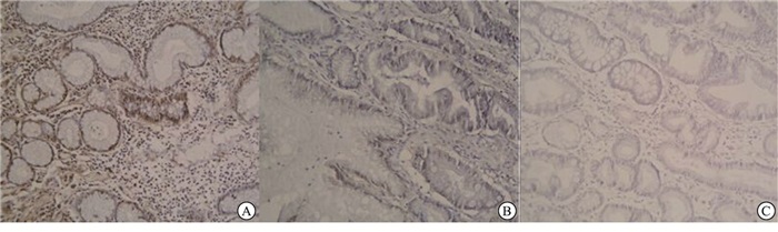
  • 胃癌组织HERG蛋白阳性表达率76.92%(60/78),明显高于癌旁组织(0/78),差异有统计学意义(χ2=20.35, P<0.01)(见图 1)。

  • HERG蛋白阳性表达率在胃癌病人年龄、性别、肿瘤浸润、TNM分期及肿瘤大小方面差异无统计学意义(P>0.05),与分化程度和淋巴结转移有关(P<0.05和P<0.01)(见表 1)。

    临床病理特征 n HERG蛋白
    阳性表达
    χ2 P
    年龄/岁
      ≤50 38 31(81.58) 0.91 >0.05
      >50 40 29(72.50)
    性别
      男 41 32(78.05) 0.06 >0.05
      女 37 28(75.68)
    肿瘤浸润
      侵及浆膜 51 40(78.43) 0.19 >0.05
      未侵及浆膜 21 20(74.07)
    TNM分期
      Ⅰ期 21 14(66.67) 3.81 >0.05
      Ⅱ期 35 26(74.29)
      Ⅲ期 22 20(90.91)
    肿瘤直径/cm
      <5 51 41(80.39) 1.00 >0.05
      ≥5 27 19(70.37)
    分化程度
      高分化 33 21(63.64) 5.69 <0.05
      中低分化 45 39(86.67)
    淋巴结转移
      有 42 39(92.86) 13.02 <0.01
      无 36 21(58.33)
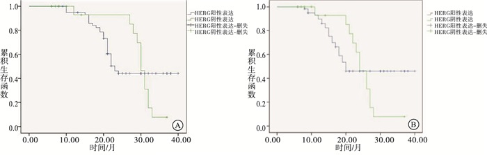
  • HERG蛋白阳性表达病人中位总生存时间和无进展生存期分别为23个月和20个月,HERG蛋白阴性表达病人中位总生存时间和无进展生存期分别为30个月和24个月,经Log-rank检验比较,HERG蛋白阳性表达病人和阴性表达病人中位总生存时间(χ2=0.11,P>0.05)和无进展生存期(χ2=0.50,P>0.05),差异无统计学意义(见图 2)。

3.   讨论
  • 胃癌是临床上常见的消化道的恶性肿瘤之一,具有高发病率和高死亡率的特点,其发病率高居恶性肿瘤的第四位,是世界范围内恶性肿瘤性死亡的第二大原因,尤其我国是胃癌的高发地区,严重威胁人们正常的生活和生命健康,成为临床关注的焦点[9-10]。目前胃癌的发病机制尚不清楚,化疗是临床治疗胃癌的重要手段,可以清除术后残留的癌细胞、防止复发以及改善预后,但更多的事实表明一线治疗的多种药物和多种给药方案效果不理想,生物治疗方案效果不佳,因此对胃癌治疗仍是临床上较为棘手的问题,对胃癌的发病机制的研究也成为学界关注的焦点[11]。近年来研究发现,钾离子通道与癌症的发病关系密切,HERG蛋白则是构成延迟整流性钾通道的α亚单位,且在多种恶性肿瘤可以检测到其阳性表达[12]。HERG基因筛选自人的海马cDNA文库,定位于人的7号染色体长臂,含16个外显子,可编码1 159个氨基酸的多肽,而HERG通道结构特点(六次跨膜片段)同外向钾离子通道家族类似,但却表现为内向整流和细胞外钾浓度依赖性等内向整流钾离子通道的特点,从而产生的细胞膜HERG电流,具有依赖去极化的活化门控开放、对Ⅲ型抗心律失常药物敏感以及增大电导的细胞外钾浓度依赖性等特点,而这种门控特点与细胞周期控制机制相关,在恶性肿瘤细胞中存在这样的电流[13]。相关研究[14-15]表明,HERG基因和HERG蛋白在恶性肿瘤高度表达,从而产生的HERG电流维持癌细胞去极化的静息电位,这也是肿瘤细胞的特征之一,因此HERG蛋白表达与胃癌的相关性,在胃癌机制的研究和临床治疗中具有重大研究价值。

    HERG蛋白可形成HERG通道,产生HERG电流,从而维持癌细胞去极化的静息电位,抑制细胞的分化,增加细胞癌变的概率。本研究中,胃癌组织HERG蛋白阳性表达率明显高于癌旁组织,表明HERG蛋白表达与胃癌的发病具有一定的相关性。进一步的研究显示,HERG蛋白阳性表达与胃癌病人年龄、性别、肿瘤浸润、TNM分期及肿瘤大小无明显相关性,但与分化程度和淋巴结转移有关,表明HERG蛋白阳性表达与癌细胞的分化和转移关系密切,与癌细胞的恶性程度和转移性具有相关性。本研究经Log-rank比较发现, HERG蛋白阳性和阴性表达病人总生存时间和无进展生存期差异均无统计学意义,这一结果与前人工作不符,可能受限于本研究样本量,因此可扩大样本量继续研究,以获得更为准确的结果。

    综上所述,HERG蛋白表达与胃癌的发病具有相关性,且与癌细胞的恶性程度和转移性具有相关性,但与胃癌的浸润、分期以及预后没有相关性,这可能受限于本研究的样本量,需扩大样本量继续研究,总体来看对HERG蛋白在胃癌癌细胞的阳性表达进行研究,具有重大研究价值,可为临床提供更多理论依据。

Reference (15)

Catalog

    /

    DownLoad:  Full-Size Img  PowerPoint
    Return
    Return