• 中国科技论文统计源期刊
  • 中国科技核心期刊
  • 中国高校优秀期刊
  • 安徽省优秀科技期刊
Volume 44 Issue 4
Apr.  2019
Article Contents
Turn off MathJax

Citation:

Clinical application value of 3D printing technology in the mini-invasive treatment of unstable distal radius fractures

  • Corresponding author: ZHOU Jian-sheng, zhoujs12399@163.com
  • Received Date: 2017-12-26
    Accepted Date: 2018-07-05
  • ObjectiveTo investigate the clinical application value of 3D printing technology in the mini-invasive treatment of unstable distal radius fractures.MethodsTwenty-five patients with unstable distal radius fracture were divided into the MIS group(12 cases) and ORIF group(13 cases).The MIS group and ORIF group were treated with mini-invasive surgery reduction(close reduction or limited incision reduction) combined with external fixation assisted by 3D printing technology, and open reduction combined with internal fixation, respectively.The operation time, incision size and postoperative following-up of the activity of wrist motion(palmarflexion, dorsiflexion, ulnardeviation, radialdeviation, pronation, supinaion) were compared between two groups.The recovery of palmar inclination, ulnar deviation, radial length and joint surface in two groups were observed using the positive and lateral wrist radiographs.According to Gartland-Werley scoring criteria, the wrist joint function between two groups was evaluated and compared.ResultsThe operation in two groups was successfully completed, and the average operation time and incision length in MIS group were less than those in ORIF group(P < 0.01).The recovery of wrist joint function in two groups was good, and the differences of the excellent and good rate of the activity of the wrist motion, and score of wrist joint function between two groups were not statistically significant(P>0.05).ConclusionsThe mini-invasive surgery reduction(close reduction or limited incision reduction) combined and external fixation assisted by 3D printing technology combined with open reduction and internal fixation in the treatment of the unstable distal radius fractures has little incision, short operation time and good recovery of wrist joint function, which can avoid secondary surgery.
  • 加载中
  • [1] ALLURI R, LONGACRE M, PANNELL W, et al.Volar, intramedullary, and percutaneous fixation of distal radius fractures[J].J Wrist Surg, 2015, 4(4):292. doi: 10.1055/s-00000183
    [2] MINEGISHI H, DOHI O, AN S, et al.Treatment of unstable distal radius fractures with the volar locking plate[J].Ups J Med Sci, 2011, 116(4):280. doi: 10.3109/03009734.2011.594183
    [3] CHIRPAZ-CERBAT JM, RUATTI S, HOUILLON C, et al.Dorsally displaced distal radius fractures treated by fixed-angle volar plating:Grip and pronosupination strength recovery.A prospective study[J].Orthop Traumatol Surg Res, 2011, 97(5):465. doi: 10.1016/j.otsr.2011.01.016
    [4] ZENKE Y, SAKAI A, OSHIGE T, et al.Clinical results of volar locking plate for distal radius fractures:conventional versus minimally invasive plate osteosynthesis[J].J Orthop Trauma, 2011, 25(7):425. doi: 10.1097/BOT.0b013e3182008c83
    [5] RICHARD MJ, WARTINBEE DA, RIBOH J, et al.Analysis of the complications of palmar plating versus external fixation for fractures of the distal radius[J].J Hand Surg Am, 2011, 36(10):1614. doi: 10.1016/j.jhsa.2011.06.030
    [6] BERGLUND LM, MESSER TM.Complications of volar plate fixation for managing distal radius fractures[J].J Am Acad Orthop Surg, 2009, 17(6):369. doi: 10.5435/00124635-200906000-00005
    [7] MARGALIOT Z, HASSE SC, KOTSIS SV, et al.A meta-analysis of outcomes of external fixation versus plate osteosynthesis for unstable distal radius fractures[J].J Hand Surg, 2005, 30(6):1185.
    [8] 赵继荣, 李红专, 慕向前.桡骨远端骨折的非手术治疗进展[J].中国中医骨伤科杂志, 2015, 23(12):77.
    [9] 巩海贤, 刘永涛.桡骨远端骨折治疗进展[J].滨州医学院学报, 2013, 36(1):52. doi: 10.3969/j.issn.1001-9510.2013.01.017
    [10] LICHTMAN DM, BIUDRA RR, BOYER MI, et al.American Academy of Orthopaedic Surgeons Clinical practice guideline on:the treatment of distal radius fractures[J].J Bone Joint Surg Am, 2011, 93(8):775. doi: 10.2106/JBJS.938ebo
    [11] NESBITT KS, FAIDIA JM, LES C.Assessment 0f instability factors in adult distal radial fractures[J].J Hand Surg Am, 2004, 29(6):1128. doi: 10.1016/j.jhsa.2004.06.008
    [12] COSTA ML, ACHTEN J, PLANT C, et al.UK DRAFFT:a randomised controlled trial of percutaneous fixation with Kirschner wires versus volar locking-plate fixation in the treatment of adult patients with a dorsally displaced fracture of the distal radius[J].Health Technol Assess, 2015, 19(17):1. doi: 10.3310/hta19170
    [13] 李德胜, 李皓恒.掌侧锁定钢板内固定与外固定支架修复桡骨远端不稳定性骨折:随机对照[J].中国组织工程研究, 2015, 19(9):1429.
    [14] 张计成, 张辉, 刘向阳.外固定架联合交叉克氏针固定治疗桡骨远端不稳定性骨折[J].实用骨科杂志, 2012, 18(5):473. doi: 10.3969/j.issn.1008-5572.2012.05.035
    [15] BAJWA AS, RAMMAPPA M, LEE L, et al.Treatment of unstable distal radius fractures:non-invasive dynamic external xator versus volar locking plate-functional and radiological outcome in a prospective case-controlled series[J].SICOT J, 2015, 1(34):1.
    [16] STAROSOLSKI ZA, KAN JH, ROSENFELD SD, et al.Application of 3-D printing (rapid prototyping) for creating physical models of pediatric orthopedic disorders[J].Pediatr Radiol, 2014, 44(2):216. doi: 10.1007/s00247-013-2788-9
  • 加载中
通讯作者: 陈斌, bchen63@163.com
  • 1. 

    沈阳化工大学材料科学与工程学院 沈阳 110142

  1. 本站搜索
  2. 百度学术搜索
  3. 万方数据库搜索
  4. CNKI搜索

Figures(7) / Tables(2)

Article views(7913) PDF downloads(13) Cited by()

Related
Proportional views

Clinical application value of 3D printing technology in the mini-invasive treatment of unstable distal radius fractures

    Corresponding author: ZHOU Jian-sheng, zhoujs12399@163.com
  • Department of Orthopedics, The First Affiliated Hospital of Bengbu Medical College, Laboratory of Tissue and Transplant in Anhui Province, Bengbu Medical College, Bengbu Anhui 233004, China

Abstract: ObjectiveTo investigate the clinical application value of 3D printing technology in the mini-invasive treatment of unstable distal radius fractures.MethodsTwenty-five patients with unstable distal radius fracture were divided into the MIS group(12 cases) and ORIF group(13 cases).The MIS group and ORIF group were treated with mini-invasive surgery reduction(close reduction or limited incision reduction) combined with external fixation assisted by 3D printing technology, and open reduction combined with internal fixation, respectively.The operation time, incision size and postoperative following-up of the activity of wrist motion(palmarflexion, dorsiflexion, ulnardeviation, radialdeviation, pronation, supinaion) were compared between two groups.The recovery of palmar inclination, ulnar deviation, radial length and joint surface in two groups were observed using the positive and lateral wrist radiographs.According to Gartland-Werley scoring criteria, the wrist joint function between two groups was evaluated and compared.ResultsThe operation in two groups was successfully completed, and the average operation time and incision length in MIS group were less than those in ORIF group(P < 0.01).The recovery of wrist joint function in two groups was good, and the differences of the excellent and good rate of the activity of the wrist motion, and score of wrist joint function between two groups were not statistically significant(P>0.05).ConclusionsThe mini-invasive surgery reduction(close reduction or limited incision reduction) combined and external fixation assisted by 3D printing technology combined with open reduction and internal fixation in the treatment of the unstable distal radius fractures has little incision, short operation time and good recovery of wrist joint function, which can avoid secondary surgery.

  • 桡骨远端骨折约占四肢骨折病人的15%,发病率高且易累及关节面[1]。此类骨折常具有以下特点:桡骨长度短缩、累及关节面、粉碎性骨折及骨折断端明显的成角畸形。对桡骨远端不稳定性骨折进行保守治疗,容易发生骨折复位后位置丢失,且很难实现关节面的解剖复位,造成骨折畸形愈合、腕关节创伤性关节炎,导致顽固性腕关节疼痛及功能受限。因此,对桡骨远端不稳定性骨折进行手术干预是目前多数临床医生的选择。切开复位锁定钛板内固定具有直接复位骨折,稳定性良好,功能恢复快的优点[2-4],但亦可引起腱鞘炎、肌腱断裂,且需要再次手术取出内固定[5-7]。我院采用3D打印辅助下闭合或有限切开复位外固定支架外固定方法治疗桡骨远端不稳定性骨折,临床疗效满意。现作报道。

1.   资料与方法
  • 选取2015年1月至2017年2月经手术治疗的桡骨远端不稳定性骨折病人25例,其中男12例,女13例;年龄25~73岁。所有病人均为闭合性骨折,不伴有神经、血管损伤。受伤原因:摔伤15例;车祸伤4例;高处坠落伤6例。

    纳入标准:(1)确诊为桡骨远端不稳定性骨折的成年病人;(2)闭合性骨折;(3)无腕关节结核、炎症、肿瘤等疾病;(4)同意并能配合研究及随访。排除标准:(1)病理性骨折;(2)开放性骨折;(3)陈旧性骨折;(4)伴精神病或其他全身条件不佳,不宜进行手术的病人;(5)无法联系随访,不配合研究的病人。

    将病人分为3D打印技术辅助下微创(闭合或有限切开)复位克氏针内固定外固定支架外固定组(MIS组)12例和切开复位接骨板内固定组(ORIF组)13例。其中MIS组男6例,女6例;年龄31~67岁;骨折AO分型:A型骨折6例,B型骨折2例,C型骨折4例。ORIF组男10例,女11例;年龄45~73岁;骨折AO分型:A型骨折6例,B型骨折2例,C型骨折5例。2组病人性别、年龄、骨折分型均具有可比性。

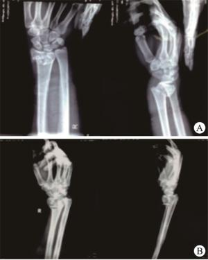
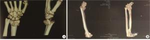
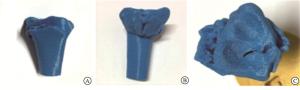
  • 所有病人入院后首先进行骨科专科体检,常规行腕关节正侧位片检查(见图 1),行CT平扫三维重建进一步明确桡骨远端关节面粉碎及塌陷程度(见图 2),MIS组打印3D模型(见图 3)。病人入院检查后立即行石膏托临时固定制动,腕关节明显肿胀者给予20%甘露醇治疗,待水肿减退后再行手术治疗。

  • MIS组病人根据3D打印模型设计复位方法及克氏针固定位置。臂丛或全身麻醉满意后,病人平卧位于手术台上,患肢外展于手术侧台上。向近端桡骨及第2掌骨各置入2枚Schanz钉,在C臂机辅助下手法复位或结合直径约2.0 mm的克氏针经皮撬拨复位,复位满意后2枚克氏针经皮交叉固定后安装外固定架。若复位不满意,行小切口切开复位。显露骨折端及关节面,直视下整复骨折,根据骨块大小选择直径1.5 mm或2.0 mm克氏针经皮交叉固定。C臂机透视见桡骨长度、掌倾角、尺偏角恢复,关节面平整,克氏针位置良好,安装外固定支架。

    ORIF组病人臂丛或全身麻醉满意后,平卧位于手术台上,患肢外展于手术侧台上。上臂根部捆充气式止血带。掌侧入路:自腕横纹水平作长约6~8 cm的“S”形切口。逐层切开,于桡侧屈腕肌和拇长屈肌腱间隙进入,保护好正中神经,剥离或适当剥离旋前方肌。显露骨折端及关节面, 直视下整复骨折后选择直径1.5 mm或2.0 mm克氏针临时固定,选取合适长度的接骨板,远近端至少3枚螺钉固定。反复冲洗切口,旋前方肌覆盖在接骨板表面并缝合,依次缝合皮下组织及皮肤,内置引流管一根以资引流。包扎切口,松止血带。石膏托临时固定并悬吊胸前。背侧入路:自桡骨茎突旁腕关节水平作长约5~6 cm的纵形切口,于桡侧腕长、短伸肌腱与拇长伸肌腱两者间隙进入,暴露骨折部位,直视下整复骨折,根据骨块大小选择直径1.5 mm或2.0 mm克氏针经皮临时固定,选取合适长度的接骨板,远近端至少3枚螺钉固定。冲洗、止血后逐层缝合,内置引流管一根以资引流。包扎切口,松止血带。石膏托临时固定并悬吊胸前随访。

  • 术后当天给予抗生素预防感染、消肿、镇痛对症治疗,术后第2天换药,并观察患肢末梢循环、感觉、运动情况,拍摄腕关节标准正侧位片,进行各手指各关节功能锻炼。MIS组病人术后10 d至2周,调整外固定架至腕关节轻度背伸尺偏位,术后3周开始每天定期松开外固定架关节,在医师指导下逐渐进行腕关节功能锻炼;术后4~6周时复查腕关节正侧位片,明确有明显骨痂形成后逐步拆除外固定架,加强手指各关节及腕关节的功能锻炼;腕关节功能恢复良好后拔出克氏针。ORIF组术后3周拆除石膏托外固定,在医师指导下行指间关节、掌指关节及腕关节的功能锻炼,循序渐进,活动次数及幅度逐步加大;嘱咐病人于术后定期来门诊随访。

  • 记录并比较2组病人手术时间和切口长度。术后6个月,随访病人腕关节掌屈、背伸、尺偏、桡偏、旋前、旋后六个方向的活动度,复查腕关节正侧位片,观察对掌倾角、尺偏角、桡骨长度及关节面恢复情况,并应用Gartland-Werley评分表对2组病人腕关节功能进行评分并比较。

  • 采取t(或t′)检验和Fisher′s确切概率法。

2.   结果
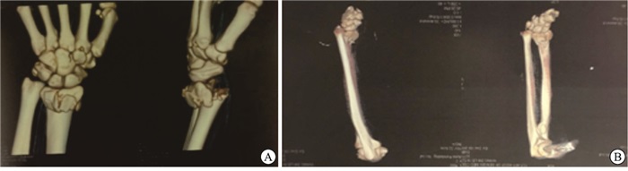
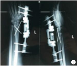
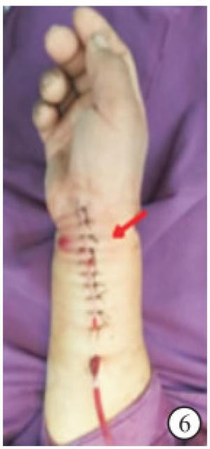
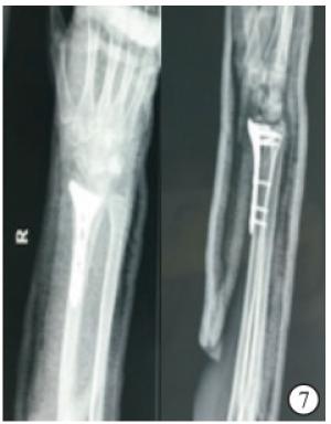
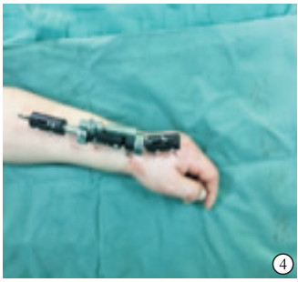
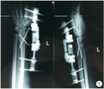
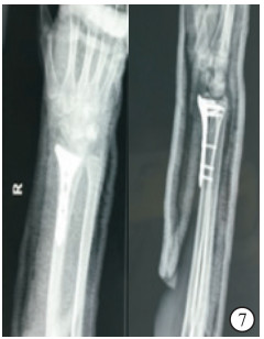
  • 2组病人均顺利完成手术。MIS组病人手术时间明显短于ORIF组(P<0.01),手术切口长度亦明显短于ORIF组(P<0.01)(见表 1)。本组25例术后均获得随访。MIS组骨折复位良好,关节面平整,桡骨长度、掌倾角、尺偏角均恢复,克氏针按术前设计位置固定良好(见图 4~5)。ORIF组病人骨折复位良好,关节面平整,桡骨长度、掌倾角、尺偏角均恢复,接骨板及螺钉位置良好(见图 6~7)。

    分组 n 手术时间/min 切口长度/cm
    MIS组 12 49.02±7.29 2.92±1.88
    ORIF组 13 75.79±3.98 6.61±1.12
    t 11.52* 5.44
    P <0.01 <0.01
    *示t′值
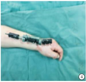
  • 2组病人术后腕关节掌屈、背伸、尺偏、桡偏、旋前、旋后六个方向的活动度差异均无统计学意义(P>0.05)(见表 2)。MIS组病人根据腕关节功能评分,优、良、可病人例数分别为9、2、1例,ORIF组病人优、良、可例数分别为8、4、1例,2组均无评分差的病人。MIS组病人评分优良率为91.67%(11/12),与ORIF组的92.31%(12/13)差异无统计学意义(Fisher′s确切概率法,P>0.05)。

    分组 n 掌屈 背伸 旋前 旋后 桡偏 尺偏
    MIS组 12 50.63±1.67 50.40±1.92 80.93±2.11 79.55±1.47 24.47±1.96 30.39±1.31
    ORIF组 13 52.26±2.20 51.08±2.02 80.68±2.08 80.45±2.66 25.44±2.05 30.11±3.03
    t 0.47 0.86 0.30 1.04 1.21 0.30
    P >0.05 >0.05 >0.05 >0.05 >0.05 >0.05
3.   讨论
  • 桡骨远端不稳定性骨折的治疗目标为:(1)恢复桡骨长度和掌倾角、尺偏角;(2)达到关节面的解剖复位;(3)牢固稳定的固定[8]。解剖复位的标准是指完全恢复桡骨长度和掌倾角、尺偏角,桡腕关节面及下尺桡关节面光滑平整;功能复位的标准是复位后骨折背侧成角小于10°,尺偏角大于15°,桡骨缩短不超过5 mm,关节面分离或移位不超过2 mm[9]

    传统的手法复位石膏或夹板外固定因难以对骨折进行持续有效的牵引,难以实现关节面的解剖复位,且不能提供足够的稳定性,从而造成骨折畸形愈合、腕关节创伤性关节炎,顽固性腕关节疼痛及功能受限等。美国矫形外科学会桡骨远端骨折治疗临床指南指出,桡骨远端不稳定性骨折即使初始复位位置良好,仍无法确定石膏外固定是否适合作为其最终治疗[10]。有学者[11]报道保守治疗50例桡骨远端不稳定性骨折的病人,4周后仅有46%病人能维持复位,54%病人复位丢失。

    切开复位接骨板内固定术目前广泛应用于治疗桡骨远端不稳定性骨折。该方法可以处理多数桡骨远端不稳定性骨折,尤其是锁定板的角稳定性、排钉技术、锁定机制,可处理复杂的关节内骨折、老年骨质疏松骨折,具有并发症少、可以早期功能锻炼、临床疗效满意等优势,但其亦存在屈肌腱断裂、螺钉穿过皮质激惹软组织、手僵硬等并发症。有研究[12]指出,相对于无创和微创的治疗方法,切开复位掌侧锁定板内固定被过度应用于治疗桡骨远端骨折,给病人带来更多的手术风险。对于桡骨远端不稳定性骨折,采用外固定支架或掌侧锁定钢板治疗均可获得满意的临床疗效,但是外固定治疗具有创伤较小、住院时间较短、并发症轻微、骨折愈合较快等优点[13]。张计成等[14]应用外固定支架外固定结合交叉克氏针固定,一期进行植骨治疗30例桡骨远端不稳定性骨折,临床疗效良好。BAJWA等[15]运用病例随机对照方法,采用无创外固定架和切开复位内固定治疗桡骨远端不稳定性骨折50例,术后3个月和1年无创外固定架组DASH评分明显优于切开复位内固定组。

    3D打印技术可打印出骨折部位的立体模型,在术前进行准确诊断和分型,通过模型进行模拟骨折复位和内固定的选择,并且可以进行不同治疗方案的反复模拟,来选择最合适的手术方案,有效节省实际手术时间、减少术中出血等,并提高手术的安全性[16]。应用3D打印技术辅助下微创(闭合或有限切开)复位克氏针内固定外固定支架外固定治疗桡骨远端不稳定性骨折优点有:(1)3D打印技术优化术前设计,缩短手术时间;(2)克氏针内固定联合外固定架外固定能为骨折断端端供更好的力学稳定性,允许病人在拆除外固定架后可继续进行功能锻炼;(3)微创手术对骨折端局部血运破坏少、切口小、出血少,有利于骨折愈合;(4)经济实用,避免内固定耗材,克氏针留在皮外,容易取出,外固定架可拆卸,不需要二次手术取出内固定;(5)并发症少,克氏针内固定对肌腱无干扰,方便功能锻炼。

    3D打印技术辅助下微创(闭合或有限切开)复位克氏针内固定外固定支架外固定和切开复位接骨板内固定两种手术方法治疗桡骨远端不稳定性骨折都可以达到满意的治疗效果,前者具有微创、手术时间短、稳定可靠、经济适用、避免二次手术等优点,是治疗桡骨远端不稳定性骨折的一种安全有效方法。但本组临床资料例数较少,且随访时间较短,长期疗效有待进一步观察研究。

Reference (16)

Catalog

    /

    DownLoad:  Full-Size Img  PowerPoint
    Return
    Return