• 中国科技论文统计源期刊
  • 中国科技核心期刊
  • 中国高校优秀期刊
  • 安徽省优秀科技期刊
Volume 49 Issue 1
Jan.  2024
Article Contents
Turn off MathJax

Citation:

The expression and clinical significance of TLR2 and TLR4 in peripheral blood myeloid dendritic cells of patients with systemic lupus erythematosus

  • Corresponding author: LI Zhijun, lizhijundr@126.com
  • Received Date: 2022-03-14
    Accepted Date: 2023-07-06
  • ObjectiveTo detect the expression of Toll-like receptor 2 (TLR2) and Toll-like receptor 4 (TLR4) on the surface of myeloid dendritic cells (mDC) and the expression levels of serum IL-10 and IL-17A in the peripheral blood of patients with systemic lupus erythematosus (SLE), and to investigate their role in the pathogenesis of SLE and their clinical significance.MethodsA total of 54 patients with SLE were included as study subjects, and divided into stable and active groups according to the SLEDAI-2000, while 30 healthy volunteers were selected as the control group.Flow cytometry was applied to detect the expression of TLR2 and TLR4 on the surface of mDC in the peripheral blood of SLE patients and healthy volunteers, and ELISA was used to detect the expression of IL-10 and IL-17A in the serum of SLE patients and healthy volunteers.ResultsThe expression rate of mDC-TLR2 in SLE patients in the active group was higher than that in the stable group and control group (P < 0.01).The expression rate of mDC-TLR4 was higher in the active group than that in the control group (P < 0.01).The expression levels of IL-10 and IL-17A in the serum of SLE patients were higher than those in the control group (P < 0.01).mDC-TLR2 expression was positively correlated with anti-Sm antibody (P < 0.05), and negatively correlated with serum IgG and 24h urine protein quantification (P < 0.05).The expression of mDC-TLR4 was positively correlated with platelet count (P < 0.05).ConclusionsThe expression rates of mDC-TLR2 and TLR4 are increased in the active SLE group, and both may play a role in the pathogenesis or progression of SLE.
  • 加载中
  • [1] GORDON C, AMISSAH-ARTHUR M B, GAYED M, et al. The British Society for Rheumatology guideline for the management of systemic lupus erythematosus in adults[J]. Rheumatology (Oxford), 2018, 57(1): e1. doi: 10.1093/rheumatology/kex286
    [2] 王涛, 江超, 赵萍, 等. 系统性红斑狼疮病人外周血血清TGF-β1的表达及临床意义[J]. 蚌埠医学院学报, 2019, 44(7): 868.
    [3] KAEWRAEMRUAEN C, RITPRAJAK P, HIRANKARN N. Dendritic cells as key players in systemic lupus erythematosus[J]. Asian Pac J Allergy Immunol, 2020, 38(4): 225.
    [4] YAMAUCHI M, MORIYAMA M, HAYASHIDA J N, et al. Myeloid dendritic cells stimulated by thymic stromal lymphopoietin promote Th2 immune responses and the pathogenesis of oral lichen planus[J]. PLoS One, 2017, 12(3): e0173017. doi: 10.1371/journal.pone.0173017
    [5] GUIDUCCI C, GONG M, XU Z, et al. TLR recognition of self nucleic acids hampers glucocorticoid activity in lupus[J]. Nature, 2010, 465(7300): 937. doi: 10.1038/nature09102
    [6] CELHAR T, FAIRHURST AM. Toll-like receptors in systemic lupus erythematosus: potential for personalized treatment[J]. Front Pharmacol, 2014, 5: 265.
    [7] CLANCY RM, MARKHAM AJ, BUYON JP. Endosomal Toll-like receptors inclinically overt and silent autoimmunity[J]. Immunol Rev, 2016, 269(1): 76. doi: 10.1111/imr.12383
    [8] GLADMAN DD, IBANEZ D, UROWITZ MB. Systemic lupus erythematosus disease activity index 2000[J]. J Rheumatol, 2002, 29(2): 288.
    [9] TEKTONIDOU MG, LEWANDOWSKI LB, HU J, et al. Survival in adults and children with systemic lupus erythematosus: a systematic review and Bayesian meta-analysis of studies from 1950 to 2016[J]. Ann Rheum Dis, 2017, 76(12): 2009. doi: 10.1136/annrheumdis-2017-211663
    [10] JORGE AM, LU N, ZHANG Y, et al. Unchanging premature mortality trends in systemic lupus erythematosus: a general population-based study (1999-2014)[J]. Rheumatology (Oxford), 2018, 57(2): 337. doi: 10.1093/rheumatology/kex412
    [11] DORNER T, FURIE R. Novel paradigms in systemic lupus erythematosus[J]. Lancet, 2019, 393(10188): 2344. doi: 10.1016/S0140-6736(19)30546-X
    [12] DURCAN L, O'DWYER T, PETRI M. Management strategies and future directions for systemic lupus erythematosus in adults[J]. Lancet, 2019, 393(10188): 2332. doi: 10.1016/S0140-6736(19)30237-5
    [13] TSAO JT, HSIEH SC, CHIANG BL, et al. Altered IL-10 and TNF-α production in peripheral blood mononuclear cells of systemic lupus erythematosus patients after Toll-like receptor 2, 4, or 9 activation[J]. Clin Exp Med, 2012, 12(3): 153. doi: 10.1007/s10238-011-0161-6
    [14] CHEN JQ, SZODORAY P, ZEHER M. Toll-like receptor pathways in autoimmune diseases[J]. Clin Rev Allergy Immunol, 2016, 50(1): 1. doi: 10.1007/s12016-015-8473-z
    [15] PATRA MC, ACHEK A, KIM GY, et al. A novel small-molecule inhibitor of endosomal TLRs reduces inflammation and alleviates autoimmune disease symptoms in murine models[J]. Cells, 2020, 9(7): 1648. doi: 10.3390/cells9071648
    [16] DOMINGUES R, DE CARVALHO GC, AOKI V, et al. Activation of myeloid dendritic cells, effector cells and regulatory T cells in lichen planus[J]. J Transl Med, 2016, 14(1): 171. doi: 10.1186/s12967-016-0938-1
    [17] DOMINGUES R, DE CARVALHO GC, DA SILVA OLIVEIRA LM, et al. The dysfunctional innate immune response triggered by Toll-like receptor activation is restored by TLR7/TLR8 and TLR9 ligands in cutaneous lichen planus[J]. Br J Dermatol, 2015, 172(1): 48. doi: 10.1111/bjd.13214
    [18] JIN MS, KIM SE, HEO JY, et al. Crystal structure of the TLR1-TLR2 heterodimer induced by binding of a triacylated lipopeptide[J]. Cell, 2007, 130(6): 1071. doi: 10.1016/j.cell.2007.09.008
    [19] PARK BS, SONG DH, KIM HM, et al. The structural basis of lipopolysaccharide recognition by the TLR4-MD-2 complex[J]. Nature, 2009, 458(7242): 1191. doi: 10.1038/nature07830
    [20] UENO H, KLECHEVSKY E, MORITA R, et al. Dendritic cell subsets in health and disease[J]. Immunol Rev, 2007, 219: 118. doi: 10.1111/j.1600-065X.2007.00551.x
    [21] KIRCHNER M, SONNENSCHEIN A, SCHOOFS S, et al. Surface expression and genotypes of Toll-like receptors 2 and 4 in patients with juvenile idiopathic arthritis and systemic lupus erythematosus[J]. Pediatr Rheumatol Online J, 2013, 11(1): 9. doi: 10.1186/1546-0096-11-9
    [22] 吕迎霞, 杨俊梅, 郑莉娟, 等. SLE患儿外周血TLR-4、HMGB1的表达及临床意义[J]. 检验医学, 2020, 35(11): 1143.
    [23] 刘昱. Toll样受体2(TLR2)在系统性红斑狼疮发病机制中的作用及其分子机制[D]. 长沙: 中南大学, 2014.
    [24] 张文兰, 胡同平, 王慧, 等. 系统性红斑狼疮患者单核细胞中TLR-4、IRAK-M的表达及意义[J]. 免疫学杂志, 2013, 29(6): 510.
    [25] 黄莉, 盛伟松. 高迁移率族蛋白B1及Toll样受体4在儿童系统性红斑狼疮中的临床意义[J]. 中华风湿病学杂志, 2019, (6): 365.
    [26] WANG X, WONG K, OUYANG W, et al. Targeting IL-10 family cytokines for the treatment of human diseases[J]. Cold Spring Harb Perspect Biol, 2019, 11(2): a028548. doi: 10.1101/cshperspect.a028548
    [27] 吕甜甜. 白细胞介素-10与系统性红斑狼疮相关性的分子流行病学研究[D]. 合肥: 安徽医科大学, 2018.
    [28] 沈茹, 李艳君, 林丽佳. 系统性红斑狼疮患儿外周血IL-6、IL-10、IL-17水平变化及其在单个核细胞中的表达研究[J]. 分子诊断与治疗杂志, 2017, 9(6): 371.
    [29] 杨依骏, 杜建钢, 朱琴芳, 等. 系统性红斑狼疮患者血清IL-10和IL-17水平检测的临床意义[J]. 现代检验医学杂志, 2017, 32(3): 71.
  • 加载中
通讯作者: 陈斌, bchen63@163.com
  • 1. 

    沈阳化工大学材料科学与工程学院 沈阳 110142

  1. 本站搜索
  2. 百度学术搜索
  3. 万方数据库搜索
  4. CNKI搜索

Figures(1) / Tables(3)

Article views(2220) PDF downloads(5) Cited by()

Related
Proportional views

The expression and clinical significance of TLR2 and TLR4 in peripheral blood myeloid dendritic cells of patients with systemic lupus erythematosus

    Corresponding author: LI Zhijun, lizhijundr@126.com
  • 1. Department of Rheumatology, The First Affiliated Hospital of Bengbu Medical University, Bengbu Anhui 233004
  • 2. Anhui Province Key Laboratory of Immunology in Chronic Diseases, Bengbu Medical University, Bengbu Anhui 233004, China

Abstract: ObjectiveTo detect the expression of Toll-like receptor 2 (TLR2) and Toll-like receptor 4 (TLR4) on the surface of myeloid dendritic cells (mDC) and the expression levels of serum IL-10 and IL-17A in the peripheral blood of patients with systemic lupus erythematosus (SLE), and to investigate their role in the pathogenesis of SLE and their clinical significance.MethodsA total of 54 patients with SLE were included as study subjects, and divided into stable and active groups according to the SLEDAI-2000, while 30 healthy volunteers were selected as the control group.Flow cytometry was applied to detect the expression of TLR2 and TLR4 on the surface of mDC in the peripheral blood of SLE patients and healthy volunteers, and ELISA was used to detect the expression of IL-10 and IL-17A in the serum of SLE patients and healthy volunteers.ResultsThe expression rate of mDC-TLR2 in SLE patients in the active group was higher than that in the stable group and control group (P < 0.01).The expression rate of mDC-TLR4 was higher in the active group than that in the control group (P < 0.01).The expression levels of IL-10 and IL-17A in the serum of SLE patients were higher than those in the control group (P < 0.01).mDC-TLR2 expression was positively correlated with anti-Sm antibody (P < 0.05), and negatively correlated with serum IgG and 24h urine protein quantification (P < 0.05).The expression of mDC-TLR4 was positively correlated with platelet count (P < 0.05).ConclusionsThe expression rates of mDC-TLR2 and TLR4 are increased in the active SLE group, and both may play a role in the pathogenesis or progression of SLE.

  • 系统性红斑狼疮(systemic lupus erythematosus,SLE)是一种全身性自身免疫性疾病,以自身免疫失衡、产生多种自身抗体为特征,通过形成免疫复合物,激活多种炎症反应,进而导致多器官损伤[1-2]。树突状细胞(dendritic cells, DCs)作为重要的抗原递呈细胞(antigen-presenting cell, APCs)之一,被认为在SLE疾病的起始、进展和延续中发挥关键作用,DCs依据功能来源不同可分为不同的DC亚群,包括髓样树突状细胞(mDC)和浆细胞样DC (plasmacytoid dendritic cells,pDC)[3-4]。Toll样受体(Toll-like receptors,TLRs)是模式识别受体的主要类型之一,在天然免疫中起重要作用。越来越多的证据表明,TLRs通过病原体相关分子模式(Pathogen-associated molecular patterns, PAMPs)识别自身分子,参与了SLE等自身免疫性疾病的发病过程。而以往较多研究[5-7]关注于细胞内TLRs如TLR7、TLR9等的表达对SLE发病的影响, 对于TLR2、TLR4的报道相对较少,特别是对于髓系来源的专职抗原递呈细胞mDC上TLR2、TLR4的表达水平与SLE病情的关系尚乏研究,故本实验旨在通过检测mDC膜表面TLR2、TLR4的表达情况及血清白细胞介素(IL)-10、IL-17A表达水平进一步探讨其在SLE发病机制中的作用。

1.   资料与方法
  • 选取2021年3月至2022年1月于蚌埠医科大学第一附属医院风湿免疫科治疗的54例SLE病人作为研究对象,纳入标准:(1)符合1997年美国风湿病学会制定的SLE分类标准;(2)除外其他结缔组织病以及肿瘤、结核等传染病。54例SLE病人中男7例,女47例;年龄15~74岁,平均(42.15±13.93)岁。根据SLEDAI-2000评分标准[8]将其分为稳定组(≤4分)21例和活动组(≥5分)33例。同时选取本院30名健康志愿者为对照组,其中男8例,女22例,年龄22~69岁,平均(38.73±15.51)岁。SLE病人组与对照组在年龄和性别上差异无统计学意义(P>0.05), 具有可比性。所有研究对象均签署知情同意书。

  • 流式抗体包括Lin1(CD3/14/16/19/20/56)-FITC、HLA-DR-APC-CY7、CD11c-APC、TLR4-PE、IgG2a-PE、TLR2-PE-Cy7和IgG2a-PE-Cy7均购自美国BioLegend公司;红细胞裂解液购自Biosharp公司;Human TruStain FcXTM购自美国BioLegend公司;其他试剂如磷酸盐缓冲溶液(PBS)、染色缓冲液(SB)、2%多聚甲醛溶液(PFA)等均为自行配制;IL-10、IL-17A检测试剂盒购自武汉华美生物工程有限公司;流式细胞仪为DxP AthenaTM流式细胞仪,酶标仪为科华KHB-ST-360型。

  • 所有研究对象均于清晨空腹状态下采集外周静脉血5 mL, 置于离心机中以4 ℃,1 500 r/min离心5 min后留取上清液并于-80 ℃冰箱中保存备用。然后向剩余血液中加入约6~10倍细胞体积的红细胞裂解液,室温裂解5 min后,再置于离心机中以4 ℃,1 500 r/min离心5 min,弃去上清液(若细胞裂解不完全可重复上述步骤1~2次),加入约5倍体积PBS洗涤,再以上述条件离心后,弃去上清液,加入适当PBS混匀,并移入相应流式管中,向各管细胞悬液中加入5 μL FcR封闭剂,置于4 ℃冰箱中封闭10 min后向各管中加入相应流式抗体,于冰上孵育30 min,向各流式管加入1 mL SB洗涤并离心,弃去上清液,加入200~300 μL 2%PFA固定,而后上机检测。

  • 将流式细胞仪电压及补偿调节完毕后,在FSC/SSC散点图上设门,然后通过FSC/FSC-W去除粘连细胞,再以Lin1/HLA-DR设十字门,得到Lin1-HLA-DR+细胞群,在此基础上以CD11c/CD123设十字门得到CD11c+CD123-细胞群即为mDC细胞群,通过同型对照组设置每组TLR2/4细胞群位置。每组样本同型对照阳性率在5%以内。

  • 采用ELISA法检测SLE病人和健康志愿者血清中IL-10、IL-17A表达水平,所有实验操作严格按照各试剂盒的说明书进行。

  • 采用t检验、单因素方差分析和多重比较(Bonferroni法)、秩和(Mann-Whitney和Kruskal-Walli)检验和Pearson相关性分析。

2.   结果
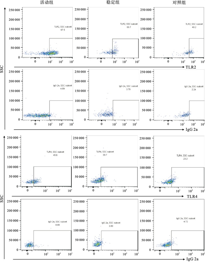
  • SLE活动组mDC-TLR2的表达率高于稳定组和对照组(P < 0.01);mDC-TLR4的表达率高于对照组(P < 0.01),与稳定组比较差异无统计学意义(P>0.05)(见表 1图 1)。

    分组 n mDC-TLR2表达率
    (x±s)/%
    mDC-TLR4表达率
    [M (P25, P75)]/%
    对照组 30 40.4±17.36 14.35(10.05,31.11)
    SLE稳定组 21 45.2±20.21 35.75(14.63,51.90)
    SLE活动组 33 59.4±18.58**△△ 38.87(15.57,64.36)*
    F 8.83 10.93#
    P < 0.01 < 0.01
    MS组内 345.210
    多重比较:与对照组比较*P < 0.05,** P < 0.01;与SLE稳定组比较△△P < 0.01。#示H
  • SLE活动组和稳定组IL-10、IL-17A表达水平均高于对照组(P < 0.01),活动组与稳定组间差异无统计学意义(P>0.05)(见表 2)。

    分组 n IL-10 IL-17A
    SLE活动组 33 193.59(39.07, 316.75)** 93.35(34.21, 150.38)**
    SLE稳定组 21   64.236(31.00, 178.27)** 47.58(15.40, 115.35)**
    对照组 30 6.58(0.60, 12.57) 4.64(4.36, 5.57)
    H 43.7 33.59
    P < 0.01 < 0.01
    与对照组比较**P < 0.01
  • 在各项实验室指标中,SLE病人外周血mDC-TLR2表达率与血清IgG、24 h尿蛋白定量均呈负相关(r=-0.270、-0.310,P < 0.05),而与抗Sm抗体呈正相关(r=0.340,P < 0.05)。mDC-TLR4表达率与血小板计数呈正相关(r=0.340,P < 0.05),与其余临床指标均无相关性(见表 3)。

    实验室指标 mDC-TLR2
    阳性率/%
    mDC-TLR4
    阳性率/%
    白细胞计数/(109/L) 0.177 -0.019
    中性粒细胞计数/(109/L) 0.195 -0.057
    淋巴细胞计数/(109/L) 0.044 0.107
    血红蛋白/(g/L) -0.014 0.181
    血小板计数/(109/L) -0.223 0.340*
    红细胞沉降率/(mm/h) -0.157 -0.026
    C反应蛋白/(mg/L) -0.089 -0.171
    IgG/(g/L) -0.270* 0.088
    IgA/(g/L) -0.116 0.180
    IgM/(g/L) -0.252 0.014
    补体C3/(g/L) -0.230 0.083
    补体C4/(g/L) -0.111 -0.052
    ANA 0.039 -0.011
    抗-SSA 0.263 0.109
    抗-SSB 0.100 0.043
    抗-Sm 0.340* 0.091
    抗-RNP 0.105 -0.137
    抗dsDNA -0.046 -0.046
    抗中性粒细胞胞质抗体 -0.009 0.179
    抗CCP抗体 0.013 -0.084
    RF 0.238 0.038
    ASO -0.010 0.140
    25-羟-VD 0.100 -0.129
    24 h尿蛋白定量 -0.310* -0.022
    *示P < 0.05
3.   讨论
  • SLE是一种慢性自身免疫性疾病,以体内自身抗体产生为特征,可通过形成免疫复合物等激活炎症细胞,促进炎症因子的表达和释放,导致免疫失衡。尽管在过去几十年里对于SLE的早期诊断和治疗有明显进步,但SLE的发病风险和死亡率仍在持续增加[9-12]。因此探索和研究SLE的确切发病机制对于SLE病人的治疗和预后有着十分重要的意义。

    TLRs作为重要的模式识别受体,在天然免疫中发挥着重要作用。研究[13-14]报道,TLRs在活化状态下可能通过调节细胞因子的产生而影响SLE疾病活动性,且TLR2和/或TLR4的缺乏使自身抗体的产生减少,延缓了SLE症状的发展。另外,近期一项研究[15]显示使用TLR阻断剂及其衍生物能够显著抑制活化B细胞NF-κB的激活,抑制炎症因子的分泌,并且对SLE小鼠的疾病进展有一定的预防作用,提示TLR2和TLR4在SLE发病中可能起到重要作用。作为较早发现的重要的胞外TLRs,TLR2和TLR4可以识别细菌、支原体、真菌和病毒的各种成分,包括脂蛋白、肽聚糖等,其中TLR2以与TLR1或TLR6形成异源二聚体的形式来识别其配体,而TLR4则通过与细胞膜表面的骨髓分化蛋白2(myeloid differentiation 2,MD2)结合形成复合物而参与配体识别,并招募特定信号分子,包括髓样分化因子88和β-干扰素TIR结构域衔接蛋白,启动下游信号事件,激活转录因子NF-kB,调节炎症细胞因子(如IL-10、IL-17A等)分泌,从而参与免疫反应[16-19]。DCs作为目前已知抗原提呈能力最强的细胞之一,在免疫激活和免疫耐受过程中起到重要作用,依据功能来源不同可分为不同的DC亚群,包括CD11c+CD123- mDC和CD11c-CD123+pDC。mDC可通过提呈抗原、协同信号传递和分泌细胞因子, 激活T细胞, 从而参与免疫反应。人外周血mDC膜表达TLR2、4, 可能通过识别PAMPs,激活前述TLR2/4-MyD88/TRIF通路,释放炎性细胞因子,进而参与SLE的疾病进展[20]

    近几年国内外有关不同细胞膜表面TLR2、TLR4的研究结果不尽相同。有研究[21]报道,SLE病人单核细胞TLR4表达水平显著低于健康对照组,TLR2表达水平与健康对照组相比差异无统计学意义。另有研究[22]显示,SLE病人活动期、稳定期血清TLR4表达水平显著高于健康对照组。而本研究旨在通过流式细胞术探究TLR2、TLR4在SLE病人外周血mDC膜表面的表达情况,及其与SLE病人各项临床指标之间的关联以及血清IL-10、IL-17A表达水平,进一步探讨TLR2、TLR4在SLE发病机制中的作用。

    本研究发现SLE活动组mDC-TLR2表达率显著高于对照组和稳定组,提示TLR2可能在SLE疾病发病与病情进展中起到重要作用,这一结果与刘昱[23]在SLE病人CD4+T细胞TLR2表达水平研究中的结果一致。本研究中SLE活动组mDC-TLR4表达率显著高于对照组, 差异有统计学意义;稳定组mDC-TLR4表达率高于对照组,但差异无统计学意义,与张文兰等[24-25]的研究结果基本一致,不同之处是本研究中活动组mDC-TLR4表达率高于稳定组,但差异无统计学意义,分析其原因可能是检测方法不同及所测细胞种类不同。本次研究结果提示mDC-TLR2、mDC-TLR4可能在SLE发病过程中起到重要作用,并可能作为SLE药物治疗的潜在靶点,为研制TLRs阻断剂治疗SLE提供一定理论支持。IL-10具有刺激免疫反应和抑制免疫反应的双重作用,在调节免疫反应过程中起到重要作用[26]。IL-17A是IL-17家族具有代表性的成员,其可以诱导多种促炎细胞因子产生,后者可募集B细胞等多种效应细胞,诱导产生大量自身抗体,激活免疫反应。在本研究中,我们发现SLE活动组及稳定组血清IL-10、IL-17A水平显著高于对照组,与以往多项研究[27-29]结果一致,提示IL-10、IL-17A可能参与SLE发病。笔者将SLE病人mDC-TLR2、mDC-TLR4表达率与实验室指标进行相关性分析发现,SLE病人血小板计数与mDC-TLR4表达率呈正相关; 血清IgG水平、24 h尿蛋白定量与mDC-TLR2表达率呈负相关; 抗Sm抗体与mDC-TLR2表达率呈正相关,表明TLR2在SLE发病中起到一定作用。

    综上所述,SLE是一种多因素参与的全身性自身免疫病,可引起多器官损害,研究mDC-TLR2、mDC-TLR4在SLE病人中的表达情况有利于对SLE发病机制的进一步探索,有望提供研制TLRs阻断剂治疗SLE的理论可能性,对完善SLE病人的靶向治疗、改善预后,具有重要的临床意义。

Reference (29)

Catalog

    /

    DownLoad:  Full-Size Img  PowerPoint
    Return
    Return