• 中国科技论文统计源期刊
  • 中国科技核心期刊
  • 中国高校优秀期刊
  • 安徽省优秀科技期刊
Volume 45 Issue 10
Nov.  2020
Article Contents
Turn off MathJax

Citation:

Expression of STC2 protein in endometrial cancer and its relationship with prognosis

  • Received Date: 2019-05-05
    Accepted Date: 2019-10-12
  • ObjectiveTo investigate the expression levels of stanniocalcin-2(STC2) protein in endometrial cancer and its relationship with prognosis.MethodsA total of 78 samples of endometrial cancer were collected.The expression level of STC2 protein in all cases were measured using immunohistochemistry, and the correlation of which with clinical characteristics was analyzed.The relationship between STC2 expression level and relapse-free survival rate was compared.ResultsThe results of immunohistochemistry showed that the tumor cells with positive STC2 staining in 61 samples were found, which accounted for 65.38%.Based on staining score system, the non-or low-expression of STC2 in 35 samples(44.87%) and high-expression of STC2 in 43 samples (55.13%) were identified.There was no statistical significance in the age between two groups(P>0.05), but the FIGO staging, lymphatic vascular invasion, myometrium invasion and cervical involvement in high-expression group significantly aggravated compared with the non-expression and low-expression group, and the difference of which was statistically significant(P < 0.05 to P < 0.01).All patients were followed up for 12 months.During the following-up, the tumor recurrence in 14 cases(17.95%) found, which included 2 cases(5.71%) in non-or low-expression group and 12 cases(27.91%) in high-expression group.The relapse-free survival rate in high-expression group was lower than that in non-or low-expression group(P < 0.05).ConclusionsThere is light-moderate STC2 expression in most cases of endometrial cancer, and its high expression level is associated with the poor prognosis.STC2 may be a prediction marker for the prognosis of endometrial cancer.
  • 加载中
  • [1] 赵宏喜, 李艳红, 谢丽, 等.子宫内膜癌的流行病学特点及其转移的危险因素分析[J].现代肿瘤医学, 2017, 25(23):3803.
    [2] 张坤, 郭红燕, 韩劲松.22例子宫内膜癌复发病例的临床特点分析[J].中国微创外科杂志, 2017, 17(6):495.
    [3] 李志鹏, 杨颖, 贾春丽, 等.STC2在肝细胞癌组织中的表达及临床意义[J].中华全科医学, 2018, 16(6):868.
    [4] 李连秀, 刘巍, 谭文华.STC2在恶性肿瘤中的研究进展[J].国际遗传学杂志, 2019, 42(1):80.
    [5] AYDIN HA, TOPTAS T, BOZKURT S, et al.Stanniocalcin-2 may be a potentially valuable prognostic marker in endometrial cancer:a preliminary study[J].Pathol Oncol Res, 2019, 25(2):751. doi: 10.1007/s12253-018-00576-y
    [6] 李小毛, 叶辉霞, 叶敏娟, 等.广东省部分医院10081例子宫内膜癌流行趋势分析[J].中国医师杂志, 2018, 20(3):367.
    [7] 王芳.子宫内膜癌复发的影响因素分析及相应治疗方案[J].实用癌症杂志, 2017, 32(5):820.
    [8] 钟睿, 李慧, 张爽, 等.如何认识和处理肿瘤异质性[J].中国肺癌杂志, 2018, 21(9):712.
    [9] LAW AY, WONG CK.Stanniocalcin-2 is a HIF-1 target gene that promotes cell proliferation in hypoxia[J].Exp Cell Res, 2010, 316(3):466. doi: 10.1016/j.yexcr.2009.09.018
    [10] 于冰冰, 曹丽群, 曲保清.STC2蛋白在胃癌组织和胃正常黏膜组织中的表达及与临床病理参数的关系[J].广东医学, 2017, 38(s1):35.
    [11] 刘荣强, 林国桢, 代天星, 等.基于Oncomine数据库分析STC 2在肝细胞癌中的表达及意义[J/CD].中华肝脏外科手术学电子杂志, 2019, 8(1): 76.
    [12] 曹蕾, 李磊, 胡明珠, 等..STC2促进人肝癌细胞HepG2增殖和EMT相关的迁移[J].中国病理生理杂志, 2017, 33(6):1000.
    [13] CHENG H, WU Z, WU C, et al.Overcoming STC2 mediated drug resistance through drug and gene co-delivery by PHB-PDMAEMA cationic polyester in liver cancer cells[J].Mater Sci Eng C Mater Biol Appl, 2018, 83:210. doi: 10.1016/j.msec.2017.08.075
    [14] 赵宏喜, 潘珍妮, 郭琳, 等.STC2在宫颈癌组织中的表达及其临床意义[J].医学临床研究, 2019, 36(4):764.
    [15] YANG S, JI Q, CHANG B, et al.STC2 promotes head and neck squamous cell carcinoma metastasis through modulating the PI3K/AKT/Snail signaling[J].Oncotarget, 2017, 8(4):5976. doi: 10.18632/oncotarget.13355
  • 加载中
通讯作者: 陈斌, bchen63@163.com
  • 1. 

    沈阳化工大学材料科学与工程学院 沈阳 110142

  1. 本站搜索
  2. 百度学术搜索
  3. 万方数据库搜索
  4. CNKI搜索

Figures(1) / Tables(2)

Article views(3553) PDF downloads(15) Cited by()

Related
Proportional views

Expression of STC2 protein in endometrial cancer and its relationship with prognosis

  • Department of Obstetrics and Gynecology, Shenzhen Nanshan People's Hospital, Shenzhen Gaungdong 518052, China

Abstract: ObjectiveTo investigate the expression levels of stanniocalcin-2(STC2) protein in endometrial cancer and its relationship with prognosis.MethodsA total of 78 samples of endometrial cancer were collected.The expression level of STC2 protein in all cases were measured using immunohistochemistry, and the correlation of which with clinical characteristics was analyzed.The relationship between STC2 expression level and relapse-free survival rate was compared.ResultsThe results of immunohistochemistry showed that the tumor cells with positive STC2 staining in 61 samples were found, which accounted for 65.38%.Based on staining score system, the non-or low-expression of STC2 in 35 samples(44.87%) and high-expression of STC2 in 43 samples (55.13%) were identified.There was no statistical significance in the age between two groups(P>0.05), but the FIGO staging, lymphatic vascular invasion, myometrium invasion and cervical involvement in high-expression group significantly aggravated compared with the non-expression and low-expression group, and the difference of which was statistically significant(P < 0.05 to P < 0.01).All patients were followed up for 12 months.During the following-up, the tumor recurrence in 14 cases(17.95%) found, which included 2 cases(5.71%) in non-or low-expression group and 12 cases(27.91%) in high-expression group.The relapse-free survival rate in high-expression group was lower than that in non-or low-expression group(P < 0.05).ConclusionsThere is light-moderate STC2 expression in most cases of endometrial cancer, and its high expression level is associated with the poor prognosis.STC2 may be a prediction marker for the prognosis of endometrial cancer.

  • 子宫内膜癌是一种发病率较高的女性妇科恶性肿瘤,近年来该病的发病率和死亡率在不断上升[1]。子宫内膜癌病人术后复发的风险较高,寻找合适的预测指标识别高复发风险的病人是目前子宫内膜癌临床诊治过程中的难点[2]。斯钙素2(stanniocalcin-2,STC2)是一种调控钙磷代谢的蛋白。近年来的研究[3-4]发现,STC2的表达与很多恶性肿瘤的疾病发展、预后都有很大的关系。但目前为止,国内外的报道对STC2在子宫内膜癌中的研究极少。因此,本次研究旨在探讨STC2在子宫内膜癌中的表达以及其与病人预后的关系。现作报道。

1.   资料与方法
  • 收集2016年1月至2017年10月在我院接受治疗的子宫内膜癌病人的手术标本78例纳入本次研究,病人年龄34~65岁。纳入标准:(1)病人首次诊断为子宫内膜癌,并接受手术治疗;(2)病人术前临床资料及术后随访资料完整。排除标准:(1)病人术前接受过放疗、化疗等新辅助治疗;(2)合并有其他器官、系统严重疾病者;(3)既往有恶性肿瘤疾病史。78例病人中行广泛子宫切除+双侧附件切除术18例,广泛子宫切除+双侧附件切除+盆腔和腹主动脉旁淋巴结清扫术60例;[4]子宫颈累及9例;国际妇产科联合会(International Federation of Gynecology and Obstetrics,FIGO)分级1级41例,2级16例,3级21级;病理分期Ⅰ期47例,Ⅱ期22例,Ⅲ期9例;术后辅助治疗:放疗17例,盆腔外束放射治疗+近距离放疗19例,序贯放化疗9例。

  • 将标本置于4%福尔马林中固定,石蜡包埋后进行连续切片,组织切片厚度为5 μm。将石蜡切片浸入二甲苯中脱蜡,乙醇复水。采用链霉亲合素-生物素酶复合物(Streptavidin-Peroxidase,SP)法进行免疫组织化学染色,试剂盒采用由上海钰博生物科技有限公司提供的即用型SP免疫组织化学试剂盒以及DAB显色试剂盒;一抗采用兔抗人STC2抗体(1:50,上海科敏生物科技有限公司),实验中以PBS液作为一抗的空白对照。所有操作均遵循试剂盒内的操作手册。

  • 所有染色组织切片由2位擅长妇科诊断的病理学主治医生进行评估和打分,医生在诊断前对病人的临床资料一无所知。强阳性定义为细胞膜或细胞质内有棕色染色信号;中度阳性为颜色呈黄褐色;弱阳性的定位为染色信号呈淡黄色。染色强度分级评分如下:0分为无染色;1分为弱阳性;2分为中度阳性;3分为强阳性。根据以下标准对肿瘤细胞染色百分率进行分级:0分为没有染色;1分为阳性肿瘤细胞≤10%;2分阳性肿瘤细胞11%~50%;3分为阳性肿瘤细胞51%~80%;4分为阳性肿瘤细胞≥81%。STC2染色结果根据染色强度和染色细胞百分率计算所得,分数范围为0~7分。0分为无染色,或阳性肿瘤细胞 < 10%;计算所得分数0分即为0,1~3分为1+, 4~5分为2+, 6~7分为3+。根据所有样本的STC2染色得分进行样品STC2表达分级,将0和1+的样品纳入无表达+低表达组;2+和3+的样品纳入高表达组。

  • 采用t检验、χ2检验、Kaplan-Meier生存曲线法和Cox比例风险回归模型。

2.   结果
  • 对所有手术标本进行STC2免疫组织化学染色,发现有STC2阳性的肿瘤细胞的样本共61件,占所有样本的65.38%。根据STC2染色结果评分标准,STC2无表达+低表达组样本35件(44.87%),STC2高表达组样本43件(55.13%)(见表 1)。

    表达特征 n 百分比/%
    肿瘤细胞STC2染色阳性 61 65.38*
    染色强度
       无染色 17 21.79
       弱阳性 31 39.74
       中度阳性 21 26.92
       强阳性 9 11.54
    染色细胞百分率
       无染色 17 21.79
       ≤10% 8 10.26
       11%~50% 21 26.92
       51%~80% 24 30.77
       ≥81% 22 28.21
    染色分数
       0 23 29.49
       1+ 12 15.38
       2+ 21 26.92
       3+ 23
    染色分组
       无表达组 23 29.49
       低表达组 12 15.38
       高表达组 43 55.13
       *示阳性率
  • 将无表达+低表达组与高表达组病人的临床特征进行比较,结果显示,2组病人年龄差异无统计学意义(P>0.05),但高表达组的FIGO分期、侵犯淋巴血管、侵犯子宫肌层以及累及子宫颈情况较无表达+低表达组明显加重,差异均有统计学意义(P < 0.05~P < 0.01)(见表 2)。

    分组 n 年龄(x±s)/岁 FIGO分期 侵犯淋巴血管 侵犯子宫肌层 累及子宫颈
    1级 2~3级
    无表达+低表达组 35 48.96±7.49 24(68.57) 11(31.42) 6(17.14) 9(25.71) 3(8.57)
    高表达组 43 49.98±6.91 16(37.21) 27(62.79) 16(37.21) 15(34.88) 8(18.60)
    χ2 0.62* 7.60 3.15 2.32 1.97
    P > 0.05 < 0.01 < 0.05 < 0.05 < 0.05
      *示t
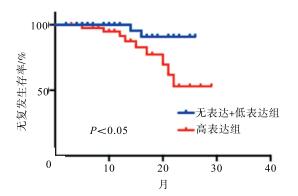
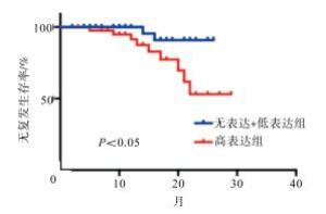
  • 对所有病人进行术后随访,术后随访时间为1~24个月,中位数为15个月。随访期间共有14例(17.95%)病人出现肿瘤复发,其中无表达+低表达组2例(5.71%),高表达组12例(27.91%)。2组病人随访期间的无复发生存率STC2高表达组低于无表达+低表达组,差异有统计学意义(χ2=6.45,P < 0.05)(见图 1)。

3.   讨论
  • 子宫内膜癌是一种发生于子宫内膜的上皮性恶性肿瘤,多发于围绝经期和绝经后的妇女,是仅次于卵巢癌和宫颈癌的第三大妇科恶性肿瘤[6]。肿瘤的组织类型、分期、分级、大小、淋巴血管累及情况、年龄等都是目前公认的影响肿瘤复发的常规危险因素[7-8]。但临床实践发现,即使是临床特征相似的病人,其预后亦存在很大区别;研究[8]认为,造成这一预后差异的原因是因为肿瘤侵袭转移的分子表型。近年来,分子标志物的研究已经成为肿瘤研究领域的热点之一。STC2是一种通过调控钙、磷代谢的蛋白,其可以促进无机磷酸盐诱导的钙化并抵制异位钙化。近期的研究[9]报道指出STC2基因是低氧诱导因子HIF-1的靶基因,能够在低氧状态下促进肿瘤细胞的增殖、侵袭、迁移和上皮间质转化。STC2在胃癌、肝癌等研究中都被发现其表达与疾病的预后有一定的关系[10-11]。本次研究我们探讨了STC2表达在子宫内膜癌中的表达以及与疾病预后的关系。

    本次研究结果显示子宫内膜癌病人中STC2阳性的比例较高,为65.38%,但在STC2染色中,大部分样品的染色强度为弱-中度(66.66%);该结果与既往的研究[5]结果基本相似。进一步比较不同STC2表达程度的样品的临床特征,结果发现,STC2高表达的样品其FIGO分期也较晚,提示STC2高表达可能是子宫内膜癌预后不佳的潜在标志物。由于本次研究的随访时间较短,因此我们比较了STC2无表达+低表达组和STC2高表达组的无复发生存率,结果显示,STC2高表达病人的无复发生存率显著低于STC低表达的病人。这一结果,进一步证实了STC2是可以用于预测子宫内膜癌预后的分子标志物。

    在其他类型癌症中,STC2高表达也已经被证实与预后不良有极为密切的联系[12-13]。其他研究者显示宫颈癌组织中的STC2表达较正常组织显著增高,且STC2表达与病人的总生存率及无复发生存率呈负相关[14]。而本次研究中我们并未发现子宫内膜癌组织中的STC2表达与年龄有关。因此,将外周血中STC2水平应用对子宫内膜癌预后的预测,还需更多的研究进一步确认。此外,STC2作用于子宫内膜癌发生发展的分子机制还需要进一步研究,有学者认为其可能与PI3K/AKT信号通路有关[15],但具体机制有待进一步挖掘。

    综上所述,大部分子宫内膜癌均表达有轻-中度的STC2蛋白,而STC2高表达与疾病的预后不良有很大的关系,其可能可以作为子宫内膜癌预后的预测指标。

Reference (15)

Catalog

    /

    DownLoad:  Full-Size Img  PowerPoint
    Return
    Return