

 下载:
下载:
-
数字化断层融合成像(digital tomosynthesis,DTS)是近年来发展起来的一项新的X射线断层融合成像技术,可以在低剂量照射后获得被扫描物体的多角度断层数据,经后处理和重构后获得任意方位、较高质量的图像信息,不受周围组织重叠影响,提高了组织分辨能力,清晰地显示被检部位组织结构以及与周围组织毗邻关系,DTS能克服常规直接数字化X射线摄影系统(digital radiography,DR)摄影二维重叠影像组织干扰,尤其在对移位不明显的骨折或细微的骨裂敏感、显示隐匿性骨折[1],提高了病变检出率,根据辐射防护优化原则,应用最小辐射剂量来保护受检者,相对螺旋CT具有辐射剂量较低、对骨性结构有较高空间分辨率、操作方便、快捷、经济及内外固定伪影少等优点[2]。在对创伤性骨关节病变诊断方面有着明显优势, 能够满足临床需要, 近年来逐渐受到临床重视。本研究对DTS在我院创伤性骨关节病变检查中的临床应用进行评价分析。
-
收集我院2015年5月至2017年12月创伤性骨关节病人44例,男28例,女16例,年龄9~76岁。其中寰枢椎关节4例、骶尾骨3例,踝关节4例,肘关节4例,手、腕关节7例,肩关节3例,髋关节5例,膝关节5例,腰椎5例,足跗骨4例。行普通DR摄影检查后:DR平片疑似骨折或骨折线显示欠清、以及骨折经内外固定术后图像显示欠佳者,均征得病人同意后再进行连续断层融合成像检查。
-
常规X线摄影使用德国西门子摄片机,DR摄影检查,按照不同部位、体厚选择不同的曝光条件。体层融合成像是基于岛津的Sonialvision SafireⅡ数字大板多功能全动态摄影成像系统。选择系统的TOMOS应用程序进行DTS检测,并根据部位不同选择相应的拍摄条件、适当增减参数。先输入病人信息并保存,病人仰卧检查床上,受检部位放置于照射野中心,设置断层融合位置中心、具体部位层厚,从而确定球管和平板起始相应位置,按下控制台SET键,透视下定位适当位置,然后进入TOMOS曝光程序,有时根据具体检查部位及临床要求,在透视下旋转至最佳合适体位,必要时加扫任意斜位断层,要求病人保持完全制动,并按下曝光制动器直到检查结束。
-
采集74张不间断曝光的原始图像,并传送到岛津Side Station工作站,在TOMOS应用程序中,调节中心位置、层厚、窗宽窗位,经后处理重建,获得容积扫描内的连续断面图像。通过在工作站上连续播放,选出其中最佳清晰度,满足诊断要求的图像进行多幅打印供临床诊断。两种成像方法获得的图像质量由两名经验丰富的放射科高年资医师分析研究。如果两名医师对评估结果有不同看法,则经过协商获得一致意见。图像评判质量的优劣如下:全面显示靶部位整体关节解剖结构、骨折线、骨折内外固定后的图像清楚、无明显干扰伪影,良好对比度,可满足临床诊断图像为优;完全显示靶部位的整体关节解剖,骨折线、骨折内外固定后的图像显示欠清晰、有少许伪影存在,对比度欠佳,但能达到临床要求诊断的为良好;不能充分显示靶部位的整个关节解剖结构、骨折线、骨折内外固定后的图像显示不清晰、明显伪影干扰、对比度较差,不能满足临床诊断的图像定为差。
-
采用χ2检验。
-
两名有经验医师的读片评价有较好一致性(Kappa=10.603)。44例病人中, 断层融合成像优41例,良好3例,差0例;44例常规DR片中优29例,良好10例,差5例;断层融合成像图像优良率100.00%(44/44),高于常规DR图像的88.64%(39/44)(χ2=5.30,P < 0.05)。
-
DTS对44例创伤性骨关节检查均明确地发现骨折,DR检查阴性但病人临床体征明显显示隐匿性骨折5例,分别是枢椎椎体骨折1例,腕骨舟状骨骨折2例,大多角骨骨折1例,腰椎体骨折1例;明确地排除DR检查疑是骨折7例,分别是尺骨茎突1例,腕骨1例,寰椎1例,骶尾骨2例,股骨颈1例,肘关节1例。
-
DTS重建图像能清晰地显示整体骨关节解剖结构和骨折线,可以去除伪影干扰(如骨折金属内固定伪影和石膏、夹板、绷带外固定制动伪影);新鲜性骨折均能清晰显示骨折线;陈旧性骨折显示骨痂形成、断端愈合情况清楚。DR显示骨折线模糊,较难显示隐匿性骨折。典型图片见图 1。
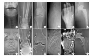
图 1 DR与DTS重建图像
-
随着多层螺旋CT的应用广泛,影像后处理技术的快速发展,单纯传统X线断层摄影技术逐渐被淘汰。多层螺旋CT虽然能获得良好的对比度和清晰度以及高空间分辨率的容积数据,对病灶检出率大大提高和作出准确性的定性诊断,但随着公众自身防护意识加强和卫生放射学发展,临床也关注到CT扫描时产生大量的辐射剂量对人体的影响, 国际放射防护委员会和世界卫生组织研究表明,常规胸部CT扫描剂量约为90~100倍于DR平片辐射剂量[3];联合国原子辐射效应科学委员会认为医疗机构剂量最大辐射来源于CT扫描,其产生的辐射剂量与诱发癌症风险或发生其他疾病相关联[4]。因此,随着低剂量原则的广泛提倡,放射剂量尽可能减少到最低限度且在不影响病变诊断前提下,影像学研究的热点已逐步转向低辐射剂量扫描技术的探究方面[3-5]。以上检查影像方法在疾病诊断方面虽有相应的应用价值,但考虑到辐射剂量,检查费用、病变检出率等因素,都有着或多或少的不足之处。
近年来由于计算机和动态大平板探测器的发展,出现了DTS,DTS是一种计算机后处理与连续断层相结合的新兴的体层摄影技术[6], 一次低剂量曝光可以获得多向扫描物体的体积图像数据,并且可以由图像重建出任意数目和任意平面与探测器平行的图像。颠覆了以往传统的X射线体层成像,一次曝光只能得到单个目标水平的图像,具有较CT、常规DR空间分辨率高,明显减少了受检者吸收的辐射剂量。扫描速度快且价格经济,易于被大众接受。遵循正当化、最优化(最小剂量、最小代价和最佳图像)放射诊疗原则,DTS可以说经济简便且用最小射线剂量获取最佳扫描图像,保护受检者,受到临床及影像医生重视并得以广泛应用。
-
DTS较多应用于胸部、骨科、乳腺及口腔科等方面[6-9]。在骨折诊断方面,常规X线照射条件得到的图像对比度低,清晰度欠佳,尤其对复杂且解剖结构重叠的骨折难以显示,不能满足临床医生判断骨折复位愈合的要求。我们对DTS在创伤性骨关节病变中应用研究结果表明,DTS较常规DR图像优良率比较差异有明显统计学意义。当骨折对位对线一致时,DR检查阴性或骨折线显示欠清,DTS检查均明确地发现骨折。骨折内固定术后和/或石膏外固定后DR片显示伪影干扰,骨折断端或其骨痂形成模糊,临床医师不能精确地判断骨折端的位置及邻近周围骨质改变及愈合的情况。虽然提高常规DR照射条件能够得到相对良好的图像,另外作为常规X线的补充检查方法CT扫描能够去除石膏重叠影的影响,提高图像的对比度,但以上两者均会使得病人接受的辐射剂量增加,再者由于CT的检查费用高,一般不作为骨折复位、愈后评价的常规检查手段。内固定金属物放射伪影会对图像质量造成一定的影响,DTS可以通过后处理重建,能够去除CT扫描金属植入产生的较大伪影,消除了复杂解剖结构、石膏外固定等重叠的影响,清晰地显示骨折断端骨痂形成情况,显示图像清晰、得到与CT断层图像相似的影像而利于诊断[10-11]。对于如平片难以确定骨关节病变的骨折、金属固定物植入后的摄片检查,具有很大的优越性。此外,使用大型平板探测器和非晶态硒作为直接转换介质,降低了感兴趣区周围图像的失真率,提高并保存了图像存储信息。重建图像显示骨折线清晰,能够检出些深在和复杂部位的组织隐匿性骨折,本研究显示出DR模糊的隐匿性骨折5例,分别是枢椎椎体骨折1例,腕骨舟状骨骨折2例,大多角骨骨折1例,腰椎体骨折1例;尤其是枢椎、腕骨解剖结构复杂且重叠,腰椎骨折有时由于肠管气体及内容物伪影存在,DR片骨折往往难以显示,DTS克服了组织结构重叠以及去除肠气及内容物伪影给医师诊断带来的不确定性,对枢椎、腕骨、腰椎骨折的检出更加凸显DTS优势;明确地排除DR检查疑似骨折7例,分别是尺骨茎突1例,腕骨1例,寰椎1例,骶尾骨2例,股骨颈1例,肘关节1例,避免了临床骨折误诊、漏诊等,因而解决了困扰医学影像科诊断医师这一医疗纠纷难点问题,对临床诊断和治疗具有重要意义。临床急重症创伤性骨关节病人,往往需要制动,而常规DR摄片由于局部骨质结构的相互重叠或者病人被动体位,不能够完全配合临床要求体位进行摄影,导致细微骨折不能及时发现或断端有无移位的病例,从而对被检者的及时治疗、预后带来了不良影响,若被迫搬至DR需要“合适体位”,这势必会造成病人二次损伤,导致损伤后遗症,甚至危及生命,DTS操作简单易行,可以使病人采取自由体位等优点,避免搬动而加剧病人的痛苦以及其他损伤,从而避免了漏诊[12]。本研究中寰枢关节损伤病人,采取严格制动检查,规避二次医源性损伤,使影像科医生及临床医生对创伤性骨关节病变作出较为准确的判断,清晰地检出寰枢关节半脱位3例,DR未发现枢椎椎体骨折1例。
DTS由于采用了特殊数据算法,因而获得的数据可重建出任意层面的断层图像,满足病人的需求,可轻松完成放射科大流量、大范围的检查工作,并且图像质量较传统断层摄影有飞跃性提高,是对现今常规DR摄像技术的提升和发展。DTS还具有价格经济、连续扫描时间短、适合长期随访等优点,断层融合技术在隐匿性骨折[1]、骨折石膏固定后复位情况的判断[10]、评估关节炎方面[7]、人工置换关节的随访[11],具有良好的应用价值,在肺结节的显示中,具有很大的临床价值[6]。在静脉肾盂造影方面[13],由于克服了组织结构重叠,当病人检查时,不需要肠道准备,且可以观察到输尿管的动态蠕动。较DR图像清晰且能明确地诊断病变,DTS是普通X线平片及螺旋CT、MRI检查有利的重要延伸和补充,随着临床医生对图像质量要求的进一步提高和科技的进步,X线设备将普及全平板化,快速、高质量的图像采集及后处理已日趋成熟,DTS作为一项有很高的实用价值的成像技术,辐射剂量较小且图像清晰度高,必然成为未来医学发展的趋势,将会被广泛地应用于临床。
X线数字化断层融合成像在创伤性骨关节病变中的临床应用
Clinical value of X-ray digital tomosynthesis imaging in the diagnosis of traumatic osteoarthritis
-
摘要:
目的探讨X线数字化断层融合成像(digital tomosynthesis,DTS)在创伤性骨关节病变中的临床应用价值。 方法对44例创伤性骨关节病人行普通直接数字化X射线摄影系统(digital radiography,DR)摄影检查后,发现DR平片疑似骨折或骨折线显示欠清,以及骨折经内外固定术后图像显示欠佳者,均征得病人同意后再进行DTS成像检查,获得扫描范围内连续断层图像。对比分析DTS与常规DR图像质量,评价DTS临床应用价值。 结果DTS重建图像均能清晰显示骨关节结构,去除骨内外固定伪影,新鲜性骨折均能清晰显示骨折线;陈旧性骨折显示骨痂形成、断端愈合情况清楚,显示隐匿性骨折5例,排除骨折7例。 结论DTS在创伤性骨关节病变检查中操作简便、易行,诊断准确率高、辐射剂量较低,在复杂结构部位的骨折以及经内外固定术后骨折的诊断与复查有着极其重要的价值,值得临床进一步推广使用。 Abstract:ObjectiveTo investigate the clinical value of digital tomosynthesis(DTS) in the diagnosis of traumatic osteoarthritis. MethodsThe results of digital radiography(DR) examination in 44 patients with traumatic osteoarthritis found that the suspected fracture or fracture line was not clear, and the image of internal and external fixation of fracture was not well.All cases were detected using DTS to obtain the consecutive tomographic images within the scanning range.The image quality between DTS and conventional DR was compared, and the clinical application value of DTS was evaluated. ResultsThe reconstructed DTS images could display the bone and joint structure, the internal and external fixation artifacts could be removed, the fracture line in fresh fracture could be clearly showed, the callus formation in old fracture could be showed, the healing of broken end was clear.The occult fracture in 5 cases was found, and the fracture was excluded in 7 cases. ConclusionsDTS is simple and easy to operate in the examination of traumatic osteoarthropathy, and has high diagnostic accuracy and low radiation dose.It has extremely important value in the diagnosis and review of complex structure fractures after internal and external fixation, and is worthy of further promotion. -
Key words:
- fracture /
- traumatic osteoarthropathy /
- X-ray /
- digitalization /
- tomosynthesis imaging
-
[1] KIM W, ORAVEC D, NEKKANTY S, et al.Digital tomosynthesis (DTS) for quantitative assessment of trabecular microstructure in human vertebral bone[J].Med Eng Phys, 2015, 37(1):109. doi: 10.1016/j.medengphy.2014.11.005 [2] ALMOKHTAR N, SHAH J, MARSON B, et al.Initial clinical experience of the use of digital tomosynthesis in the assessment of suspected fracture neck of femur in the elderly[J].Eur J Orthop Surg Traumatol, 2015, 25(5):941. doi: 10.1007/s00590-015-1632-3 [3] 冉姗姗, 綦维维, 张淼, 等.多参数设置对低剂量胸部CT扫描图像质量及辐射剂量的影响[J].中国医学影像技术, 2018, 34(1):113. [4] DONG WOOK K, WEON KUU C, Myonggeun Y.Imaging doses and secondary cancer risk from kilovoltage cone-beam CT in radiation therapy[J].Health Physics, 2013, 104(5):499. doi: 10.1097/HP.0b013e318285c685 [5] AFAT S, BROCKMANN C, NIKOUBASHMAN O, et al.Diagnostic accuracy of simulated low-dose perfusion ct to detect cerebral perfusion impairment after aneurysmal subarachnoid hemorrhage:a retrospective analysis[J].Radiology, 2018, 287(2):643. doi: 10.1148/radiol.2017162707 [6] HEE LK, MO GJ, MIN LS, et al.Digital Tomosynthesis for evaluating metastatic lung nodules:nodule visibility, learning curves, and reading times[J].Korean J Radiol, 2015, 16(2):430. doi: 10.3348/kjr.2015.16.2.430 [7] SON CN, SONG Y, KIM SH, et al.Digital tomosynthesis as a new diagnostic tool for assessing of chronic gout arthritic feet and ankles:comparison of plain radiography and computed tomography[J].Clin Rheumatol, 2017, 36(9):1. [8] POPLACK SP, TOSTESON TD, KOGEL CA, et al.Digital breast tomosynthesis:initial experience in 98 women with abnormal digital screening mammography[J].Ajr Am J Roentgenol, 2007, 189(3):616. doi: 10.2214/AJR.07.2231 [9] 邢海源, 郭雪松, 翟玉涛.数字化断层融合在颌骨疾病诊断中的应用[J].放射学实践, 2014, 29(6):704. [10] 丁昌懋, 卢振威, 王博, 等.X线断层融合摄影在桡骨远端骨折复位石膏固定后随访中的应用价值[J].河南外科学杂志, 2015, 21(4):40. [11] GILLET R, TEIXEIRA P, BONARELLI C, et al.Comparison of radiographs, tomosynthesis and CT with metal artifact reduction for the detection of hip prosthetic loosening[J].Eur Radiol, 2019, 29(3):1258. doi: 10.1007/s00330-018-5717-3 [12] XIA W, YIN XR, WU JT, et al.Comparative study of DTS and CT in the skeletal trauma imaging diagnosis evaluation and radiation dose[J].Eur J Radiol, 2013, 82(2):e76. doi: 10.1016/j.ejrad.2012.09.008 [13] LIU S, WANG H, FENG W, et al.The value of X-ray digital tomosynthesis in the diagnosis of urinary calculi[J].Exp Ther Med, 2018, 15(2):1749. -

 点击查看大图

图(1)
计量
- 文章访问数: 4426
- HTML全文浏览量: 2477
- PDF下载量: 16
- 被引次数: 0
