


-
子宫内膜癌是发生于子宫内膜的一种上皮性恶性肿瘤,是最常见的女性生殖系统肿瘤之一[1]。脆性组氨酸三联体(fragile histidinetriad, Fhit)基因是活跃的第一位脆性位点的抑癌基因,它对于肿瘤的发生及进展起着重要的负性调控作用,在多种肿瘤中都有不同程度的表达缺失且具有相关性[2]。RNA激活(RNA activation,RNAa)系将靶向目的基因启动子区域的双链RNA分子(dsRNA)导入肿瘤细胞,RNAa是小双链RNA引导的Argonaute蛋白参与的转录基因激活机制,这种小双链RNA被称为小激活RNA(saRNA)[3]。本研究检测Fhit基因在子宫内膜癌中的表达及其与临床病理因素的相关性,拟运用RNA激活上调人子宫内膜癌细胞株(Ishikawa, ISK)中Fhit基因的表达,探讨其对子宫内膜癌细胞增殖和侵袭、迁移能力的影响。现作报道。
-
实验组为子宫内膜癌组织,术后均经常规病理组织检查证实为Ⅰ型子宫内膜癌(即子宫内膜样腺癌)。选自2018年1月至2019年7月就诊的135例病人,年龄27~81岁。根据2000年FIGO标准,临床病理分期为Ⅰ~ Ⅱ期70例,Ⅲ~Ⅳ期65例。根据2003年WHO标准,病理分级为高分化44例,中分化58例,低分化33例。肌层浸润>50%者69例,≤50%者66例。有淋巴结转移者43例,无转移者92例。术前未接受抗癌性治疗,具有完整临床资料。对照组为正常子宫内膜组织,选自妇科活检内膜组织或因其他良性疾病而行子宫切除的40例病人。所有组织均来自蚌埠医学院第一附属医院病理科。
-
自动切片机将蜡块切成0.4 μm厚度的切片,脱蜡后在121 ℃枸橼酸盐溶液中进行3 min的抗原修复,自然冷却后在3%的H2O2溶液中静置10 min,加抗Fhit兔抗人一抗60 ℃ 1 h,PBS冲洗3×5 min,二抗37 ℃ 30 min,PBS冲洗后DAB显色,镜下观察。Fhit蛋白主要定位于细胞质, 以抗体在细胞质内出现黄色颗粒为阳性信号,对细胞染色强度及阳性细胞百分比综合评分[4]。按染色强度打分:0分为无色,1分为浅黄色,2分为棕黄色,3分为棕褐色,按阳性细胞所占百分比打分:无阳性细胞为0分,阳性细胞≤10%为1分,阳性细胞11%~50%为2分,阳性细胞51%~75%为3分,阳性细胞>75%为4分;染色强度与阳性细胞百分比的乘积≥2分为免疫反应阳性, < 2分为表达下降或免疫反应阴性。用Image-Pro Plus软件进行图像分析,积分吸光度(integrated optical density,IOD)/面积值作为判断Fhit蛋白表达情况。
-
人子宫内膜癌细胞株ISK、构建Fhit-saRNA表达载体、逆转录试剂盒、PCR试剂盒(上海吉玛);DMEM(高)培养液、PBS平衡液、胎牛血清(Hyclone);Fhit抗体(Abcam, ab181004);β-actin抗体(Abcam, ab8229);脂质体转染试剂LipofectamineTM2000、Trizol试剂(Invitrogen);CCK-8试剂、Transwell小室(碧云天);结合基质胶(Matrigel,BD)。
-
将ISK细胞置于37 ℃、5%CO2培养箱中培养,细胞贴壁生长,每2~3天传代一次,传代时弃去旧培养液,用PBS洗涤2~3次,加入胰酶消化,加入培养液吹打细胞,重置细胞液接种于培养瓶。实验取处于对数生长期的细胞。
-
saRNA序列为dsFhit:5′-CGA ATT CGC CCT TGC TTA TTA-3′;5′-AAT TGG CCA TTA GCC GCG GCG-3′;Control:5′-GGC CTT AAG GCC TAA TGC TGC-3′;5′-TTA ACC GGG ATT AGC CGG CAT-3′。
-
转染前按2×10⁵个细胞/孔接种6孔板,待细胞密度达70%~80%时开始转染,转染步骤按lipofentamineTM2000试剂说明书进行,dsFhit的终浓度为50 nmol/L,孵育48 h,将dsFhit转染入ISK细胞。实验分组:实验组(转染dsFhit)、阴性对照组(转染无序RNA)、空白对照组(不转染)。
-
转染48 h后收集各组细胞提取蛋白,用10%聚丙烯胺凝胶分离蛋白。配制分离胶和积层胶,每孔加入等量蛋白样本电泳、封闭。将膜放入稀释的一抗(1:3 000稀释)中,温和振荡2 h后置于4 ℃冰箱中过夜,置于TBST溶液中洗膜3×10 min,在二抗中温和振荡2 h,置于TBST溶液中洗膜3×10 min。将膜用显影溶液处理后曝光,显影,通过凝胶成像系统进行灰度值分析。
-
细胞转染48 h后用Trizol试剂提取各组细胞总RNA,用紫外分光光度计准确定量。取5 μg总RNA进行逆转录反应。Fhit基因上游引物:5′-AGG ACT CCG AAG AGG TAG CAT-3′,下游引物:5′-TCA CTG AAA GTA GAC CCG CAG-3′;β-actin上游引物:5′-AAC AAG ATG AGA TTG CCA TGC-3′,下游引物:5′-AGT GGG GTG GCT TTT AGG ATA-3′;PCR扩增反应条件为:95 ℃变性3 min后,按下列参数循环35次:95 ℃变性30 s,58 ℃退火30 s,72 ℃延伸45 s,最后72 ℃孵育10 min。取5 μL PCR产物进行凝胶电泳30 min,在紫外灯下观察DNA条带,拍照,并用凝胶图像处理系统进行灰度值分析。
-
取对数生长期的细胞,调整细胞密度为5×10⁴个/毫升,接种于96孔板中,每孔种150 μL细胞液,置于细胞培养箱中孵育24 h后转染,于24、48、72、96、120 h向各孔内分别加入20 μL的CCK-8试剂继续培养4 h。酶标仪在450 nm处测定各孔的吸光度(OD)值。实验重复3次。
-
侵袭实验将结合基质胶每孔50 μL均匀地铺在Transwell小室膜上,细胞转染48 h后,将各组细胞消化成细胞悬液,调整细胞密度为4×10⁵个/毫升,取200 μL细胞悬液至Transwell小室中,将小室置于24孔板的培养基中,孵育24 h后取出Transwell小室,用棉签擦掉滤膜上层细胞,将滤膜用甲醇固定,加适量结晶紫溶液染色,于400倍目镜下计数每个膜随机不同视野透过膜的细胞数,取平均值,每组平行设3个小室,实验重复3次。迁移实验Transwell小室膜上不铺结合基质胶,细胞转染48 h后,其余步骤同侵袭实验。
-
采用t检验、χ2检验、方差分析和q检验。
-
结果显示,正常子宫内膜组织中的细胞质内有阳性染色表达,子宫内膜癌组织中的细胞质内阳性染色表达减弱甚至缺失,2组差异有统计学意义(P < 0.01)(见图 1和表 1)。

图 1 Fhit在不同子宫内膜组织中的表达
组织学类型 n 阴性 阳性 χ2 P 正常子宫内膜组织
子宫内膜癌组织40
1357
10633
2950.22 < 0.01 表 1 Fhit在不同组织中的表达
-
子宫内膜癌中Fhit表达阳性率FIGO分期Ⅲ+Ⅳ高于FIGO分期Ⅰ+Ⅱ、肌层浸润>1/2高于肌层浸润≤1/2、有淋巴结转移高于无淋巴结转移,差异均有统计学意义(P < 0.05~P < 0.01),阳性率与年龄无明显相关性(P>0.05)(见表 2)。
因素 n 阴性 阳性 χ2 P 年龄/岁 < 50
≥5026
10916
6510
440.03 >0.05 组织学分级 G1 44 26 18 3.03 >0.05 G2 58 42 16 G3 33 25 8 FIGO分期 Ⅰ+Ⅱ
Ⅲ+Ⅳ70
6540
5030
155.93 < 0.05 肌层浸润 ≤1/2
>1/266
6939
5327
164.88 < 0.05 淋巴结转移 无
有92
4355
3637
77.64 < 0.01 表 2 Fhit表达与子宫内膜癌临床病理特征的联系
-
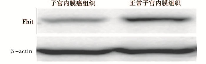
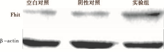
与子宫内膜癌相比,正常子宫内膜中Fhit蛋白表达显著增高(见图 2)。2组间差异有统计学意义(P < 0.01)(见表 3)。细胞转染48 h后与阴性对照组和空白对照组相比,实验组Fhit蛋白表达显著增高(见图 3),各组间差异有统计学意义(P < 0.01)(见表 4)。

图 2 Western blotting检测Fhit基因在不同子宫内膜组织中的表达
分组 Fhit相对表达量 t P 子宫内膜癌组织
正常子宫内膜组织0.44±0.02
0.80±0.0211.06 < 0.01 表 3 子宫内膜癌组织与正常子宫内膜组织中Fhit的表达比较(x±s)

图 3 Western blotting检测Fhit基因蛋白表达
分组 Fhit相对表达量 F P 空白对照组 0.21±0.02 阴性对照组 0.23±0.01 14.78 < 0.01 实验组 1.04±0.11 表 4 RNAa转染后ISK细胞中Fhit的表达比较(x±s)
-
细胞转染48 h后实验组mRNA表达水平为0.86±0.15,显著高于阴性对照组的0.34±0.01和空白对照组的0.33±0.01(P < 0.01)。
-
CCK-8检测结果显示,细胞转染24、48、72、96、120 h抑制率分别为5.3%、15.6%、37.2%、43.3%和54.8%,与阴性对照组相比,实验组细胞增殖减慢,生长明显受抑制,差异有统计学意义(P < 0.05)(见图 4)。

图 4 CCK-8检测ISK增殖
-
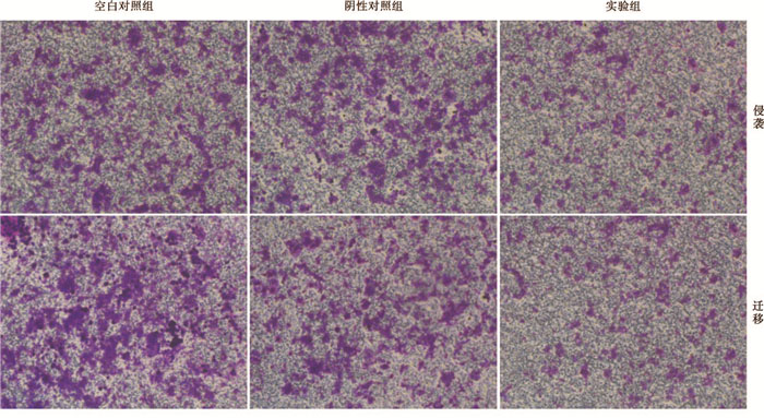
Transwell小室实验显示,与空白对照组和阴性对照组相比,实验组细胞的侵袭和迁移能力有明显下降(P < 0.01)(见表 5、图 5)。
分组 穿膜细胞数量(侵袭) 穿膜细胞数量(迁移) 空白对照组 179.0±6.9 111.3±7.1 阴性对照组 162.7±8.9 108.3±7.8 实验组 41.2±8.1 38±7.2 F 265.20 94.95 P < 0.01 < 0.01 表 5 Fhit对ISK细胞侵袭和迁移能力的影响(x±s)

图 5 Fhit对ISK细胞侵袭和迁移能力的影响
-
子宫内膜癌在全世界发病率高及治愈率低,科学家们也一直致力于此肿瘤的发病机制研究,希望从分子水平为其诊断、治疗及预后提供帮助。肿瘤的发生、发展与癌基因异常激活、抑癌基因失活及DNA错配修复基因异常密切相关[5]。而RNAa为近年来基因功能和治疗研究领域最热门的技术之一,RNAa通过选择性的激活或增强某个抑癌基因的表达来抑制肿瘤生长,而不需要找到肿瘤特定的致癌基因,RNAa效应不涉及对任何靶序列的降解,而是通过募集转录激活因子导致基因转录过程的活化并引起染色质修饰的激活。因此,RNAa具有几乎无限的靶基因,可以提高靶基因mRNA的总丰度,同时保留mRNA天然拼接异构体的多样性,在转录及表观遗传水平发挥作用,因此能够持久激活靶基因,但不改变基因组。这一发现将开拓性地扩展治疗肿瘤、代谢及遗传性等疾病[6]。
Fhit基因是1996年OHTA等[7]用差异显示分析探针及外显子捕获法在3p14.2区分离鉴定出的一个抑癌基因,全长1 095 bp,由10个外显子组成,外显子5~ 9构成开放阅读框,编码1个由147个氨基酸组成的、相对分子质量16 800,起始外显子位于t(3;8)异断裂处的着丝粒侧,t(3;8)的移位可干扰Fhit基因表达。Fhit基因存在于上皮性组织中,主要通过参与细胞周期调控和凋亡发挥作用,它可使细胞受阻于增殖周期的S期,并激活Caspase酶介导的凋亡级联反应[8]。综合目前的研究,该基因在正常组织如子宫内膜、卵巢、肺等有不同水平的Fhit mRNA表达,在人类部分上皮性肿瘤中表达降低或缺失,且与肿瘤预后差密切相关[9]。
在本研究中,我们通过对135例子宫内膜癌组织和40例正常子宫内膜组织应用免疫组织化学及Western blotting检测,结果显示Fhit蛋白在子宫内膜癌中阳性表达率为21.5%,与正常子宫内膜组82.5%相比,阳性表达率明显降低,Fhit蛋白在2种不同子宫内膜组织中表达差异有统计学意义(P < 0.01),说明随着子宫内膜组织向癌组织进展,Fhit蛋白的阳性表达率明显降低,Fhit蛋白与子宫内膜癌的发生及发展有明显相关性;子宫内膜癌中Fhit基因表达与肿瘤组织学分级、临床分期、肌层浸润和淋巴结转移明显相关(P < 0.05~P < 0.01)。CCK-8试剂盒用于检测人子宫内膜癌细胞中Fhit基因RNA激活后细胞的增殖情况,结果显示实验组细胞生长与阴性组相比较明显减慢,说明Fhit基因被激活后子宫内膜癌细胞倍增时间明显延长。Transwell小室模型实验是研究肿瘤细胞侵袭迁移的一种较好方法, 它能模拟肿瘤细胞消化基质并穿越屏障侵袭迁移的过程。实验结果表明Fhit表达激活后,能够穿越屏障的子宫内膜癌细胞与阴性对照组相比较明显减少,这表明子宫内膜癌细胞侵袭和迁移能力受到抑制,进一步证实了Fhit基因与人子宫内膜癌细胞的增殖和侵袭转移存在密切关系。
本文研究发现Fhit基因的表达缺失与子宫内膜癌的发生和发展密切相关,检测其表达水平对子宫内膜癌的早期诊断、临床进展及估计预后有一定的参考价值[10]。通过多因素分析,探讨Fhit基因在人子宫内膜癌中的抑制作用和子宫内膜癌转移的危险因素,为病情监测和高危人群的筛查提供更为确切的手段,它的检测将有助于提高诊断的准确性和客观性,对于子宫内膜癌的预后评估、病情随访及合理治疗具有一定的指导意义[11]。
Fhit基因在子宫内膜癌增殖、侵袭中的作用和临床意义
Role and clinical significance of Fhit gene in the proliferation and invasion of endometrial carcinoma
-
摘要:
目的探讨Fhit基因在子宫内膜癌中的表达情况,并通过RNA激活上调Fhit表达后观察其对子宫内膜癌细胞增殖和侵袭、迁移的影响。 方法收集135例子宫内膜癌组织和40例正常子宫内膜组织,使用免疫组织化学和Western blotting检测Fhit基因的表达情况并分析Fhit表达与临床病理特征的联系。构建Fhit-saRNA表达载体,转染至子宫内膜癌ISK细胞株,建立上调Fhit基因表达的子宫内膜癌细胞株。Western blotting和RT-PCR验证转染效果。通过CCK-8和Transwell试验分析转染前后增殖、迁移、侵袭能力的差异。 结果免疫组织化学和Western blotting结果均显示在子宫内膜癌组织中Fhit基因表达与正常子宫内膜组织相比明显降低甚至缺失(P < 0.01);CCK-8结果显示Fhit基因表达上调后子宫内膜癌细胞生长明显减慢,与未转染组比较差异有统计学意义(P < 0.01);Transwell试验结果显示Fhit基因表达上调后子宫内膜癌细胞迁移和侵袭能力显著减弱,空白对照组的穿膜细胞数显著高于转染组(P < 0.01)。 结论相比于正常子宫内膜组织,Fhit基因在子宫内膜癌组织中表达较低且与临床病理特征相关。在子宫内膜癌细胞株中通过saRNA上调Fhit后,其增殖、迁移、侵袭能力受到明显抑制。 Abstract:ObjectiveTo explore the expression level of Fhit gene in endometrial carcinoma, and observe its effects on the proliferation, invasion and migration of endometrial carcinoma cells through the RNA activation up-regulating the expresson of Fhit. MethodsThe expression levels of Fhit protein in 135 endometrial carcinoma tissues and 40 normal endometrial tissues were detected using immunohistochemistry and Western blotting.The correlation between Fhit expression and clinicopathological features was analyzed.The Fhit-saRNA expression vector was constructed, and transfected into endometrial carcinoma ISK cell line, and the endometrial carcinoma cell line with Fhit up-regulating expression was established.The transfection effects were verified using Western blotting and RT-PCR.The differences of the proliferation, migration and invasion abilities between before and after transfection were analyzed using CCK-8 and Transwell assays. ResultsThe results of immunohistochemistry and Western blotting showed that the expression levels of Fhit in endometrial carcinoma tissue significantly reduced or even missed compared with normal endometrial tissue(P < 0.01).The results of CCK-8 showed that the growth of endometrial carcinoma significantly slowed down after the up-regulating of Fhit expression, and the difference of which between transfection group and non-transfection group was statistically significant(P < 0.01).The results of Transwell assay showed that the migration and invasion abilities of endometrial carcinoma cells after the level of Fhit up-regulating significantly weakened, and the number of transmembrane cells in blank control group were significantly higher than that in transfection group(P < 0.01). ConclusionsCompared with normal endometrial tissues, the expression level of Fhit in endometrial carcinoma tissues is lower, and related to the clinicopathological features.After up-regulating the Fhit expression by RNA activation in endometrial carcinoma cell line, the abilities of proliferation, migration and invasion of cells are significantly inhibited. -
Key words:
- endometrial carcinoma /
- fragile histidinetriad /
- RNA activation
-
表 2 Fhit表达与子宫内膜癌临床病理特征的联系
因素 n 阴性 阳性 χ2 P 年龄/岁 < 50
≥5026
10916
6510
440.03 >0.05 组织学分级 G1 44 26 18 3.03 >0.05 G2 58 42 16 G3 33 25 8 FIGO分期 Ⅰ+Ⅱ
Ⅲ+Ⅳ70
6540
5030
155.93 < 0.05 肌层浸润 ≤1/2
>1/266
6939
5327
164.88 < 0.05 淋巴结转移 无
有92
4355
3637
77.64 < 0.01  下载: 导出CSV
表 3 子宫内膜癌组织与正常子宫内膜组织中Fhit的表达比较(x±s)
分组 Fhit相对表达量 t P 子宫内膜癌组织
正常子宫内膜组织0.44±0.02
0.80±0.0211.06 < 0.01
下载: 导出CSV
表 4 RNAa转染后ISK细胞中Fhit的表达比较(x±s)
分组 Fhit相对表达量 F P 空白对照组 0.21±0.02 阴性对照组 0.23±0.01 14.78 < 0.01 实验组 1.04±0.11
下载: 导出CSV
表 5 Fhit对ISK细胞侵袭和迁移能力的影响(x±s)
分组 穿膜细胞数量(侵袭) 穿膜细胞数量(迁移) 空白对照组 179.0±6.9 111.3±7.1 阴性对照组 162.7±8.9 108.3±7.8 实验组 41.2±8.1 38±7.2 F 265.20 94.95 P < 0.01 < 0.01
下载: 导出CSV
-
[1] TORRE LA, BRAY F, SIEGEL RL, et al.Global cancer statistics, 2012[J].Cancer J Clin, 2015, 65(2):87. [2] SEVINC ED, CECENER G, AK S, et al.Expression and clinical significance of mi RNAs that may be associated with the FHIT gene in breast cancer[J].Gene, 2016, 590(2):278. [3] LI LC, OKINO ST, ZHAO H, et al.Small ds RNAs induce transcriptional activation in human cells[J].Proc Natl Acad Sci U S A, 2006, 103(46):17337. [4] ISHⅡ H, MIMORI K, INAGETA T, et al.Fhit, Components of DNA damage checkpoint pathway regulate UV exposure dependent alterations of gene expression of FHIT and WWOX at chromosome fragile sites[J].Mol Cancer Res, 2005, 3(3):130. [5] JIANG E, XU Z, WANG M, et al.Tumoral microvesicle-activated glycometabolic reprogramming in fibroblasts promotes the progression of oral squamous cell carcinoma[J].FASEB J, 2019, 33(4):5690. [6] JANOWSKI BA, YOUNGER ST, HARDY DB, et al.Activating gene expression in mammalian cells with promoter targeted duplex RNAs[J].Nat Chem Biol, 2007, 3(3):166. [7] OHTA M, INOUE H, COTTICELLI MG, et al.The FHIT gene, spanning the chromosome 3p14.2 fragile site and renal carcinoma-associated t(3;8)breakpoint, is abnormal in digestive tract cancers[J].Cell, 1996, 84(4):587. [8] KISS DL, BAEZ W, HUEBNER K, et al.Impact of FHIT loss on the translation of cancer-associated mRNAs[J].Mol Cancer, 2017, 16(1):179. [9] FASSAN M, RUSEV B, CORBO V, et al.Fhit downregulation is an early event in pancreatic carcinogenesis[J].Virchows Arch, 2017, 470(6):647. [10] SILVEIRA ZAVALHIA L, WEBER MEDEIROS A, OLIVEIRA SILVA A, et al.Do FHIT gene alterations play a role in human solid tumors[J].Asia Pac J Clin Oncol, 2018, 14(5):214. [11] URAL S, SIMON R, KRUSHKAL J.Correlation of gene expression and associated mutation profiles of APOBEC3A, APOBEC3B, REV1, UNG, and FHIT with chemosensitivity of cancer cell lines to drug treatment[J].Hum Genomics, 2018, 12(1):20. -

 点击查看大图

图(5)表(5)
计量
- 文章访问数: 4088
- HTML全文浏览量: 2240
- PDF下载量: 9
- 被引次数: 0
