

-
糖尿病足是糖尿病最常见的并发症之一,其致残、致死率极高[1]。糖尿病足溃疡(diabetic foot ulcer, DFU)是由周围神经、末梢血管等不同程度的病变而导致的足部感染、溃疡及深层组织的破坏[2]。一般认为,难愈性DFU在经2~4周常规处理后,足部溃疡恶化或仍未见好转,可能增加截肢风险[3],由难愈性DFU引起的下肢截肢率可高达85%[4]。难愈性DFU创面感染相对较重,坏死组织、细菌及富含蛋白质的分泌物和窦道较多,常规清创及外科换药的治疗效果并不理想,既增加了病人的经济负担,也严重影响着病人的生活质量。目前报道显示,自体富血小板凝胶(autologous platelet-rich gel,APG)已被国外广泛地应用于难愈性DFU的治疗[5]。为观察APG治疗难愈性DFU的疗效,笔者对我院2018年1月至2019年6月进行APG治疗的难愈性DFU住院病人的溃疡面愈合情况进行分析,现作报道。
-
我院内分泌科2018年1月至2019年6月收治的共50例难愈性DFU病人,所有病例均按照《糖尿病足诊治指南》(2017版)中的DFU的诊断标准筛选。入组标准:(1)年龄、性别不限,明确1型或2型糖尿病病史;(2)溃疡面>1 cm2, 没有坏死,且经其他治疗手段至少4周以上无好转或持续恶化;(3)未使用免疫抑制剂;(4)血红蛋白>110 g/L;(5)DFU分级为Wagner糖尿病足分级标准Ⅱ~Ⅲ级。排除标准:(1)哺乳期、妊娠期妇女;(2)有血管重建、坏疽等明确手术指征;(3)恶性肿瘤导致的溃疡;(4)血红蛋白 < 110 g/L,血小板 < 150×109/L;(5)伴有肾功能衰竭、血液疾病、严重心血管疾病;(6)近期服用过免疫抑制剂、抗凝剂等影响凝血功能或者影响正常获取富血小板血浆的药物。按照随机数字表的方法,随机分为观察组(25例)和对照组(25例)。观察组中男15例,女10例;年龄45~85岁;对照组中男13例,女12例,年龄42~82岁。2组病人及痊愈病人在年龄、糖尿病病程、糖化血红蛋白、血小板计数、溃疡面积等方面差异均无统计学意义(P>0.05)(见表 1、2)。
分组 n 年龄/岁 糖尿病病程/年 血小板计数/(×109/L) 糖化血红蛋白/% 溃疡面积/cm2 观察组 25 66±3.1 10.3±3.7 219±53 9.3±0.94 2.32±0.61 对照组 25 64±4.2 11.19±2.3 221±46 9.4±0.81 2.21±0.72 t — 1.92 1.02 0.14 0.40 0.58 P — >0.05 >0.05 >0.05 >0.05 >0.05 表 1 2组病人基本资料(x±s)
分组 n 年龄/岁 糖尿病病程/年 血小板计数/(×109/L) 糖化血红蛋白/% 溃疡面积/cm2 观察组 20 63±2.9 11.5±3.3 207±65 9.0±0.98 2.27±0.65 对照组 6 62±3.5 12.3±2.1 211±57 9.1±0.87 2.19±0.81 t — 0.71 0.56 0.14 0.22 0.25 P — >0.05 >0.05 >0.05 >0.05 >0.05 表 2 2组痊愈病人基本资料(x±s)
-
根据病人溃疡面大小,用含枸橼酸钠的采血管抽取一定量病人的静脉血,在超净工作台内,离心机660 g离心力离心10 min,弃下层红细胞层,混匀后再以660 g离心力离心10 min,弃上层约2/3贫血小板血浆(platelet poorplasma,PPP),静置15 min,混匀5 min,即为富血小板血浆(platelet richplasma,PRP),每10 mL静脉血可制备约1 mL PRP。将凝血酶冻干粉与10%葡萄糖酸钙按100(U):1(mL)的比例混合成激活剂,在使用之前将PRP与激活剂按照10:1的比例混合形成APG。
-
2组病人均给予降糖、抗感染、改善微循环等对症支持治疗。足部溃疡面均给予常规的清创处理,无脓性分泌物后,观察组将APG均匀地外敷在溃疡面或注射入窦道,再用凡士林油纱覆盖封闭创面,外用纱布敷料包扎;对照组病人常规外科换药处理溃疡面,外用纱布敷料包扎。每隔2~3 d换一次药,同时测定溃疡面的长度和宽度。若敷料表面破损或脱落,及时更换。观察组应用APG前及5 d后分别取溃疡面相同的部位做病理切片。
-
临床观察时间为10周,以病人痊愈即为结束治疗(痊愈:溃疡面上皮化完全愈合)。愈合率=痊愈人数/总人数×100%;愈合时间,即从病人接受治疗到溃疡面完全愈合所需要的时间。
-
临床观察周期内,溃疡面完全愈合的病人。溃疡面缩小速率=接受治疗时溃疡面的面积/溃疡面愈合的时间(cm2/w)。
-
采用χ2检验和t检验。
-
观察组愈合率为80.00%(20/25),高于对照组愈合率24.00%(6/25)(χ2=15.71, P < 0.01)。
-
临床观察周期内,观察组溃疡面积缩小速率与对照组差异有统计学意义(P < 0.05), 愈合时间明显少于对照组(P < 0.01)(见表 3)。
分组 n 溃疡面积缩小速率/(cm2/w) 愈合时间/d 观察组 20 0.48±0.56 26.7±3.0 对照组 6 0.29±0.09 50.3±3.7 t — 1.46 16.05 P — < 0.05 < 0.01 表 3 2组间溃疡面积缩小速率及愈合时间的比较(x±s)
-
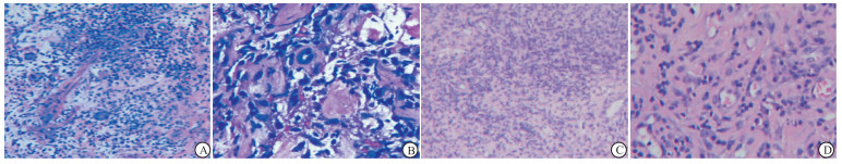
应用APG后,炎性渗出明显改善,血管和肉芽组织有少量的生成(见图 1)。

图 1 APG治疗前后溃疡面病组织理图片
-
相对于一般的创面,DFU创面由于受机体持续高血糖、周围神经血管病变、创面炎症及感染、细胞过度凋亡[6]等相关代谢因素的影响,导致难以愈合;另局部生长因子大量消耗导致的生长因子匮乏也是影响DFU愈合的一个重要因素。PRP是从自体来源的全血中提取的富含血小板的血浆,血小板含量是普通全血的3~5倍。PRP在凝血酶或钙剂的激活下形成的凝胶状物质即为APG。APG中的血小板激活后可释放大量促进创面愈合的细胞因子和生物因子,包括血小板源性生长因子、转化生长因子、血管内皮生长因子、表皮生长因子、胰岛素样生长因子-Ⅰ等。这些生长因子接触创面后,因子之间相互协同,可以促进新生血管的形成,使DFU创面新生血管形成增加,血供充足,促进新生肉芽组织的生成,在溃疡愈合中发挥重要的作用。近年来,PRP已被广泛应用于DFU的治疗,并取得较好的临床疗效[6-7]。另一方面,APG中所含的高浓度的白细胞及抑菌肽被激活后,能够对创面起到很好的抗感染作用,可抑制炎症反应[8]。同时,APG中含有的纤维蛋白可作为新生表皮的攀爬支架,促进创面收缩,并网罗住血小板、白细胞等生物活性物质,让其能够在溃疡局部发挥较长时间功效[9]。
近年来,国内、外的多项实验已将证实了APG在治疗慢性溃疡、创面方面发挥着十分确定的积极作用[10]。APG用于DFU的临床治疗也已取得一定的临床疗效。本研究中,PRP组的愈合率为80%,传统方法组的愈合率为24%,PRP组的愈合率与COBOS等[11]在PRP治疗DFU创面效果与效率评估研究中发现的73%~80%一致,而传统方法组的愈合率显著高于COBOS等研究的传统方法组的愈合率20%,这可能与选取病人的糖尿病病程、血糖控制水平、血小板计数、溃疡面的清创程度等方面有一定的差异,本文所选取的样本量较少也是一个可能因素。在愈合时间方面,本次治疗过程中,观察组愈合的平均时间为(26.7±3.0)d,对照组为(50.3±3.7)d,相较于对照组,观察组的愈合时间显著短,差异有统计学意义(P < 0.05)。在COBOS等[11]的临床研究中,APG治疗后创面愈合时间为15周,传统方法组为35.5周,SHAILENDRA等[12]对72例病人随机应用APG与传统方法治疗,结果也显示APG治疗的痊愈时间较传统治疗组显著缩短,本研究结果与上述文章报道的一致。但本次研究中2组的愈合时间与上述报道相比均缩短,这可能与本研究所选取的病人的溃疡面积较小有关。国内袁南兵等[13]采用APG治疗)也取得了不错的临床效果。在评估创面修复方面,本研究选择溃疡面积缩小速率作为评价指标。在10周的临床观察周期内,观察组的溃疡面积缩小速率为(0.48±0.56)cm2/w,较之对照组的(0.29±0.09)cm2/w, 差异有统计学意义(P < 0.05),这方面的结果与BABAEI等[13]报道的一致。另外,本研究在APG应用前后的病理组织切片显示,炎性细胞减少,生成少量的肉芽组织及新生血管,说明PRP在抑制炎症、促进新生血管及肉芽组织的生成方面具有一定的疗效,但由于本研究只在应用一次APG之后做了病理,所以效果并不十分显著,在以后的研究中会加强病理方面的研究。
本研究所选取病人的溃疡面积虽然较小,但均是糖尿病引起的长期迁延不愈合的溃疡面,同样增加病人的痛苦和经济负担,本次研究APG在DFU应用中的疗效也得到了证实,值得广泛的应用于临床。另外,APG能够缩短溃疡的愈合时间,既减少了病人的痛苦,缩短了住院周期,减轻病人的经济负担,也提高了医院的病床周转率。因本院自体PRP治疗开展时间较短,病例较少,对于创面愈合相关的评价指标应在下一步的实验和研究中进行相应的完善和补充。据报道,APG中生长因子的浓度和促进创面愈合的作用并不呈现绝对的正相关[14],适当的“有效剂量”更能促进细胞的修复作用。如何制定APG治疗创面的最适浓度仍然是我们下一步需要研究和探讨的问题。
自体富血小板凝胶治疗难愈性糖尿病足溃疡的临床疗效观察
Clinical observation of autogenous platelet-rich gel in the treatment of refractory diabetic foot ulcer
-
摘要:
目的观察自体富血小板凝胶(APG)治疗难愈性糖尿病足溃疡(DFU)的临床疗效。 方法住院的50例难愈性DFU病人随机分为观察组(25例)和对照组(25例),观察组治疗采用APG,对照组采用常规外科换药,对2组治疗后的溃疡愈合率、溃疡面缩小速率及愈合时间进行比较,并对观察组病例APG治疗前后的溃疡面组织进行病理切片。 结果观察组愈合率为80.00%,溃疡面缩小速率为(0.48±0.56)cm2/w,愈合时间为(26.7±3.0)d;对照组愈合率为24.00%,溃疡面缩小速率为(0.29±0.09)cm2/w,愈合时间为(50.3±3.7)d。观察组的愈合率与愈合时间均优于对照组(P < 0.01),溃疡面缩小速率与对照组差异无统计学意义(P>0.05)。观察组在应用APG之后溃疡面的病理切片结果显示,新生血管和肉芽组织有少量的生成。 结论与传统方法相比,APG对难愈性DFU的愈合具有较好的疗效,能够提高愈合率和缩短溃疡面的愈合时间。 Abstract:ObjectiveTo observe the clinical efficacy of autologous platelet-rich gel(APG) in the treatment of refractory diabetic foot ulcers(DFU). MethodsForty-five patients with refractory DFU were randomly divided into the observation group(25 cases) and control group(20 cases).The observation group were treated with, and the control group were treated with traditional surgical dressing change.The ulcer healing rate, ulcer surface reduction rate and healing time between two groups were compared after treatment, and the pathological sections of ulcer tissue in observation group before and after APG were made. ResultsThe healing rate, reduction rate of ulcer surface and healing time in observation group were 80.00%, (0.48±0.56)cm2/w and (26.7±3.0)d, respectively.The healing rate, reduction rate of ulcer surface and healing time in control group were 24.00%, (0.29±0.09)cm2/w and (50.3±3.7)d, respectively.The healing rate and time in observation group were better than those in control group(P < 0.01), and the difference of reduction rate of ulcer surface between two groups was not statistically significant(P>0.05).After APG was applied in the observation group, the results of pathological section of ulcer surface showed that a small amount of neovascularization and granulation tissue generated. ConclusionsCompared with traditional surgical dressing change, APG has a good effects on the healing of refractory DFU, which can significantly improve the healing rate, and shorten the healing time of ulcer surface. -
Key words:
- diabetic foot ulcer /
- autogenous platelet-rich gel /
- ulcer healing
-
表 1 2组病人基本资料(x±s)
分组 n 年龄/岁 糖尿病病程/年 血小板计数/(×109/L) 糖化血红蛋白/% 溃疡面积/cm2 观察组 25 66±3.1 10.3±3.7 219±53 9.3±0.94 2.32±0.61 对照组 25 64±4.2 11.19±2.3 221±46 9.4±0.81 2.21±0.72 t — 1.92 1.02 0.14 0.40 0.58 P — >0.05 >0.05 >0.05 >0.05 >0.05  下载: 导出CSV
表 2 2组痊愈病人基本资料(x±s)
分组 n 年龄/岁 糖尿病病程/年 血小板计数/(×109/L) 糖化血红蛋白/% 溃疡面积/cm2 观察组 20 63±2.9 11.5±3.3 207±65 9.0±0.98 2.27±0.65 对照组 6 62±3.5 12.3±2.1 211±57 9.1±0.87 2.19±0.81 t — 0.71 0.56 0.14 0.22 0.25 P — >0.05 >0.05 >0.05 >0.05 >0.05
下载: 导出CSV
表 3 2组间溃疡面积缩小速率及愈合时间的比较(x±s)
分组 n 溃疡面积缩小速率/(cm2/w) 愈合时间/d 观察组 20 0.48±0.56 26.7±3.0 对照组 6 0.29±0.09 50.3±3.7 t — 1.46 16.05 P — < 0.05 < 0.01
下载: 导出CSV
-
[1] MOXEY PW, GOGALNICEANU P, HINCHLIFFE RJ, et al.Lower extremity amputations-a review of global variability in incidence[J].Diabet Med, 2011, 28(10):1144. doi: 10.1111/j.1464-5491.2011.03279.x [2] GONCHAR IV, LIPUNOV AR, AFANASOV IM, et al.Platelet rich plasma and growth factors cocktails for diabetic foot ulcers treatment:state of art developments and future prospects[J].Diabetes Metab Syndr, 2018, 12(2):189. doi: 10.1016/j.dsx.2017.09.007 [3] BABAEI V, AFRADI H, GOHARDANI HZ, et al.Management of chronic diabetic foot ulcers using platelet-rich plasma[J].J Wound Care, 2017, 26(12):784. doi: 10.12968/jowc.2017.26.12.784 [4] 杨文英.中国糖尿病的流行特点及变化趋势[J].中国科学, 2018, 48(8):812. [5] ETULAIN J.Platelets in wound healing and regenerative medicine[J].Platelets, 2018, 29(6):556. doi: 10.1080/09537104.2018.1430357 [6] YAZDANPANAH L, SHAHBAZIAN H, NAZARI I, et al.Incidence and risk factors of diabetic foot ulcer:a population-based diabetic foot cohort (ADFC study) two year follow-up study[J].Int J Endocrinol, 2018, 10(3):1. [7] DOUPIS J, VEVES A.Classification, diagnosis, and treatment of diabetic foot ulcers[J].Wounds, 2008, 20(5):117. [8] SURESH DH, SURYANARAYAN S, SARVAJNAMURTHY S, et al.Treatment of a non-healing diabetic foot ulcer with platelet-rich plasma[J].J Cutan Aesthet Surg, 2014, 7(4):229. doi: 10.4103/0974-2077.150786 [9] MOHAMMADI MH, MOLAVI B, MOHAMMADI S, et al.Evaluation of wound healing in diabetic foot ulcer using platelet-rich plasma gel:a single-arm clinical trial[J].Transfus Apher Sci, 2017, 56(2):160. doi: 10.1016/j.transci.2016.10.020 [10] 吴开泽, 陈献聪, 康禹, 等.富血小板血浆在膝关节骨关节炎治疗中的应用[J].国际骨科学杂志, 2015, 36(6):414. doi: 10.3969/j.issn.1673-7083.2015.06.008 [11] COBOS R, AIZPURU F, PARRAZA N, et al.Effectiveness and efficiency of platelet rich plasma in the treatment of diabetic ulcers[J].Curr Pharm Biotechnol, 2015, 16(7):630. doi: 10.2174/138920101607150427111926 [12] SINGH SP, KUMAR V, PANDEY A, et al.Role of platelet-rich plasma in healing diabetic foot ulcers:a prospective study[J].J Wound Care, 2018, 27(9):550. doi: 10.12968/jowc.2018.27.9.550 [13] 袁南兵, 王椿, 冉兴无.自体富血小板凝胶在糖尿病难治性皮肤溃疡中的初步应用[J].四川大学学报(医学版)2007, 38(5):900. doi: 10.3969/j.issn.1672-173X.2007.05.041 [14] SALARINIA R, SADEGHNIA H, ALAMDARI D, et al.Platelet rich plasma:effective treatment for repairing of spinal cord injury in rat[J].Acta Orthop Traumatol Turc, 2017, 51(3):254. doi: 10.1016/j.aott.2017.02.009 -

 点击查看大图
图(1)表(3)
计量
- 文章访问数: 3693
- HTML全文浏览量: 2353
- PDF下载量: 19
- 被引次数: 0
