

-
近年来,随着超声检查在人群健康体检以及临床中的普及,甲状腺结节的检出率较前明显增加[1],虽然超声是首选筛查手段,但其在鉴别结节良恶性方面作用相对有限[2]。如何在初筛时准确地对结节进行风险预测,是学者们研究的热点[3]。超声弹性成像是一种新型成像技术,能在组织生物力学层面评估组织硬度,操作易实施且无创[4]。超声弹性对比指数(elasticity contrast index,ECI)是在弹性成像的基础上,以颈动脉的搏动作为内部压力源,通过测量感兴趣区域(region of interest,ROI)内组织应变的差异性,反映病灶内组织均一性[5]。但在临床实际操作中,ECI的测量方法多样,目前尚无统一标准,本研究从横切、纵切及横切面结节内不均质区测定3种方法测量ECI,探讨每种方法的诊断效能。现作报道。
-
选择2018-2020年在我院就诊的甲状腺结节病人31例共36个结节,所有病人在甲状腺细针穿刺或手术切除前均接受常规超声弹性成像,分别以横切、纵切及横切面结节内不均质区测定3种方法测量ECI值。所有病人均获得细胞学或病理诊断。排除因颈动脉搏动过强ECI测量干扰过大的病例。
-
采用Samsung RS80A型超声诊断仪,选择LA4-18B高频探头,具有ECI模式。常规多切面扫查,观察并记录甲状腺结节的具体位置、数目、大小、边界、内部回声、纵横比、钙化等特征及颈部淋巴结情况。选择标准ECI横切扫查切面,启动程序,嘱病人屏住呼吸3~5 s,等待弹性压力指示棒由黄色全部变为绿色时立即冻结图像,标记感兴趣区ROI,勾勒出横切面结节边界,仪器自动计算出ECI值,记录数值;勾勒横切面结节内不均质区,记录ECI值;探头纵切扫查,重复上述操作步骤,记录勾勒结节边界处测得的ECI值,每个结节测量两次,取平均值。该步骤由能熟练行ECI操作的同一高年资超声医师完成。
-
采用t检验和ROC曲线分析。
-
本研究共纳入病人31例,其中男11例,女20例,常规超声共检出结节36个,经FNA或手术病理证实,其中良性结节11个,恶性结节25个。
-
横切、纵切及横切面结节内不均质区测定3种测量法中恶性结节的ECI值均明显高于良性结节,差异均有统计学意义(P < 0.05~P < 0.01)(见表 1、图 1~3)。3种ECI测量方法对诊断甲状腺结节的敏感性、特异性和准确性、阳性预测率及阴性预测率见表 2。ROC曲线分析显示,横切结节内不均质区测量法鉴别甲状腺结节良恶性的最佳界值为2.93;当ECI值≥2.93时,考虑为恶性;ECI值< 2.93时,考虑为良性;对应的灵敏度为82.0%,特异度为76.4%,准确率为75.3%;AUC为0.82,95%CI为0.69~0.87。
ECI测量方法 ECI值 t′ P 恶性 良性 横切结节边界 2.95±1.00 1.63±0.52 4.90 < 0.01 横界结节内不均质区 3.02±2.01 1.74±0.46 2.50 < 0.05 纵切结节边界 2.82±1.51 1.61±0.76 2.99 < 0.01 表 1 3种ECI测量方法对甲状腺良恶性结节的诊断结果

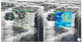
图 1 横切结节边界描记测量

图 2 横切结节内不均质区描记测量

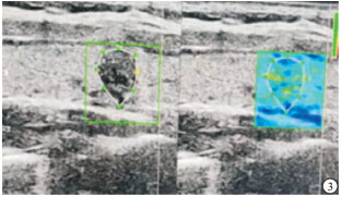
图 3 纵切结节边界描记测量
ECI测量方法 敏感性/% 特异性/% 准确性/% 阳性预测值/% 阴性预测值/% 横切结节边界 77.7 72.7 72.0 80.5 73.9 横界结节内不均质区 82.0 76.4 75.3 81.3 82.8 纵切结节边界 79.4 71.1 71.4 77.6 76.9 表 2 3种ECI测量方法对甲状腺良恶性结节的诊断效能
-
近年来,甲状腺结节的检出率逐年提高[6],其原因是多方面的,其中超声技术在临床的广泛应用是很重要的因素之一[7]。但近来有研究发现随着甲状腺癌发病率明显增加,与其相关的死亡率并没有明显升高[8],其中大部分的甲状腺癌为分化型,预后较好,10年生存期可达90%以上[9],因此有学者提出,目前甲状腺结节诊治领域存在着过度诊断与过度治疗的问题[10]。如何选择治疗时机及方式,术前的精准诊断尤为重要,目前单纯常规超声已不能满足临床的要求。
RAGO等[11]于2007年将反应组织间硬度差异的弹性成像技术引入甲状腺超声检查,在此基础上将颈动脉的搏动作为来自体内的压力源,检测ROI内应变的差异性,计算内应变均一性,并定义为ECI值[12-13]。有研究[14]表明,ECI除可反映病灶组织硬度,还可反映ROI内应变的均匀性,一般ECI值越高,认为结节质地越硬,当同时伴有内部成分不均质时,判断为恶性的风险进一步提高。
本研究应用常规超声及联合ECI技术对36个结节进行恶性风险评估,发现对有恶性风险的甲状腺结节有明显的检出率,表现在恶性及良性结节的ECI值有明显的统计学差异。其中横切结节内不均质法ECI测值,甲状腺恶性结节的ECI值为(3.02±2.01),明显高于良性结节的(1.74±0.46)(P < 0.05)。
但在临床实际操作中,对ECI的测量尚无统一操作标准,仪器厂家推荐的测量方法要求在横切扫查时,将颈动脉和甲状腺结节最大径同时显示,标记ROI,勾勒的测量区为结节的边界。有研究[15-16]报道,从甲状腺长轴切面或从短轴切面分别获取结节的ECI值,取2次的平均值作为结节的ECI值。也有学者利用横切面结节内(横内)、横切面结节与周围组织交界处(横界)、纵切面结节内(纵内)、纵切面结节与周围组织交界处(纵界)四种方法测量ECI,其中在横界法时描记结节及其旁同样面积的组织,二者各取一半[17]。笔者认为,在临床实际操作中,对于较小的深在的、不临近气管的甲状腺结节用此法尚可行,而结节较大的、近靠近包膜区者则无法行有效标记。参考上述学者的经验,结合恶性肿瘤组织侵袭性生长过程中不断坏死、钙盐沉积导致结节内均一性差的病理基础,本研究在横切、纵切测量基础上加入横切面结节内不均质区测定ECI,利用ROC曲线进行分析,该方法AUC最大,为0.82,95%CI为0.69~0.87。对应的灵敏度为82.0%,特异度为76.4%,准确率为75.3%,与葛喜凤等[18]学者的观点吻合。
综上所述,ECI能较好地评价病灶的硬度和均一性,联合常规超声在甲状腺结节诊断有较高的临床价值,其中横切面结节内不均质区测量法有较高的诊断效能。
不同方法测量超声弹性对比指数诊断甲状腺结节效能比较
Comparison of the diagnostic efficacy of different methods measuring ultrasonicity elastic contrast index in the diagnosis of thyroid nodule
-
摘要:
目的探讨超声弹性对比指数(elasticity contrast index,ECI)在诊断甲状腺结节中的应用价值,同时比较不同测量方法的效能。 方法选取甲状腺结节病人31例共36个结节,分别以勾勒横切面结节边界、横切面结节内不均质区、纵切面结节边界3种方法测量ECI,绘制受试者工作特征(ROC)曲线,比较不同测量方法对甲状腺结节的诊断效能。 结果3种方法中,横切面结节内不均质区测量法ROC曲线下面积最大,为0.82,对应ECI界值2.93,其评价甲状腺结节恶性风险的敏感性、特异性、准确性分别为82.0%、76.4%、75.3%,以横切面结节内不均质区法测定,恶性结节的ECI值高于良性结节(P < 0.05)。 结论横切面结节内不均质区测量ECI法对甲状腺结节有较高的诊断效能。 Abstract:ObjectiveTo explore the application value of ultrasonic elasticity contrast index(ECI) in the diagnosis of thyroid nodules, and compare the efficacy among different measurement methods. MethodsA total of 31 patients with thyroid nodules(36 nodules) were investigated, and the ECI was measured using the sketching the cross-sectional junction boundary, measurement of inhomogeneous areas within nodules in cross section and cross-sectional junction boundary, respectively.The ROC curve was drawn, and the diagnostic effects of thyroid nodules were compared among different measurement methods. ResultsAmong the three methods, the area under the ROC curve of the measurement of inhomogeneous areas within nodules in cross section was the largest(0.82), and the corresponding ECI cut-off value was 2.93.The sensitivity, specificity and accuracy of the measurement of inhomogeneous areas within nodules in cross section in evaluating the malignant risk of thyroid nodules were 82.0%, 76.4% and 75.3%, respectively.Using the measurement of inhomogeneous areas within nodules in cross section, the ECI value of malignant nodules is high than that of benign nodules (P < 0.05). ConclusionsThe measurement of inhomogeneous areas within nodules in cross section has a high diagnostic effect on thyroid nodules. -
Key words:
- thyroid nodule /
- ultrasonic elasticity contrast index /
- ultrasound
-
表 1 3种ECI测量方法对甲状腺良恶性结节的诊断结果
ECI测量方法 ECI值 t′ P 恶性 良性 横切结节边界 2.95±1.00 1.63±0.52 4.90 < 0.01 横界结节内不均质区 3.02±2.01 1.74±0.46 2.50 < 0.05 纵切结节边界 2.82±1.51 1.61±0.76 2.99 < 0.01  下载: 导出CSV
表 2 3种ECI测量方法对甲状腺良恶性结节的诊断效能
ECI测量方法 敏感性/% 特异性/% 准确性/% 阳性预测值/% 阴性预测值/% 横切结节边界 77.7 72.7 72.0 80.5 73.9 横界结节内不均质区 82.0 76.4 75.3 81.3 82.8 纵切结节边界 79.4 71.1 71.4 77.6 76.9
下载: 导出CSV
-
[1] 孙芳, 石岩, 刘菲菲, 等. 基于超声影像构建机器学习模型预测甲状腺良恶性结节[J]. 国际医学放射学杂志, 2021, 44(4): 392. [2] KANT R, DAVIS A, VERMA V. Thyroid nodules: advances in evaluation and management[J]. Am Fam Physician, 2020, 102(5): 298. [3] TIAN C, WANG Z, HOU X, et al. The diagnostic accuracy of superb microvascular imaging in distinguishing thyroid nodules: A protocol for systematic review and meta analysis[J]. Medicine (Baltimore), 2020, 99(40): e22350. doi: 10.1097/MD.0000000000022350 [4] SIGRIST RMS, LIAU J, KAFFAS AE, et al. Ultrasound elastography: review of techniques and clinical applications[J]. Theranostics, 2017, 7(5): 1303. doi: 10.7150/thno.18650 [5] 孙佩璇, 童宇洋, 苏蕾, 等. 超声弹性对比指数在甲状腺结节鉴别诊断中的应用[J]. 肿瘤影像学, 2018, 27(2): 92. doi: 10.3969/j.issn.1008-617X.2018.02.006 [6] KIM J, GOSNELL JE, ROMAN SA. Geographic influences in the global rise of thyroid cancer[J]. Nat Rev Endocrinol, 2020, 16(1): 17. doi: 10.1038/s41574-019-0263-x [7] 张博. 超声弹性对比指数在甲状腺结节诊断中的应用价值分析[J]. 影像研究与医学应用, 2019, 3(23): 151. [8] 李阳, 郭婕, 苏蕾, 等. 超微血管三维立体超声成像在甲状腺良恶性结节鉴别诊断中的应用价值[J]. 蚌埠医学院学报, 2020, 45(4): 507. [9] LI M, BRITO JP, VACCARELLA S. Long term declines of thyroid cancer mortality: an international age-period-cohort analysis[J]. Thyroid, 2020, 30(6): 838. doi: 10.1089/thy.2019.0684 [10] COLONNA M, UHRY Z, GUIZARD AV, et al. Recent trends in incidence, geographical distribution, and survival of papillary thyroid cancer in France[J]. Cancer Epidemiol, 2015, 39(4): 511. doi: 10.1016/j.canep.2015.04.015 [11] BURKI TK. Unnecessary thyroid cancer screening in South Korea[J]. Lancet Oncol, 2017, 18(1): e6. doi: 10.1016/S1470-2045(16)30640-4 [12] SONG Q, TIAN X, JIAO Z, et al. Value of conventional ultrasonography with contrast-enhanced ultrasonography in the differential diagnosis of partial cystic thyroid nodules[J]. Ultrasound Med Biol, 2021, 47(9): 2494. doi: 10.1016/j.ultrasmedbio.2021.03.009 [13] KIM MH, LUO S, KO SH, et al. Thyroid nodule parameters influencing performance of ultrasound elastography using intrinsic compression[J]. Ultrasound Med Biol, 2015, 41(9): 2333 doi: 10.1016/j.ultrasmedbio.2015.05.002 [14] XIE X, YU Y. Effect of the location and size of thyroid nodules on the diagnostic performance of ultrasound elastography: A retrospective analysis[J]. Clinics (Sao Paulo), 2020, 75: e1720. doi: 10.6061/clinics/2020/e1720 [15] CHOI WJ, PARK JS, KOO HR, et al. Ultrasound elastography using carotid artery pulsation in the differential diagnosis of sonographically indeterminate thyroid nodules[J]. AJR Am J Roentgenol, 2015, 204(2): 396. doi: 10.2214/AJR.14.12871 [16] 盛建国, 王斌, 曹昆昆, 等. 桥本氏甲状腺炎甲状腺超声弹性对比指数与血清自身抗体、Th1/Th2型细胞因子含量的关系[J]. 海南医学院学报, 2016, 22(19): 2357. [17] 徐翠, 杨智, 石岩, 等. 超声弹性对比指数在甲状腺结节诊断中的应用及测量方法探讨[J]. 中国医师进修杂志, 2021, 44(1): 76. doi: 10.3760/cma.j.cn115455-20200522-00643 [18] 葛喜凤, 崔立刚, 李磊, 等. 超声弹性对比指数在甲状腺结节诊断中的应用价值[J]. 中国超声医学杂志, 2017, 33(10): 871. doi: 10.3969/j.issn.1002-0101.2017.10.003 -

 点击查看大图

图(3)表(2)
计量
- 文章访问数: 3873
- HTML全文浏览量: 1693
- PDF下载量: 5
- 被引次数: 0
