

-
鼻咽癌是一种起源于鼻咽部黏膜上皮的肿瘤,具有明显的区域分布特征,其发生发展是一个多阶段、多途径、多遗传变异、多机制积累的复杂过程,涉及一系列癌基因、抑癌基因及信号通路相关因子改变,寻找影响鼻咽癌发生相关基因,并研究其作用机制,对于鼻咽癌诊疗具有重要意义[1-2]。X连锁凋亡抑制蛋白(X-linked inhibitor of apoptosis protein,XIAP)是一个重要的凋亡抑制蛋白,具有明显凋亡抑制作用,有研究[3-4]表明,抑制XIAP表达可促进多种肿瘤细胞凋亡。鼻咽癌中XIAP表达升高,但其对癌细胞生物学特性影响研究尚未明确[5]。肿瘤坏死因子α(tumor necrosis factor-α,TNF-α)是一种细胞因子,参与细胞凋亡、免疫应答等多种途径的调控[6]。已有研究[7-8]表明,TNF-α可诱导包括鼻咽癌在内的多种肿瘤细胞凋亡。因此,本研究通过RNAi技术抑制鼻咽癌细胞XIAP表达,并用TNF-α处理细胞,旨在研究抑制XIAP表达是否可促进TNF-α诱导的鼻咽癌细胞凋亡及降低免疫抑制。
-
人鼻咽癌5-8F细胞购自中国科学院上海细胞库;RPMI 1640培养基和胎牛血清(FBS)均购自美国Gibco;人重组TNF-α购自美国R&D;MTT试剂盒和二甲基亚砜(DMSO)均购自美国Sigma;流式凋亡检测试剂盒和流式细胞仪均购自美国BD;XIAP、血管内皮生长因子(VEGF)、环氧化酶-2(COX-2)、Cleaved caspase3、Bax、NF-κB p65和Ikkβ抗体均购自美国CST;酶标仪购自美国Thermo。siRNA序列由上海生工设计合成。XIAP siRNA sense:5′-GGA GAU ACC GUG CGG UGC UdT dT-3′,anti-sense:5′-AGC ACC GCA CGG UAU CUC CdT dT-3′;阴性对照组siRNA sense:5′-ACA ACT GCC TGG TTG GCA A-3′,anti-sense:5′-TTG CCA ACC AGG CAG TTG T-3′。
-
5-8F细胞用含10% FBS的改良型RPMI 1640培养基,在37 ℃、5% CO2及饱和湿度培养箱内常规传代培养。
-
实验分为空白组(细胞不经特殊处理)、TNF-α组(10 ng/mL的外源性TNF-α处理5-8F细胞12 h)、si-XIAP组(si-XIAP转染5-8F细胞48 h)和TNF-α+si-XIAP组(si-XIAP转染5-8F细胞48 h后,使用10 ng/mL的外源性TNF-α处理细胞12 h)。阴性对照siRNA(NC)及XIAP特异性siRNA(si-XIAP)转染参照LipofectamineTM 2000说明书。转染前1 d以3×10⁵/孔接种5-8F细胞于6孔板,使转染前细胞达60%~70%融合,将制备的Lipo2000-siRNA复合物加入6孔板,培养箱内常规孵育6 h,更换含血清的培养基,继续培养48 h,Western blotting检测转染后的细胞XIAP表达。
-
适量RIPA细胞裂解液提取细胞总蛋白,BCA法检测总蛋白浓度,总蛋白加上样缓冲液100 ℃煮5 min变性,每孔道上样40 μg,经10%~12%的SDS-PAGE分离,电泳后276 mA转膜2.5 h,取出膜,加5%脱脂奶粉,室温条件封闭1 h,TBST洗膜,加一抗(稀释比例均为1∶1 000),4 ℃孵育过夜,加HRP标记的二抗(稀释比例1∶2 000),室温孵育2 h,TBST洗膜,ECL显色,暗室下X线胶片显影、定影。Quantity-One软件分析蛋白条带灰度值。以目的蛋白与GAPDH灰度值比值为蛋白的相对表达量。实验重复3次。
-
以5×10³/孔接种5-8F细胞于96孔板,37 ℃培养箱常规孵育过夜,按照1.3实验分组用TNF-α和si-XIAP单独或联合处理5-8F细胞,处理后的每孔细胞加MTT 20 μL,37 ℃孵育4 h,吸去培养上清,每孔加DMSO 200 μL,摇床低速震荡10 min,酶标仪测定490 nm的吸光度值(A490)。实验重复3次。
-
以5×10⁵/孔接种5-8F细胞于96孔板,培养箱内常规孵育过夜,按照1.3实验分组用TNF-α和si-XIAP单独或联合处理5-8F细胞,预冷PBS洗涤处理后,胰酶消化细胞,离心,收集细胞,加1×Binding Buffer重悬细胞,之后加5 μL Annexin V-FITC,混匀后再加5 μL PI,混匀,室温避光孵育5~15 min,1 h内通过流式细胞仪检测。实验重复3次。
-
采用方差分析和q检验。
-
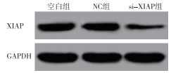
XIAP siRNA转染5-8F细胞48 h,Western blotting检测XIAP蛋白表达,结果显示,si-XIAP组XIAP相对表达量为0.097±0.010,明显低于空白组的0.602±0.057(t=15.11,P < 0.01)(见图 1)。MTT法检测各组细胞活力,结果显示,TNF-α组和si-XIAP组细胞活力均低于空白组(P < 0.05),TNF-α+si-XIAP组细胞活力低于TNF-α组和si-XIAP组(P < 0.05)(见表 1)。

图 1 转染XIAP siRNA的5-8F细胞XIAP表达
分组 n A490 F P MS组内 空白组 3 0.747±0.059 49.56 < 0.01 0.002 TNF-α组 3 0.511±0.041* si-XIAP组 3 0.547 ±0.043* TNF-α+si-XIAP组 3 0.309±0.028*#▲ q检验:与空白组比较*P < 0.05;与TNF-α组比较#P < 0.05;与si-XIAP组比较▲P < 0.05 表 1 抑制XIAP表达对TNF-α诱导的5-8F细胞活力抑制的影响(x±s)
-
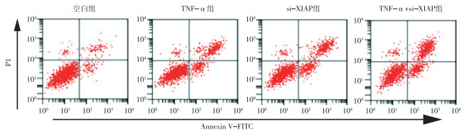
TNF-α组和si-XIAP组5-8F细胞凋亡率均高于空白组(P < 0.05),TNF-α+si-XIAP组细胞凋亡率高于TNF-α组和si-XIAP组(P < 0.05)(见图 2、表 2)。

图 2 抑制XIAP表达对TNF- a诱导的5-8F细胞凋亡的影响
分组 n 细胞凋亡率/% F P MS组内 空白组 3 3.02±0.31 343.16 < 0.01 1.824 TNF-α组 3 22.27±1.02*# si-XIAP组 3 26.82 ±1.62*# TNF-α+si-XIAP组 3 37.59±1.88*#▲ q检验:与空白组比较*P < 0.05;与TNF-α组比较#P < 0.05;与si-XIAP组比较▲P < 0.05 表 2 抑制XIAP表达对TNF-α诱导的5-8F细胞凋亡的影响(x±s)
-
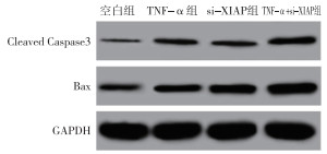
TNF-α组和si-XIAP组Cleaved caspase3和Bax表达均高于空白组(P < 0.05),TNF-α+si-XIAP组Cleaved caspase3和Bax表达均高于TNF-α组和si-XIAP组(P < 0.05)(见图 3、表 3)。

图 3 抑制XIAP表达对TNF-a诱导的5-8F细胞凋亡相关蛋白表达的影响
分组 n Cleaved caspase3 Bax 空白组 3 0.021±0.005 0.058±0.007 TNF-α组 3 0.062±0.007* 0.121±0.013* si-XIAP组 3 0.055±0.006* 0.202±0.018*# TNF-α+si-XIAP组 3 0.123±0.012*#▲ 0.509±0.045*#▲ F — 85.11 186.78 P — < 0.01 < 0.01 MS组内 — 0.000 0.001 q检验:与空白组比较*P < 0.05;与TNF-α组比较#P < 0.05;与si-XIAP组比较▲P < 0.05 表 3 抑制XIAP表达对TNF-α诱导的5-8F细胞Cleaved caspase3和Bax表达的影响(x±s)
-
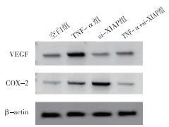
TNF-α组VEGF和COX-2表达均高于空白组(P < 0.05),si-XIAP组VEGF和COX-2表达均低于空白组(P < 0.05);TNF-α+si-XIAP组VEGF和COX-2表达均低于TNF-α组,高于si-XIAP组(P < 0.05)(见图 4、表 4)。

图 4 抑制XIAP表达对TNF-a诱导的5-8F细胞VEGF和COX-2表达的影响
分组 n VEGF COX-2 空白组 3 0.302±0.030 0.124±0.016 TNF-α组 3 0.611±0.054* 0.235±0.024* si-XIAP组 3 0.089±0.009*# 0.058±0.008*# TNF-α+si-XIAP组 3 0.216±0.023*#▲ 0.111±0.010#▲ F — 133.96 66.63 P — < 0.01 < 0.01 MS组内 — 0.001 0.000 q检验:与空白组比较*P < 0.05;与TNF-α组比较#P < 0.05;与si-XIAP组比较▲P < 0.05 表 4 抑制XIAP表达对TNF-α诱导的5-8F细胞VEGF和COX-2表达表达的影响(x±s)
-
TNF-α组NF-κB p65和Ikkβ表达均高于空白组(P < 0.05),si-XIAP组NF-κB p65和Ikkβ表达均低于空白组(P < 0.05);TNF-α+si-XIAP组NF-κB p65和Ikkβ表达均低于TNF-α组,高于si-XIAP组(P < 0.05)(见图 5、表 5)。

图 5 抑制XIAP表达对TNF-a诱导的5-8F细胞NF-κ B信号通路的影响
分组 n NF-κB p65 Ikkβ 空白组 3 0.334±0.026 0.132±0.015 TNF-α组 3 0.862±0.057* 0.321±0.031* si-XIAP组 3 0.166±0.020*# 0.075±0.010*# TNF-α+si-XIAP组 3 0.302±0.028#▲ 0.138±0.013#▲ F — 220.11 94.14 P — < 0.01 < 0.01 MS组内 — 0.001 0.000 q检验:与空白组比较*P < 0.05;与TNF-α组比较#P < 0.05;与si-XIAP组比较▲P < 0.05 表 5 抑制XIAP表达对TNF-α诱导的5-8F细胞NF-κB信号通路的影响(x±s)
-
研究[9]证实,肿瘤发生发展及演进过程伴有抑癌基因失活、原癌基因激活及参与细胞增殖、凋亡和分化的信号途径失调等。基因治疗正是针对这些特点,将正常基因或有治疗价值的其他基因引入肿瘤细胞,从而达到抑制肿瘤细胞生长的目的。因此,寻找影响鼻咽癌发生发展的基因,并研究其机制具有重要意义。凋亡抑制蛋白(inhibitor of apoptosis protein,IAPs)是一类内在的凋亡抑制因子,可通过阻断细胞凋亡所必需依赖的caspases蛋白活性,从而起到细胞凋亡抑制作用,在多种肿瘤中IAPs呈现过表达[10-11]。XIAP是IAPs家族成员之一,已有研究证实XIAP是唯一一个与caspase3、caspase9和caspase7直接结合的凋亡抑制蛋白[12]。且多项研究表明XIAP与肿瘤发生发展密切相关,如microRNA-137可通过调控XIAP促进卵巢癌细胞凋亡[13],XIAP可通过调控Bcl-2家族损伤肾癌凋亡过程中线粒体功能[14]。XIAP在鼻咽癌中高表达,提高XIAP表达可抑制鼻咽癌细胞凋亡[7]。TNF-α是一个关键的调节分子,参与细胞生长、死亡、分化、炎症等多种重要功能的调控,已被公认为重要的前凋亡因子,可诱导肿瘤细胞凋亡[15]。有研究[16]证实,一些基因可促进或抑制TNF-α诱导的肿瘤细胞凋亡,如沉默RIPK4基因表达可增加TNF-α诱导的骨肉瘤MG-63细胞凋亡敏感性。若XIAP基因可增强TNF-α诱导的鼻咽癌细胞凋亡,这将给鼻咽癌治疗提供很大帮助。本研究通过RNAi技术抑制鼻咽癌5-8F细胞XIAP表达,并用TNF-α处理细胞,发现XIAP表达抑制可增加TNF-α对5-8F细胞活力抑制及凋亡促进作用。提示XIAP同样可增强TNF-α对鼻咽癌凋亡诱导作用,可能是一个有效的鼻咽癌治疗靶点。
NF-κB有广泛的生物学效应,活化的NF-κB因子在癌症发生、免疫调节、细胞生长、炎症反应等过程中发挥重要作用[17]。有实验[18-20]证明,在鼻咽癌、乳腺癌、胶质瘤等多种实体肿瘤中都可检测到NF-κB信号通路的持续高度活化状态。有研究显示,抑制NF-κB信号通路可通过对凋亡及免疫相关因子调控,从而影响肿瘤细胞凋亡及免疫,如阻断皮肤鳞癌细胞中NF-κB信号通路,可下调抑凋亡蛋白Bcl-2表达,上调促凋亡蛋白Bax表达及提高caspase活性[21];TNF-α在HepG2细胞中能上调凋亡抑制因子VEGF表达,而黄连素能下调由其诱导的VEGF表达,可能是通过NF-κB信号通路所介导[22];黄芩素可抑制TNF-α诱导的宫颈癌细胞NF-κB信号及其下游靶基因免疫抑制因子COX-2表达[23]。肿瘤细胞对TNF-α的反应主要由NF-κB来介导,TNF-α可激活NF-κB信号通路,而活化的NF-κB信号可拮抗TNF-α诱导的细胞毒性[24]。有研究[25]显示,抑制XIAP可增强TNF-α诱导的乳腺癌细胞凋亡及下调NF-κB信号通路。本研究结果显示,抑制XIAP表达可上调TNF-α诱导的细胞Bax、Cleaved caspase3表达,下调VEGF、COX-2、NF-κB p65和IκBα表达。提示抑制XIAP表达可通过下调NF-κB信号通路诱导鼻咽癌细胞凋亡及降低免疫抑制。
综上所述,下调XIAP基因表达可增强TNF-α诱导的鼻咽癌5-8F细胞凋亡及降低VEGF和COX-2表达,机制与抑制NF-κB信号通路有关。该研究提示XIAP可能是鼻咽癌诊疗的有效靶点之一。
XIAP基因对TNF-α诱导的鼻咽癌细胞凋亡及VEGF和COX-2表达的机制研究
Study on the mechanism of XIAP gene on TNF-α-induced apoptosis and expression of VEGF and COX-2 in nasopharyngeal carcinoma cells
-
摘要:
目的探讨X连锁凋亡抑制蛋白(XIAP)基因对肿瘤坏死因子α(TNF-α)诱导的鼻咽癌细胞凋亡及血管内皮生长因子(VEGF)和环氧化酶-2(COX-2)表达的影响。 方法将人鼻咽癌5-8F细胞分为空白组、TNF-α组(10 ng/mL的外源性TNF-α处理5-8F细胞12 h)、si-XIAP组(si-XIAP转染5-8F细胞48 h)和TNF-α+si-XIAP(si-XIAP转染5-8F细胞48 h后,使用10 ng/mL的外源性TNF-α处理细胞12 h)。Western blotting检测XIAP、VEGF、COX-2、Cleaved caspase3、Bax、NF-κB p65和Ikkβ蛋白表达;MTT法及流式细胞术分别检测细胞活力和凋亡率。 结果转染si-XIAP的5-8F细胞XIAP表达明显低于空白组(P < 0.01)。TNF-α组和si-XIAP组细胞活力低于空白组(P < 0.05),细胞凋亡率及Cleaved caspase3和Bax表达均高于空白组(P < 0.05)。TNF-α组VEGF、COX-2、NF-κB p65和Ikkβ表达均高于空白组(P < 0.05)。si-XIAP组VEGF、COX-2、NF-κB p65和Ikkβ表达均低于空白组(P < 0.05)。TNF-α+si-XIAP组细胞活力低于TNF-α组和si-XIAP组(P < 0.05);VEGF、COX-2、NF-κB p65和Ikkβ表达均低于TNF-α组、高于si-XIAP组(P < 0.05);细胞凋亡率及Cleaved caspase3和Bax表达均高于TNF-α组和si-XIAP组(P < 0.05)。 结论下调XIAP基因表达增强TNF-α诱导的鼻咽癌5-8F细胞凋亡及降低VEGF和COX-2表达,机制与抑制NF-κB信号通路有关。 Abstract:ObjectiveTo investigate the effect of X-linked inhibitor of apoptosis protein(XIAP) gene on the tumor necrosis factor-α(TNF-α)-induced apoptosis, and expression of vascular endothelial growth factor(VEGF) and cyclooxygenase-2(COX-2) in nasopharyngeal carcinoma cells. MethodsHuman nasopharyngeal carcinoma 5-8F cells were divided into blank group, TNF-α group(cells treated with 10 ng/mL exogenous TNF-α for 12 h), si-XIAP group(cells transfected with si-XIAP for 48 h) and TNF-α+si-XIAP(cells transfected with si-XIAP for 48 h, then treated with 10 ng/mL exogenous TNF-α for 12 h).Western blotting was used to detect the protein expression of XIAP, VEGF, COX-2, Cleaved caspase3, Bax, NF-κB p65 and Ikkβ.MTT assay and flow cytometry were used to detect cell viability and apoptosis rate. ResultsThe expression of XIAP in 5-8F cells transfected with si-XIAP was significantly lower than that in blank group(P < 0.01).The cell viability in TNF-α group and si-XIAP group was lower than that in blank group(P < 0.05), and the apoptosis rate and the expression of Cleaved caspase3 and Bax were higher than those in blank group(P < 0.05).The expressions of VEGF, COX-2, NF-κB p65 and Ikkβ in TNF-α group were higher than those in blank group(P < 0.05).The expressions of VEGF, COX-2, NF-κB p65 and Ikkβ in si-XIAP group were lower than those in blank group(P < 0.05).The cell viability in TNF-α+si-XIAP group was lower than that in TNF-α group and si-XIAP group(P < 0.05);the expressions of VEGF, COX-2, NF-κB p65 and Ikkβ were lower than those in TNF-α group and higher than those in si-XIAP group(P < 0.05);the apoptosis rate and the expression of Cleaved caspase3 and Bax were higher than those in TNF-α group and si-XIAP group(P < 0.05). ConclusionsDown-regulating the expression of XIAP gene enhances TNF-α-induced apoptosis and reduces the expression of VEGF and COX-2 in nasopharyngeal carcinoma 5-8F cells, which is related to the inhibition of NF-κB signaling pathway. -
表 1 抑制XIAP表达对TNF-α诱导的5-8F细胞活力抑制的影响(x±s)
分组 n A490 F P MS组内 空白组 3 0.747±0.059 49.56 < 0.01 0.002 TNF-α组 3 0.511±0.041* si-XIAP组 3 0.547 ±0.043* TNF-α+si-XIAP组 3 0.309±0.028*#▲ q检验:与空白组比较*P < 0.05;与TNF-α组比较#P < 0.05;与si-XIAP组比较▲P < 0.05  下载: 导出CSV
表 2 抑制XIAP表达对TNF-α诱导的5-8F细胞凋亡的影响(x±s)
分组 n 细胞凋亡率/% F P MS组内 空白组 3 3.02±0.31 343.16 < 0.01 1.824 TNF-α组 3 22.27±1.02*# si-XIAP组 3 26.82 ±1.62*# TNF-α+si-XIAP组 3 37.59±1.88*#▲ q检验:与空白组比较*P < 0.05;与TNF-α组比较#P < 0.05;与si-XIAP组比较▲P < 0.05
下载: 导出CSV
表 3 抑制XIAP表达对TNF-α诱导的5-8F细胞Cleaved caspase3和Bax表达的影响(x±s)
分组 n Cleaved caspase3 Bax 空白组 3 0.021±0.005 0.058±0.007 TNF-α组 3 0.062±0.007* 0.121±0.013* si-XIAP组 3 0.055±0.006* 0.202±0.018*# TNF-α+si-XIAP组 3 0.123±0.012*#▲ 0.509±0.045*#▲ F — 85.11 186.78 P — < 0.01 < 0.01 MS组内 — 0.000 0.001 q检验:与空白组比较*P < 0.05;与TNF-α组比较#P < 0.05;与si-XIAP组比较▲P < 0.05
下载: 导出CSV
表 4 抑制XIAP表达对TNF-α诱导的5-8F细胞VEGF和COX-2表达表达的影响(x±s)
分组 n VEGF COX-2 空白组 3 0.302±0.030 0.124±0.016 TNF-α组 3 0.611±0.054* 0.235±0.024* si-XIAP组 3 0.089±0.009*# 0.058±0.008*# TNF-α+si-XIAP组 3 0.216±0.023*#▲ 0.111±0.010#▲ F — 133.96 66.63 P — < 0.01 < 0.01 MS组内 — 0.001 0.000 q检验:与空白组比较*P < 0.05;与TNF-α组比较#P < 0.05;与si-XIAP组比较▲P < 0.05
下载: 导出CSV
表 5 抑制XIAP表达对TNF-α诱导的5-8F细胞NF-κB信号通路的影响(x±s)
分组 n NF-κB p65 Ikkβ 空白组 3 0.334±0.026 0.132±0.015 TNF-α组 3 0.862±0.057* 0.321±0.031* si-XIAP组 3 0.166±0.020*# 0.075±0.010*# TNF-α+si-XIAP组 3 0.302±0.028#▲ 0.138±0.013#▲ F — 220.11 94.14 P — < 0.01 < 0.01 MS组内 — 0.001 0.000 q检验:与空白组比较*P < 0.05;与TNF-α组比较#P < 0.05;与si-XIAP组比较▲P < 0.05
下载: 导出CSV
-
[1] LI L, GU M, YOU B, et al. Long non-coding RNA ROR promotes proliferation, migration and chemoresistance of nasopharyngeal carcinoma[J]. Cancer Sci, 2016, 107(9): 1215. doi: 10.1111/cas.12989 [2] MAO Y, WU S, ZHAO R, et al. MiR-205 promotes proliferation, migration and invasion of nasopharyngeal carcinoma cells by activation of AKT signalling[J]. J Int Med Res, 2016, 44(2): 231. doi: 10.1177/0300060515576556 [3] WU L, ZHANG X, LIN X, et al. Inhibition of X-linked inhibitor of apoptosis protein enhances anti-tumor potency of pure total flavonoids on the growth of leukemic cells[J]. Exp Ther Med, 2018, 15(2): 2020. [4] LIU Y, ZHANG B, SHI T, et al. Inhibition of X-linked inhibitor of apoptosis protein suppresses tumorigenesis and enhances chemosensitivity in anaplastic thyroid carcinoma[J]. Oncotarget, 2017, 8(56): 95764. doi: 10.18632/oncotarget.21320 [5] YANG S, LI SS, YANG XM, et al. Embelin prevents LMP1-induced TRAIL resistance via inhibition of XIAP in nasopharyngeal carcinoma cells[J]. Oncol Lett, 2016, 11(6): 4167. doi: 10.3892/ol.2016.4522 [6] QIAO Y, HE H, JONSSON P, et al. AP-1 is a key regulator of proinflammatory cytokine TNFα-mediated triple-negative breast cancer progression[J]. J Biol Chem, 2016, 291(10): 5068. doi: 10.1074/jbc.M115.702571 [7] REN M, WANG Z, GAO G, et al. Impact of X-linked inhibitor of apoptosis protein on survival of nasopharyngeal carcinoma patients following radiotherapy[J]. Tumor Biol, 2016, 37(9): 1. [8] YAN H, XIAO F, ZOU J, et al. NR4A1-induced increase in the sensitivity of a human gastric cancer line to TNFα-mediated apoptosis is associated with the inhibition of JNK/Parkin-dependent mitophagy[J]. Int J Oncol, 2018, 52(2): 367. [9] AN Q, HAN C, ZHOU Y, et al. Matrine induces cell cycle arrest and apoptosis with recovery of the expression of miR-126 in the A549 non-small cell lung cancer cell line[J]. Mol Med Rep, 2016, 14(5): 4042. doi: 10.3892/mmr.2016.5753 [10] ABDELMAGID AF. Modulation of the inhibitors of apoptosis proteins(iaps) activities for cancer treatment[J]. Acs Med Chem Lett, 2017, 8(5): 471. doi: 10.1021/acsmedchemlett.7b00148 [11] KHAN S, SIMPSON J, LYNCH JC, et al. Racial differences in the expression of inhibitors of apoptosis(IAP) proteins in extracellular vesicles(EV) from prostate cancer patients[J]. PLoS One, 2017, 12(10): e0183122. doi: 10.1371/journal.pone.0183122 [12] CHECINSKA A, HOOGELAND B SJ, RODRIGUEZ JA, et al. Role of XIAP in inhibiting cisplatin-induced caspase activation in non-small cell lung cancer cells[J]. Exp Cell Res, 2016, 313(6): 1215. [13] LI X, CHEN W, ZENG W, et al. microRNA-137 promotes apoptosis in ovarian cancer cells via the regulation of XIAP[J]. Brit J Cancer, 2017, 116(1): 66. doi: 10.1038/bjc.2016.379 [14] CHEN C, LIU TS, ZHAO SC, et al. XIAP impairs mitochondrial function during apoptosis by regulating the Bcl-2 family in renal cell carcinoma[J]. Exp Ther Med, 2018, 15(5): 4587. [15] ZHANG G, XU M, SONG Y, et al. TNF-α produced by SEC2 mutant(SAM-3)-activated human T cells induces apoptosis of HepG2 cells[J]. Appl Microbiol Biot, 2016, 100(6): 2677. doi: 10.1007/s00253-015-7104-1 [16] 蒲彦川, 封昱辰, 王建澍, 等. 沉默RIPK4基因表达增加TNF-α诱导骨肉瘤MG-63细胞凋亡的敏感性[J]. 肿瘤, 2017, 37(12): 1268. doi: 10.3781/j.issn.1000-7431.2017.11.611 [17] 庄红, 张苏川, 蒋伟. KLF5沉默通过抑制NF-κB信号通路减少氧化应激条件下心肌细胞炎症因子分泌[J]. 免疫学杂志, 2018, 34(12): 21. [18] VERHOEVEN R JA, SHUANG T, ZHANG G, et al. NF-κB signaling regulates expression of Epstein-Barr virus BART microRNAs and long noncoding RNAs in nasopharyngeal carcinoma[J]. J Virol, 2016, 90(14): 6475. doi: 10.1128/JVI.00613-16 [19] YI Y, ANBALAGAN D, LEE LH, et al. ANXA1 inhibits miRNA-196a in a negative feedback loop through NF-kB and c-Myc to reduce breast cancer proliferation[J]. Oncotarget, 2016, 7(19): 27007. doi: 10.18632/oncotarget.8875 [20] LI F, TANG C, JIN D, et al. CUEDC2 suppresses glioma tumorigenicity by inhibiting the activation of STAT3 and NF-κB signaling pathway[J]. Int J Oncol, 2017, 51(1): 115. doi: 10.3892/ijo.2017.4009 [21] 夏永华, 刘冬, 张彩凤, 等. NF-κB信号通路的阻断对皮肤鳞癌SCL-1细胞凋亡的影响[J]. 北京大学学报(医学版), 2011, 43(2): 179. doi: 10.3969/j.issn.1671-167X.2011.02.004 [22] 李井彬, 柯善栋, 胡少明. 黄连素通过NF-κB信号通路对TNF-α诱导的VEGF表达的影响[J]. 华中科技大学学报(医学版), 2014, 43(4): 386. doi: 10.3870/j.issn.1672-0741.2014.04.004 [23] 李俊博. 黄芩素对宫颈癌细胞中NF-κB及其下游靶基因表达的影响[D]. 延吉: 延边大学, 2017. [24] LIU YP, LEE JJ, LAI TC, et al. Suppressive function of low-dose deguelin on the invasion of oral cancer cells by down-regulating TNF-α-induced NF-κB signaling[J]. Head & Neck, 2016, 38(S1): E524. [25] 史文利. 天然蛋白酶体抑制剂抑制XIAP增强TNFα抗肿瘤作用的机制研究[D]. 广州: 广州医科大学, 2014. -

 点击查看大图
图(5)表(5)
计量
- 文章访问数: 6919
- HTML全文浏览量: 3941
- PDF下载量: 25
- 被引次数: 0
