

-
输尿管结石是泌尿系统常见疾病,是引起疼痛、血尿、泌尿道感染、肾积水、肾功能损害的常见原因。既往的二十多年间,输尿管镜下碎石已逐渐成为输尿管结石的一线治疗方法[1]。输尿管镜碎石通常是中、下段输尿管结石的首选,而上段输尿管结石通常采用体外冲击波碎石或经皮肾镜碎石,但随着小口径半硬质镜、输尿管软镜、拦截/取石装置、输尿管通道鞘、碎石激光的发展,输尿管镜下钬激光碎石对于各类困难结石、上段结石的治疗成功率不断提升,从而成为治疗输尿管各位置结石安全有效的方法[2]。临床工作中经常会遇到一些结石长期卡顿于一处即所谓“嵌顿性结石”病人,其手术难度较普通结石大,且术后输尿管狭窄发生率高,处理存在复杂性。碎石术后的输尿管狭窄作为后期并发症之一,往往因为病人长期无自觉症状而耽误诊治,最终导致永久性肾功能损害,故如何减少嵌顿性结石术后输尿管狭窄的发生一直是迫切需要解决的问题[3]。输尿管镜碎石术后双J管的置入可减少术后肾绞痛、肾积水的发生,并减少粘连,从而促进输尿管的愈合,尽管既往指南指出非复杂情况的输尿管镜碎石可以尝试不放置双J管,但实践中大多数医师还是将术后留置双J管作为常规[4]。经过我们的临床研究和实践发现,在对输尿管嵌顿性结石行钬激光碎石时,通过使用N-trap拦截网篮联合术中预防性放置2根F6号双J管,能降低术后输尿管狭窄的发生并保证手术的成功率及净石率。现作报道。
-
回顾性分析2015-2019年我院因嵌顿性输尿管结石行输尿管镜下钬激光碎石后留置单根F7或单侧2根F6双J管病人临床资料,目标人群共550例,按照纳入及排除标准进行初筛研究。
纳入标准:(1)单侧、单发的输尿管嵌顿性结石,定义为结石在影像学检查上停留于同一处超过2个月,内镜下确认结石卡顿、黏膜水肿,合并或不合并细小息肉形成;(2)行输尿管硬/半硬质镜下钬激光碎石一次即成功者,术前未予其他外科手段治疗(诸如体外冲击波碎石、经皮肾镜碎石、先期留置双J管引流扩张等);(3)术中使用N-trap网篮清石,并留置单根F7或单侧2根F6双J管者;(4)年龄18~75岁,性别不限;(5)拔除双J管后我院随访满1年或未满1年即发现输尿管狭窄。
排除标准:(1)术前已明确输尿管狭窄或畸形诊断者;(2)术中输尿管镜检发现结石处已发生明显输尿管狭窄者,或术中发现大量广基、指状息肉包绕结石引起中央管腔缩窄者、或术中出现输尿管穿孔损伤、黏膜撕脱者;(3)肾盂及输尿管超上段结石(结石上端距肾盂距离 < 3 cm者);(4)合并严重基础疾病(包括精神类疾病),有严重心、肝、肾功能不全、严重凝血功能障碍者;(5)合并恶性肿瘤者。
从初筛者中使用倾向性评分法,以留置双J管情况(单根F7和2根F6)为分组变量,结石长径及嵌顿时长为匹配变量,在卡钳值0.02水平成功匹配97对(总n=194),对2组病人进行双J管拔除后输尿管狭窄发生情况、结石净石率、双J管相关尿路症状的对比分析。
-
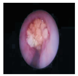
术前常规准备。斑马导丝引导,F6/F7.5或F8/F9.8半硬质镜(Wolf,德国)进镜到达结石位置后确认嵌顿情况,于结石与输尿管壁间寻找缝隙,尝试将斑马导丝自该缝隙上插,无缝隙者利用激光纤维于结石中心位置初步碎石破坏结石完整性,待结石松动后于结石周围缝隙置入N-Trap拦截网篮(Cook, 美国)成功封堵结石(见图 1),将结石从原嵌顿处推移至管壁黏膜正常位置后再行钬激光碎石,避免原位碎石。钬激光发生机(瑞科恩,中国)设定能量1.0~1.2 J, 频率15~20 Hz,365 μm光纤(瑞科恩,中国)通过工作通道将结石击碎为3 mm左右的碎片,碎石满意后调整网篮头端“勺口”的方位,拖拉网篮使其携带碎石通过结石嵌顿处并继续下行进入膀胱,于膀胱内打开网篮倾倒出碎石。再次上镜并重复上述操作数次,将输尿管内的碎石清理干净。清理完碎石后,留置单根F7双J管(BioTeq,英国)或2根F6双J管(BioTeq,英国)。如遇到阻力较大导致2根双J管推入困难时,用F8/9.8输尿管镜沿一根导丝上行,保留另一根导丝于输尿管镜鞘外,通过输尿管下“双导丝扩张法”扩张[5],经此操作可大大提高2根双J管的置入成功率。

图 1 嵌顿性结石输尿管镜下表现,中央为珊瑚形嵌顿结石,输尿管壁明显水肿,间隙消失
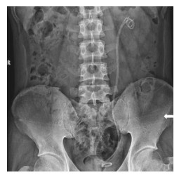
病人术后第1天复查泌尿系平片联合肾输尿管超声,或单独行输尿管CT平扫检查,明确净石(无长径3 mm以上碎石残留或逃逸)情况及双J管位置(见图 2)。于术后8~10周膀胱镜下拔除双J管,拔管后半个月复查超声,此后每2~3个月行超声检查1次,随访终点时间为1年。随访期间对于超声检查提示肾脏积水加重,考虑有发生输尿管狭窄可能者行CTU检查,影像学检查高度提示疑似狭窄者进一步行输尿管镜检明确狭窄诊断。以影像学结合输尿管镜下细窄段的长度作为输尿管狭窄长度的评定标准。

图 2 泌尿系平片显示单侧2根双J管,白色箭头处为2根平行排列的F6双J管
-
采用t检验、χ2检验和Fisher′s确切概率法。
-
2组病人年龄、性别构成、结石长径、嵌顿时长、结石侧别和结石部位差异均无统计学意义(P>0.05)(见表 1)。
分组 n 男 女 年龄/岁 结石长径/mm 嵌顿时长/周 结石侧别 结石部位 左侧 右侧 上段 中下段 单根F7双J管组 97 56 41 47.2±14.0 8.1±2.0 12.7±3.9 50 47 59 38 2根F6双J管组 97 60 37 44.3±18.1 7.8±2.1 13.3±3.4 61 36 66 31 t — 0.34* 1.25 1.02 1.14 2.55* 1.10* P — >0.05 >0.05 >0.05 >0.05 >0.05 >0.05 *示χ2值 表 1 2组病人一般情况比较(x±s)
-
术后首日复查可见在联合N-trap网篮清石后病人总体净石率达到88.1%(171/194),2组净石率差异无统计学意义(P>0.05);拔除双J管后随访过程中共发现输尿管狭窄22例,总发生率为11.3%,其中单根F7双J管组输尿管狭窄发生率明显高于2根F6双J管组(P < 0.01),预防性留置2根F6双J管能减少嵌顿性结石钬激光碎石术后狭窄的发生(RR=0.294,95%CI 0.113~0.766);2组双J管相关尿路症状(发生肉眼血尿、尿急、尿频、下腹痛、腰痛中至少一项)发生率均接近100%,差异无统计学意义(P>0.05)(见表 2)。
分组 n 输尿管狭窄 结石净石率 支架相关尿路症状发生率 单根F7双J管组 97 17(17.5) 89(91.8) 96(97.9) 2根F6双J管组 97 5(5.2) 82(84.5) 95(99.0) χ2 — 7.38 2.42 — P — < 0.01 >0.05 >0.05△ △示Fisher′s确切概率法 表 2 2组病人治疗结果比较[n;百分率(%)]
-
对22例术后发现输尿管狭窄病人进行统计,其输尿管平均狭窄长度(10.5±1.6)mm;位于左侧12例,右侧10例;上段狭窄18例,中下段狭窄4例。2组狭窄长度、狭窄侧别、狭窄部位差异均无统计学意义(P>0.05)(见表 3)。
分组 n 狭窄段长度/mm 狭窄侧别 狭窄部位 左侧 右侧 上段 中下段 单根F7双J管组 17 10.5±1.8 10(58.8) 7(41.2) 13 (76.5) 4 (23.5) 2根F6双J管组 5 10.6±1.0 2(40.0) 3(60.0) 5 (100) 0 (0.00) t — 0.12 — — P — >0.05 >0.05△ >0.05△ △示Fisher′s确切概率法 表 3 2组病人术后输尿管狭窄情况比较(x±s)
-
输尿管镜下碎石术后输尿管狭窄的发生率各中心报道不一,其影响因素众多[6-8],包括嵌顿性结石、结石部位、结石大小、内镜下碎石工具(如激光、气压弹道、超声等)、有无术中并发症(如输尿管穿孔、输尿管出血等)、输尿管镜粗细,并与操作医师经验相关。输尿管狭窄的诱因分缺血性因素和非缺血性因素,结石本身引起输尿管狭窄的机制尚未完全明了,主要与结石长期刺激引起的慢性炎症有关,结石长期嵌顿可进一步造成反应性上皮增生、间质纤维组织增生、管壁缺血及组织脆性增加,最终导致输尿管狭窄[9]。
既往很多研究[10-12]已经表明嵌顿性结石是输尿管镜碎石后狭窄发生的主要危险因素。本研究中,所有病例均为嵌顿性结石,总体术后输尿管狭窄发生率为11.3%(22/194),留置2根F6双J管组狭窄发生率仅为5.2%,比其他嵌顿性结石术后狭窄发生率低[13-15]。嵌顿性结石行钬激光碎石病人需高度警惕术后输尿管狭窄的发生。
双J管的最大作用是被动扩张、支撑及引流。在上尿路重建手术后、输尿管镜操作后、体外冲击波碎石/软镜碎石前、输尿管源性或外源性输尿管狭窄(如肿瘤、腹膜后纤维化)等有广泛应用[4]。目前非复杂性输尿管镜钬激光碎石术后常放置单根双J管或无管化(是否无管化存在争议),留置双J管的主要目的其一是预防因输尿管镜操作后水肿、结石碎片造成输尿管梗阻,并充分引流肾积水保护肾功能;其二便是起到“脚手架”的作用,让输尿管黏膜围绕管周生长,防止粘连狭窄[16]。双重双J管置入(以下称双管法)最初报道于治疗恶性肿瘤压迫引起的顽固输尿管狭窄[17],后在难治性、反复性输尿管狭窄的治疗中出现多例报道。IBRAHIM等[18]的双管法临床随机对照试验比较了输尿管狭窄激光内切开后留置双管与单管的效果,结果发现对于>1.5 cm的长段狭窄,留置双管组疗效更好。其可能机制包括:2根双J管间存在缝隙,当管内引流受阻时,双管法的管周引流更通畅;双管留置后较单管更硬,不容易受压变形,整体支撑效果更好。刘杰等[19]通过前瞻性随机对照试验比较了输尿管结石伴息肉导致输尿管狭窄者输尿管镜碎石后留置双管与单管的疗效,拔管4周后复查超声显示,双管组肾积水的缓解要明显优于单管组;CHRISTMAN等[20]认为双管法后2根双J管会产生相对运动,可以持续扩张狭窄段输尿管并可防止输尿管粘连,从而达到良好的扩张和引流效果;刘洪凯等[21]认为留置2根双J管可以形成相互支撑,有较大的空间,通畅引流可防止尿液外渗,降低瘢痕发生。本研究发现在结石大小及嵌顿时间相当时,双管组输尿管狭窄的发生率明显低于单管组(P < 0.01),相对于传统留置单根双J管,预防性留置2根双J管是减少术后输尿管狭窄发生的保护因素。
双管法也有其固有缺点,本研究中拔管时间设置在8~10周,2组病人近乎100%出现了双J管相关症状如肉眼血尿、尿频、尿急、腰痛、下腹痛等,既往观点认为双J管越粗,尿路症状更严重,但也有随机临床试验的证据表明双J管粗细型号与尿路症状无明显关联[22]。实际工作中可明显感受到长期留置双J管所导致的身心问题,部分病人可因血尿出现焦虑症状反复就诊,增加了时间和经济成本。JOSHI等[23]研究发现,约80%留置双J管的病人存在至少一项尿路症状。留置双J管时间的长短虽无共识,但较长的留置时间通常会诱发反复感染、双J管结壳。KAWAHARA等[24]报道,置管时间6~12周间双J管结壳率50%左右,超过12周时结壳率可高达75%。较遗憾本研究未记录拔除双J管时结壳的情况。
输尿管上段结石通常面临结石逃逸及清石的问题,本研究中使用N-trap网篮减少了结石的逃逸,提高了手术的成功率;同时因有网篮拦截,在碎石时可以适当加大灌注液流量,这样既可以保证碎石时视野的清晰,避免激光误伤输尿管黏膜,又能达到很好的降温作用,以减少激光对输尿管黏膜的热损伤。相较于其他只能封堵而无法取石的封堵器,N-trap网篮的取石功能在处理输尿管嵌顿性结石时显得尤为重要。由于结石嵌顿导致输尿管黏膜水肿、毛糙,这些因素都会影响碎石的排出。如对碎石进行粉末化处理,一方面大大延长了手术时间,另一方面也增加了激光损伤输尿管黏膜的风险。N-trap网篮可发挥“搬运工”样作用,将碎石套入网篮后从结石嵌顿处带离,最后将碎石倾倒入膀胱内。术中通过网篮清理干净输尿管内的碎石,尤其是黏附于输尿管黏膜的结石,对促进受损输尿管黏膜的修复尤其重要,输尿管黏膜结石残留所引发的结石肉芽肿也是输尿管狭窄的原因之一[25]。本研究中2组总净石率88.1%(171/194), 结石残留者考虑原因为中上段结石梗阻近端积水重,空间较大,故网篮张开后无法遮盖住整个管腔,部分小碎石可能随灌注液回至肾盂内。
本研究仍有不足之处。其一最终配对入组后样本量较小;其二输尿管结石碎石术后狭窄的发生有众多危险因素,本研究中均衡了结石大小及嵌顿时间2个变量,也排除了输尿管镜手术过程中发生并发症的病例,故仍需后续研究提出更精准的预测输尿管狭窄的方案。
综上,和非嵌顿性结石相比,嵌顿性结石输尿管碎石术后有着更高的输尿管狭窄发生风险。对于嵌顿性结石,输尿管镜碎石后留置双重双J管配合术中N-trap网篮的使用能降低术后输尿管狭窄的风险,该方法安全、可靠,值得临床推广。
双重双J管置入联合N-trap网篮对嵌顿性输尿管结石钬激光碎石术后狭窄的预防作用
Protective effect of two double-J stents placement combined with N-trap stone basket agains postoperative ureteral stricture after ureteroscopic Ho: YAG lithotripsy
-
摘要:
目的探讨输尿管镜钬激光碎石术后置入2根双J管联合术中N-trap网篮对术后输尿管狭窄的预防效果。 方法回顾性分析因嵌顿性结石行输尿管镜钬激光碎石术后留置单根F7双J管或2根F6双J管病人临床资料,按倾向性评分法,以结石长径及嵌顿时间匹配入组97对(n=194),比较2组病人术后狭窄发生率(置管8~10周,随访终点时间1年)、净石率及双J管相关尿路症状发生率。 结果术后首日复查病人总体净石率达到88.1%(171/194),2组净石率差异无统计学意义(P>0.05);拔除双J管后随访过程中共发现输尿管狭窄22例,总发生率为11.3%,其中单根F7双J管组输尿管狭窄发生率明显高于2根F6双J管组(P < 0.01),留置2根F6双J管是输尿管镜碎石术后输尿管狭窄发生的保护因素(RR=0.294,95%CI 0.113~0.766);2组双J管相关尿路症状(发生肉眼血尿、尿急、尿频、下腹痛、腰痛中至少一项)发生率均接近100%,差异无统计学意义(P>0.05)。 结论嵌顿性输尿管结石行输尿管镜钬激光碎石术后置入2根双J管配合术中N-trap网篮的使用能更为安全、可靠地降低术后输尿管狭窄的发生风险。 Abstract:ObjectiveTo investigate the protective effect of two double-J stents placement combined with N-trap stone basket against postoperative ureteral stricture after ureteroscopic Ho: YAG lithotripsy. MethodsInformation of patients with impacted ureteral stones who underwent single F7 or two F6 double-J stents placement during ureteroscopic Ho: YAG lithotripsy was retrospectively reviewed.A total of 97 pairs of patients (n=194) were distributed into two groups (single-F7 stents group and two-F6 stents group) by means of propensity score matching with stone size and impaction period being predictors.Postoperative ureteral stricture rate (with stents in place for 8 to 10 weeks and 1 year follow-up), stone-free rate and stent-related urinary symptom incidence were compared between two groups. ResultsOn the first day after operation, the overall stone free rate of patients reached 88.1% (171/194), and there was no significant difference between the two groups (P>0.05).During follow-up after stent removal, 22 cases of ureteral stricture (overall incidence 11.3%) were diagnosed.The incidence of ureteral stenosis in the single-F7 stents group was significantly higher than that in the two-F6 stents group (P < 0.01).Indwelling two F6 double-J stents was a protective factor for ureteral stricture after ureteroscopic lithotripsy (RR=0.294, 95%CI: 0.113-0.766).Incidence of stent-related urinary symptoms (at least one of gross hematuria, urgency, frequent urination, lower abdominal pain, flank pain) reached almost 100% in both groups, and showed no significant difference(P>0.05). ConclusionsAfter ureteroscopic Ho: YAG lithotripsy for incarcerated ureteral calculi, the placement of two double-J stents combined with N-trap stone basket can safely and reliably reduce the risk of postoperative ureteral stricture. -
表 1 2组病人一般情况比较(x±s)
分组 n 男 女 年龄/岁 结石长径/mm 嵌顿时长/周 结石侧别 结石部位 左侧 右侧 上段 中下段 单根F7双J管组 97 56 41 47.2±14.0 8.1±2.0 12.7±3.9 50 47 59 38 2根F6双J管组 97 60 37 44.3±18.1 7.8±2.1 13.3±3.4 61 36 66 31 t — 0.34* 1.25 1.02 1.14 2.55* 1.10* P — >0.05 >0.05 >0.05 >0.05 >0.05 >0.05 *示χ2值  下载: 导出CSV
表 2 2组病人治疗结果比较[n;百分率(%)]
分组 n 输尿管狭窄 结石净石率 支架相关尿路症状发生率 单根F7双J管组 97 17(17.5) 89(91.8) 96(97.9) 2根F6双J管组 97 5(5.2) 82(84.5) 95(99.0) χ2 — 7.38 2.42 — P — < 0.01 >0.05 >0.05△ △示Fisher′s确切概率法
下载: 导出CSV
表 3 2组病人术后输尿管狭窄情况比较(x±s)
分组 n 狭窄段长度/mm 狭窄侧别 狭窄部位 左侧 右侧 上段 中下段 单根F7双J管组 17 10.5±1.8 10(58.8) 7(41.2) 13 (76.5) 4 (23.5) 2根F6双J管组 5 10.6±1.0 2(40.0) 3(60.0) 5 (100) 0 (0.00) t — 0.12 — — P — >0.05 >0.05△ >0.05△ △示Fisher′s确切概率法
下载: 导出CSV
-
[1] BADER MJ, EISNER B, PORPIGLIA F, et al. Contemporary management of ureteral stones[J]. Eur Urol, 2012, 61(4): 764. doi: 10.1016/j.eururo.2012.01.009 [2] LEONE NT, GARCIA-ROIG M, BAGLEY DH. Changing trends in the use of ureteroscopic instruments from 1996 to 2008[J]. J Endourol, 2010, 24(3): 361. doi: 10.1089/end.2009.0222 [3] TEPELER A, RESORLU B, SAHIN T, et al. Categorization of intraoperative ureteroscopy complications using modified Satava classification system[J]. World J Urol, 2014, 32(1): 131. doi: 10.1007/s00345-013-1054-y [4] BEYSENS M, TAILLY TO. Ureteral stents in urolithiasis[J]. Asian J Urol, 2018, 5(4): 274. doi: 10.1016/j.ajur.2018.07.002 [5] 宋小飞, 巫嘉文. F6/7.5输尿管镜联合双管扩张法在处理输尿管结石合并狭窄患者中的疗效观察[J]. 国际泌尿系统杂志, 2018, 38(5): 705. [6] DE LA ROSETTE J, DENSTEDT J, GEAVLETE P, et al. The clinical research office of the endourological society ureteroscopy global study: indications, complications, and outcomes in 11, 885 patients[J]. J Endourol, 2014, 28(2): 131. doi: 10.1089/end.2013.0436 [7] EL-ABD AS, SULIMAN MG, ABO FARHA MO, et al. The development of ureteric strictures after ureteroscopic treatment for ureteric calculi: a long-term study at two academic centres[J]. Arab J Urol, 2014, 12(2): 168. doi: 10.1016/j.aju.2013.11.004 [8] DONG H, PENG Y, LI L, et al. Prevention strategies for ureteral stricture following ureteroscopic lithotripsy[J]. Asian J Urol, 2018, 5(2): 94. doi: 10.1016/j.ajur.2017.09.002 [9] YAMAGUCHI K, MINEI S, YAMAZAKI T, et al. Characterization of ureteral lesions associated with impacted stones[J]. Int J Urol, 1999, 6(6): 281. doi: 10.1046/j.1442-2042.1999.00067.x [10] SEITZ C, TANOVIC E, KIKIC Z, et al. Impact of stone size, location, composition, impaction, and hydronephrosis on the efficacy of holmium: YAG-laser ureterolithotripsy[J]. Eur Urol, 2007, 52(6): 1751. doi: 10.1016/j.eururo.2007.04.029 [11] DARWISH AE, GADELMOULA MM, ABDELKAWI IF, et al. Ureteral stricture after ureteroscopy for stones: a prospective study for the incidence and risk factors[J]. Urol Ann, 2019, 11(3): 276. doi: 10.4103/UA.UA_110_18 [12] TAŞS, TUǦCU V, MUTLU B, et al. Incidence of ureteral stricture after ureterorenoscopic pneumatic lithotripsy for distal ureteral calculi[J]. Arch Ital Urol Androl, 2011, 83(3): 141. [13] ROBERTS WW, CADEDDU JA, MICALI S, et al. Ureteral stricture formation after removal of impacted calculi[J]. J Urol, 1998, 159(3): 723. doi: 10.1016/S0022-5347(01)63711-X [14] BRITO AH, MITRE AI, SROUGI M. Ureteroscopic pneumatic lithotripsy of impacted ureteral calculi[J]. Int Braz J Urol, 2006, 32(3): 295. doi: 10.1590/S1677-55382006000300006 [15] FAM XI, SINGAM P, HO CC, et al. Ureteral stricture formation after ureteroscope treatment of impacted calculi: a prospective study[J]. Korean J Urol, 2015, 56(1): 63. doi: 10.4111/kju.2015.56.1.63 [16] FOREMAN D, PLAGAKIS S, FULLER AT. Should we routinely stent after ureteropyeloscopy?[J]. BJU Int, 2014, 114(Suppl 1): 6. [17] LIU JS, HREBINKO RL. The use of 2 ipsilateral ureteral stents for relief of ureteral obstruction from extrinsic compression[J]. J Urol, 1998, 159(1): 179. doi: 10.1016/S0022-5347(01)64050-3 [18] IBRAHIM HM, MOHYELDEN K, ABDEL-BARY A, et al. Single versus double ureteral stent placement after laser endoureterotomy for the management of benign ureteral strictures: a randomized clinical trial[J]. J Endourol, 2015, 29(10): 1204. doi: 10.1089/end.2015.0445 [19] 刘杰, 薛江辉, 冉光勇, 等. 同侧两根双J管引流在结石伴息肉导致输尿管狭窄患者中的应用[J/CD]. 中华腔镜泌尿外科杂志(电子版), 2019, 13(4): 251. doi: 10.3877/cma.j.issn.1674-3253.2019.04.009 [20] CHRISTMAN MS, KASTURI S, LAMBERT SM, et al. Endoscopic management and the role of double stenting for primary obstructive megaureters[J]. J Urol, 2012, 187(3): 1018. doi: 10.1016/j.juro.2011.10.168 [21] 刘洪凯, 王金清, 李焕军, 等. 输尿管镜钬激光联合球囊扩张与单纯球囊扩张治疗输尿管狭窄的疗效分析[J]. 国际泌尿系统杂志, 2016, 36(4): 567. [22] DAMIANO R, AUTORINO R, DE SIO M, et al. Does the size of ureteral stent impact urinary symptoms and quality of life?A prospective randomized study[J]. Eur Urol, 2005, 48(4): 673. [23] JOSHI HB, OKEKE A, NEWNS N, et al. Characterization of urinary symptoms in patients with ureteral stents[J]. Urology, 2002, 59(4): 511. [24] KAWAHARA T, ITO H, TERAO H, et al. Ureteral stent encrustation, incrustation, and coloring: morbidity related to indwelling times[J]. J Endourol, 2012, 26(2): 178. [25] DRETLER SP, YOUNG RH. Stone granuloma: a cause of ureteral stricture[J]. J Urol, 1993, 150(6): 1800. -

 点击查看大图
图(2)表(3)
计量
- 文章访问数: 2329
- HTML全文浏览量: 932
- PDF下载量: 4
- 被引次数: 0
