

-
肝硬化是我国常见疾病之一,其伴随的肝功能减退在临床上常表现为内分泌紊乱,可导致肝脏对雌激素的灭活作用减弱,干扰丘脑-垂体-性腺轴[1]的正常活动,使性激素水平失衡,从而引起男性乳腺的病理性发育,且中老年男性为高发人群[2]。本文旨在通过现有影像学资料的研究,探讨肝硬化与老年男性乳腺发育症是否存在相关性,从而为临床干预提供一定依据。现作报道。
-
随机选取2018年1月至2021年11月我院收治的肝硬化老年男性病人123例作为研究对象,年龄60~92岁。根据病人是否乳腺发育分为乳腺发育症组(n=53)和无乳腺发育症组(n=70)。排除标准:肥胖所致乳腺脂肪堆积者,使用肾上腺皮质激素或螺内酯等激素类相关药物以及恶性肿瘤、乳腺、内分泌或生殖系统疾病所致性激素紊乱者。测量病人身高、体质量,估算病人体表面积(BSA)(m2)=0.007 1×身高(cm)+0.013 3×体质量(kg)-0.197 1[3]。
-
根据病人治疗前总胆红素(TBil)及血清白蛋白水平、凝血酶原延长时间(PT)、影像学检查发现腹水的程度及意识状态5种指标,计分相加确定病人Child-Pugh评分[4],根据总分评估肝脏功能的储备情况,分为A级(5~6分)、B级(7~9分)、C级(≥10分)。
-
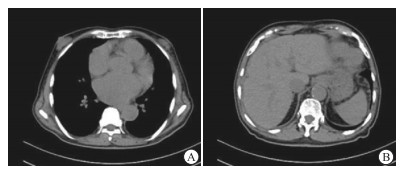
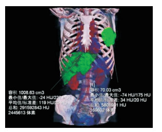
综合使用CT、MRI或超声多普勒(BUS)影像学资料,估算病人乳腺体积(V乳腺),其中BUS通过多普勒操作台距离按键直接测量病人乳腺的最大厚度a(cm)、高度b(cm)、宽度c(cm),并根据椭球型体积公式推算V乳腺(mL) =×π×a×b×c,而CT或MRI在薄层重建的图像数据基础上使用区域增长或容积测算技术直接测量V乳腺(见图 1)。以上测量区域仅包括BUS上呈中强回声的乳腺小叶组织,或在磁共振的T1WI图像上信号强度略高于胸部肌肉组织, 但显著低于高信号脂肪组织的乳腺腺体部分,或CT值介于15~30 HU的乳腺腺体组织,不包括肋骨、胸大肌及腺体周围脂肪层体积。单侧乳腺发育者测病侧乳腺体积,双侧发育者测双侧乳腺体积并求平均值。

图 1 以男性乳腺发育症病人的CT薄层图像为基础,使用区域增长技术直接测量左侧病变乳腺体积为70.03 mL, 肝脏体积为1008.63 mL
计算标准化乳腺体积(R乳/表)=V乳腺/BSA,计算正常肝脏体积(V肝脏)(cm3)=163×BSA+162.8[3]。利用CT或MRI的区域增长或容积测算技术测量病变肝脏的体积(V肝脏)(cm3)(见图 1),测量时不包括肝内血管分支、胆囊、肝圆韧带等非肝细胞结构。求得病变肝脏体积变化率(R肝)=(V肝脏-V病肝)/V肝脏。
通过涂氏肝硬化影像学分级法[5]评估各受检者肝脏的影像学资料,并根据其定义的肝硬化严重程度划分为Ⅰ~Ⅳ级。将R乳/表根据大小分为:轻度增大,1级,20~60;中度增大,2级,60~110;重度增大,3级,110~150。
-
采用方差分析、q检验、t检验、秩和检验和Spearman等级相关分析。
-
乳腺发育症组病人V乳腺为30~250 mL不等,其中40例呈丘状或锥体样隆起型乳腺,10例呈弥漫性增大型乳腺,3例呈扁平状增大。大多病人R乳/表分级1级(21/53,39.62%)或2级(23/53,43.40%)。其中19例呈单侧增大,且以左侧增大为主,34例呈双侧增大,单侧和双侧乳腺增生病人的R乳/表分级差异有统计学意义(P < 0.01)(见图 2~4、表 1)。乳腺发育症组53例中,有37例为初次入院治疗,其中20例表现为乳腺1级增大,17例为2级增大,且均为Child-Pugh分级A或B级;16例因肝硬化及其并发症多次入院治疗(3~4次),其中9例为乳腺3级增大,且Child-Pugh分级为B或C级,6例2级增大者和仅有的1例1级增大者均为Child-Pugh分级A级。

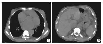
图 2 病例男,66岁,图A示双侧乳腺呈1级增大,图B示肝脏涂氏肝硬化影像学分级Ⅱ级,Child-Pugh评分5分, 为A级

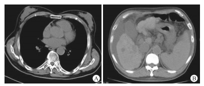
图 3 病例男,88岁,图A示双侧乳腺呈2级增大,图B示肝脏涂氏肝硬化影像学分级Ⅲ级,Child-Pugh评分8分,为B级

图 4 病例男, 73岁,图A示双侧乳腺呈3级增大,图B示肝脏涂氏肝硬化影像学分级为Ⅳ级,Child-Pugh评分12分,为C级
分组 1级 2级 3级 uc P 单侧乳腺增生 12 6 1 2.69 < 0.01 双侧乳腺增生 9 17 8 合计 21 23 9 表 1 单侧和双侧乳腺增生病人的R乳/表分级比较(n)
-
2组病人V乳腺、R乳/表、Child-Pugh分级、Child-Pugh评分和肝硬化CT分级差异均有统计学意义(P < 0.01),而V病肝、R肝差异均无统计学意义(P>0.05)(见表 2)。
分组 n V乳腺/mL R乳/表/ (mL/m3) V病肝/cm3 R肝/% Child-Pugh分级/级 Child-Pugh评分/分 肝硬化CT分级/级 A B C 0 Ⅰ Ⅱ Ⅲ 乳腺发育组 53 118.63±28.50 73.30±18.76 862.09±225.20 -28.31±6.69 12 36 5 9.58±2.15 0 15 33 5 无乳腺发育组 70 24.14±4.60 9.58±2.27 914.28±282.41 -26.33±7.24 34 32 4 7.16±2.24 0 37 30 3 t — 23.90* 24.59* 1.11 1.55 2.83* 6.04 2.76* P — < 0.01 < 0.01 >0.05 >0.05 < 0.01 < 0.01 < 0.01 *示uc值 表 2 2组病人相关影像学指标比较(x±s)
-
根据Child-Pugh分级法将2组病人分别分为A、B、C级,其中乳腺发育症组病人V乳腺、R乳/表随肝功能分级的增加均有增加趋势(P<0.05~P < 0.01),且与无乳腺发育组各对应分级比较差异均有统计学意义(P < 0.01);2组病人病变肝脏的V病肝、R肝随肝功能分级增加均有减小趋势(P < 0.01),但乳腺发育症组各肝功能分级病人与无乳腺发育症组相应肝功能分级比较差异均无统计学意义(P>0.05)(见表 3)。
分组 n V乳腺/mL R乳/表/(mL/m3) V病肝/cm3 R肝/% 乳腺发育症组 肝功能A级 12 95.93±14.46 62.37±17.63 1 081.93±274.09 -15.18±6.03 肝功能B级 36 115.13±19.94** 72.62±15.26 883.62±173.68** -30.29±6.96** 肝功能C级 5 129.70±25.76* 83.55±20.94 676.50±79.13**# -44.62±9.24**## F — 6.65 3.32 8.55 36.31 P — < 0.01 < 0.05 < 0.01 < 0.01 MS组内 — 377.408 266.465 38 143.816 48.738 无乳腺发育症组 肝功能A级 34 22.18±4.58△△ 9.15±2.72△△ 1 046.51±275.39 -14.97±5.99 肝功能B级 32 23.76±4.75▲▲ 9.93±3.08▲▲ 874.14±162.60** -29.39±7.35** 肝功能C级 4 25.20±5.33▽▽ 9.98±2.95▽▽ 663.28±83.41** -41.06±6.23**## F — 1.37 0.64 8.30 54.09 P — >0.05 >0.05 < 0.01 < 0.01 MS组内 — 22.043 8.422 49 898.235 44.405 q检验:与肝功能A级比较*P < 0.05,**P < 0.01;与肝功能B级比较#P < 0.05,##P < 0.01。与乳腺发育症组肝功能A级比较△△P < 0.01;与乳腺发育症组肝功能B级比较▲▲P < 0.01;与乳腺发育症组肝功能C级比较▽▽P < 0.01 表 3 2组病人不同肝功能分级间的相关影像学指标比较(x±s)
-
V乳腺、R乳/表与肝功能Child-Pugh分级、涂氏肝硬化CT分级均呈明显正相关关系(P < 0.01),V病肝、R肝与肝功能分级、涂氏肝硬化CT分级均呈明显负相关关系(P < 0.01)(见表 4)。
指标 Child-Pugh分级 涂氏肝硬化CT分级 V乳腺 0.592** 0.588** R乳/表 0.804** 0.794** V病肝 -0.871** -0.861** R肝 -0.895** -0.878** **P < 0.01 表 4 乳腺发育症组病人影像学指标与肝功能分级的相关性(r)
-
蜘蛛痣、肝掌和男性乳腺发育并称为肝病三联征,虽然男性乳腺增生症(指乳晕下乳腺腺体组织直径>2 cm,占男性乳腺疾病的90%以上[6])的发生率较前两者少,但由于老年男性睾丸退化后的睾酮减少以及体脂增加活化体内的芳香化酶导致雌激素增多,都对乳腺的增大起到了叠加作用,65%年龄>65岁的老年男性可发生持续存在的乳腺组织增生[7]。老年男性肝硬化病人因为体内激素水平的不同,可伴有或不伴有乳腺发育;但出现乳腺增生的老年男性,其肝功能障碍的概率明显增加,可作为一项临床体征提示筛查早期肝硬化。本研究发现单侧乳腺发育多集中于轻度乳腺发育者,肝硬化程度较轻,而双侧乳腺增生多表现为中度级以上乳房增大,肝硬化处于病程中期,故对于因单侧乳腺增生就诊的老年病人应全面检查肝功能等指标,尽早对可能存在的肝脏损伤进行干预。对于重度乳腺增大的病人,已有增生的乳腺组织已不可逆,也应积极治疗改善临床指标,防止进一步进展。尽管大部分的男性乳腺增生症本身不会对机体功能产生直接影响,但肝硬化的老年男性既受到各种门脉高压症状及肝癌威胁的困扰,亦可能受到乳腺增大的心理羞耻感的负面作用。早期及中期肝硬化可内科药物治疗改善肝脏功能,或服用抗雌激素他莫昔芬等药物[8],从而缩小甚至消除锥体样或小丘状增大的乳腺,手术移除晚期弥漫性增大的乳腺组织,并加以心理疏导,可以有效减轻病人的焦虑、恐惧情绪[9-10],提高老年男性生活质量。
本研究结果显示,乳腺发育症组和无乳腺发育症组病人V乳腺、R乳/表、肝硬化Child-Pugh分级、Child-Pugh评分和肝硬化CT分级差异均有统计学意义,但2组V病肝、R肝差异均无统计学意义。根据Child-Pugh分级法将2组病人各分为A、B、C级,其中乳腺发育症组病人V乳腺、R乳/表均有随肝功能分级增加而增加趋势,而无乳腺发育症组则无明显变化;乳腺发育症组病人V病肝、R肝随肝功能分级增加均有减小趋势,但各肝功能分级病人与无乳腺发育组相应肝功能分级组病人比较无明显差异。相关性分析显示,V乳腺、R乳/表与肝功能Child-Pugh分级、涂氏肝硬化CT分级均呈明显正相关关系,V病肝、R肝与肝功能分级、涂氏肝硬化CT分级均呈明显负相关关系。肝硬化早期与肝硬化晚期病人的雌激素水平有明显差异[11],不同周期的激素差异可能与肝硬化导致的代谢差异有关;而应用于肝硬化肝脏储备功能评估的Child-Pugh分级法,结合了临床表现和实验室检查结果,其使用广泛且可量化。既往研究[1]发现,男性肝硬化病人若有乳腺增生,其实验室检查总胆红素、白蛋白及凝血酶原时间发生明显差异,并且Child-Pugh评分越高,其雌二醇及催乳素水平随之升高,睾酮水平也随之显著降低,睾酮和雌激素能够相互转换,雌激素/睾酮比升高,进一步导致机体雌激素受体的激素结合力增高,睾丸功能损伤,男性特征减弱而女性特征增强。作为性激素的直接靶器官,乳腺组织随雌激素血清浓度的上升而诱导腺体组织的增生肥大,部分病人甚至出现泌乳现象[8]。
基于形态学的肝硬化CT分级以及肝脏体积变化率对肝硬化导致的肝脏损坏程度有一定评估价值[12],其与男性乳腺增大率有一定相关性,但正常男性的肝脏体积随着年龄增长有逐渐缩小趋势,肝细胞有正常的年龄损失率,60岁后,肝细胞数量下降明显,80~90岁的老年人肝脏体积和质量仅为50岁左右中年人的一半。所以不能仅以肝脏萎缩程度来评估肝细胞的坏死和肝纤维组织的增生,而应将实验室检查结果与肝性脑病、胃底食管静脉曲张破裂及腹水等并发症综合加以考量。秦凌云[13]发现,老年人肝脏各叶的体积成比例缩小,且与BSA呈明显正相关关系(r=0.886)。故本研究采用基于BSA计算的标准化肝脏体积以及标准化乳腺体积为指标,更加客观有效,减轻了传统意义上对肝硬化的影像学分级所受年龄因素的影响。
超声检查操作简便且避免了辐射风险,在体格检查中发现可疑乳腺增大者后应及时利用高频多普勒超声进行筛查[14];CT检查对男性乳腺发育症有明确诊断的作用[15],但因X线生物学损伤效应一般不专门应用于乳腺,往往在肺部扫描过程中偶然发现乳腺的增大,此时影像科应及时提醒临床对病人可能存在的肝脏功能损害行进一步检查;MRI检查成本昂贵,检查时间长,多用于男性单侧乳腺增生与浸润性导管癌、乳头状癌等良恶性鉴别[16]。在日常的诊疗过程中采用多种影像学方法监控老年男性肝硬化病人的乳腺病变情况是比较经济和科学的处理方式。
综上,综合使用各类影像学手段测量有老年男性肝硬化乳腺发育症病人的乳腺体积增大情况,可以作为推断和评估肝功能损害和肝脏萎缩程度的参考指标。
肝硬化老年男性病人影像指标与乳腺发育症的相关性分析
Correlation analysis between the imaging indexes and gynecomastia in elderly male patients with liver cirrhosis
-
摘要:
目的探讨综合使用各类影像学手段建立老年男性肝硬化与乳腺发育症相关性的可行性。 方法选取肝硬化老年男性病人123例为研究对象,年龄均>60岁。确定病人Child-Pugh分级,根据是否乳腺发育分为乳腺发育症组(n=53)和无乳腺发育症组(n=70)。采用CT、MRI或BUS影像学资料估算病人乳腺体积(V乳腺)、体表面积(BSA)、标准化乳腺体积(R乳/表)、正常肝脏体积(V肝脏),区域增长或容积测算技术测定病变肝脏体积(V病肝)、肝脏体积变化率(R肝),涂氏肝硬化影像学分级评估肝脏图像。 结果乳腺发育症组病人中19例呈单侧增大,34例呈双侧增大,单侧和双侧乳腺增生病人的R乳/表分级差异有统计学意义(P < 0.01)。2组病人V乳腺、R乳/表、Child-Pugh分级、Child-Pugh评分和肝硬化CT分级差异均有统计学意义(P < 0.01),而V病肝、R肝差异均无统计学意义(P>0.05)。根据Child-Pugh分级法将2组病人分别分为A、B、C级,其中乳腺发育症组病人V乳腺、R乳/表随肝功能分级的增加均有增加趋势(P < 0.05~P < 0.01),且与无乳腺发育组各对应分级比较差异均有统计学意义(P < 0.01);2组病人病变肝脏的V病肝、R肝随肝功能分级增加均有减小趋势(P < 0.01),但2组各相应肝功能分级间比较差异均无统计学意义(P>0.05)。乳腺发育症组病人的V乳腺、R乳/表与肝功能Child-Pugh分级、涂氏肝硬化CT分级均呈明显正相关关系(P < 0.01),V病肝、R肝与肝功能分级、涂氏肝硬化CT分级均呈明显负相关关系(P < 0.01)。 结论综合使用各类影像学手段测量肝硬化老年男性病人的乳腺体积增大情况,可以帮助推断和评估病人肝功能损害和肝脏萎缩程度。 Abstract:ObjectiveTo explore the feasibility of using various imaging methods to establish the correlation between the severity of cirrhosis and gynecomastia in elderly men. MethodsA total of 123 elderly male patients with liver cirrhosis aged >60 years were selected as the research object.The Child-Pugh grade of patients was determined.According to the breast development, the patients were divided into the breast development group(n=53) and non-breast development group(n=70).The CT, MRI or BUS imaging data were used to estimate the breast volume(Vbreast), body surface area(BSA), normalized breast volume(Rbreast/surface) and normal liver volume(Vliver).The regional growth or volume measurement techniques were used to measure the volume of diseased liver(Vliver disease) and rate of change of liver volume(Rliver), and the imaging grading of smear cirrhosis was used to evaluate the liver images. ResultsThere were 19 cases of unilateral enlargement and 34 cases of bilateral enlargement in the breast development group.The difference of the Rbreast/surface grade between unilateral and bilateral breast hyperplasia patients was statistically significant(P < 0.01).The differences of the Vbreast, Rbreast/surfac, Child-Pugh grade, Child-Pugh score and CT grade of liver cirrhosis between two groups were statistically significant(P < 0.01), while the differences of the Vdisease liver and Rliver were not statistically significant(P>0.05).According to the Child-Pugh grading method, the patients in the two groups were divided into the A, B and C grades, respectively.The Vbreast and Rbreast/surface of patients in the breast development group showed an increasing trend with the increase of liver function grade(P < 0.05 to P < 0.01), and the differences of which was statistically significant compared with those in the non-breast development group(P < 0.01).The Vdisease liver and Rliver of the diseased liver in two groups showed a decreasing trend with the increase of liver function grade(P < 0.01), but there was no statistical significance in the corresponding liver function grade between two groups(P>0.05).There were significant positive correlations among the Vbreast, Rbreast/surface, Child-Pugh grade of liver function and CT grade of smear cirrhosis(P < 0.01), while there were significant negative correlations among the Vdisease liver and Rliver and liver function grade and CT grade of smear cirrhosis(P < 0.01). ConclusionsThe comprehensive use of various imaging methods to measure the enlargement of breast volume in elderly male patients with cirrhosis can help to infer and evaluate the degree of liver function damage and liver atrophy. -
Key words:
- liver cirrhosis /
- gynecomastia /
- imaging
-
表 1 单侧和双侧乳腺增生病人的R乳/表分级比较(n)
分组 1级 2级 3级 uc P 单侧乳腺增生 12 6 1 2.69 < 0.01 双侧乳腺增生 9 17 8 合计 21 23 9  下载: 导出CSV
表 2 2组病人相关影像学指标比较(x±s)
分组 n V乳腺/mL R乳/表/ (mL/m3) V病肝/cm3 R肝/% Child-Pugh分级/级 Child-Pugh评分/分 肝硬化CT分级/级 A B C 0 Ⅰ Ⅱ Ⅲ 乳腺发育组 53 118.63±28.50 73.30±18.76 862.09±225.20 -28.31±6.69 12 36 5 9.58±2.15 0 15 33 5 无乳腺发育组 70 24.14±4.60 9.58±2.27 914.28±282.41 -26.33±7.24 34 32 4 7.16±2.24 0 37 30 3 t — 23.90* 24.59* 1.11 1.55 2.83* 6.04 2.76* P — < 0.01 < 0.01 >0.05 >0.05 < 0.01 < 0.01 < 0.01 *示uc值
下载: 导出CSV
表 3 2组病人不同肝功能分级间的相关影像学指标比较(x±s)
分组 n V乳腺/mL R乳/表/(mL/m3) V病肝/cm3 R肝/% 乳腺发育症组 肝功能A级 12 95.93±14.46 62.37±17.63 1 081.93±274.09 -15.18±6.03 肝功能B级 36 115.13±19.94** 72.62±15.26 883.62±173.68** -30.29±6.96** 肝功能C级 5 129.70±25.76* 83.55±20.94 676.50±79.13**# -44.62±9.24**## F — 6.65 3.32 8.55 36.31 P — < 0.01 < 0.05 < 0.01 < 0.01 MS组内 — 377.408 266.465 38 143.816 48.738 无乳腺发育症组 肝功能A级 34 22.18±4.58△△ 9.15±2.72△△ 1 046.51±275.39 -14.97±5.99 肝功能B级 32 23.76±4.75▲▲ 9.93±3.08▲▲ 874.14±162.60** -29.39±7.35** 肝功能C级 4 25.20±5.33▽▽ 9.98±2.95▽▽ 663.28±83.41** -41.06±6.23**## F — 1.37 0.64 8.30 54.09 P — >0.05 >0.05 < 0.01 < 0.01 MS组内 — 22.043 8.422 49 898.235 44.405 q检验:与肝功能A级比较*P < 0.05,**P < 0.01;与肝功能B级比较#P < 0.05,##P < 0.01。与乳腺发育症组肝功能A级比较△△P < 0.01;与乳腺发育症组肝功能B级比较▲▲P < 0.01;与乳腺发育症组肝功能C级比较▽▽P < 0.01
下载: 导出CSV
表 4 乳腺发育症组病人影像学指标与肝功能分级的相关性(r)
指标 Child-Pugh分级 涂氏肝硬化CT分级 V乳腺 0.592** 0.588** R乳/表 0.804** 0.794** V病肝 -0.871** -0.861** R肝 -0.895** -0.878** **P < 0.01
下载: 导出CSV
-
[1] 汪维艳, 杨京, 陆爽, 等. 男性肝硬化患者乳腺发育症与血清性激素及肝功能的相关性[J]. 中华消化杂志, 2015, 35(11): 734. doi: 10.3760/cma.j.issn.0254-1432.2015.11.004 [2] 贺科文, 刘兆芸, 于志勇. 男性乳腺癌与男性乳房发育症的鉴别及相关研究进展[J]. 中国肿瘤外科杂志, 2016, 8(3): 163. doi: 10.3969/j.issn.1674-4136.2016.03.005 [3] 黎一鸣, 吕凡, 吉鸿, 等. 肝脏体积变化与病肝储备功能的关系研究[J]. 中华普通外科杂志, 2003, 18(2): 79. doi: 10.3760/j.issn:1007-631X.2003.02.005 [4] 赵首捷, 杨振宇, 雷世雄, 等. Child-Pugh评分和ALBI分级对BCLC-B期肝癌生存预后预测价值比较[J/CD]. 中华肝脏外科手术学电子杂志, 2021, 10(1): 38. [5] 涂蓉, 张阳德, 伍保忠, 等. 肝硬化CT分级方法研究[J]. 放射学实践, 2003, 18(9): 621. doi: 10.3969/j.issn.1000-0313.2003.09.001 [6] FAGERLUND A, LEWIN R, RUFOLO G, et al. Gynecomastia: a systematic review[J]. J Plast Surg Hand Surg, 2015, 9(7): 1. [7] WU FC, TAJAR A, PYE SR, et al. Hypothalamic- pituitary- testicularaxis disruptions in older men are differentially linked to age and modifiable risk factors: the european male aging study[J]. J Clin Endocrinol Metab, 2008, 93(7): 2737. doi: 10.1210/jc.2007-1972 [8] 张爱玲, 秦贵军, 孟海阳, 等. 药源性男性乳腺发育症研究进展[J]. 药物不良反应杂志, 2015, 17(5): 357. [9] 韩景健, 晏文华. 男性乳腺发育症的病因及发病机制研究进展[J]. 中国美容整形外科杂志, 2020, 31(2): 89. doi: 10.3969/j.issn.1673-7040.2020.02.009 [10] 叶敏, 张爱霞, 王爱琳, 等. 男性乳腺发育症患者诊疗期间心理体验的质性研究[J]. 国际护理学杂志, 2016, 35(19): 2686. doi: 10.3760/cma.j.issn.1673-4351.2016.19.033 [11] 岳春燕. 肝硬化患者并发肝掌、蜘蛛痣的相关因素分析[J]. 中华现代护理杂志, 2019, 25(20): 2604. doi: 10.3760/cma.j.issn.1674-2907.2019.20.022 [12] 曾艳妮, 韩军, 刘良进, 等. 不同Child-Pugh分级的血吸虫病性肝硬化患者肝脏CT灌注成像参数分析[J]. 实用肝脏病杂志, 2020, 23(5): 691. doi: 10.3969/j.issn.1672-5069.2020.05.022 [13] 秦凌云. 正常老年人肝脏CT与年龄变化的相关性研究[D]. 南宁: 广西医科大学, 2012. [14] 杨伟. 高频超声应用于男性乳腺发育症的诊断价值与临床应用[J/CD]. 临床医药文献电子杂志, 2020, 7(17): 171. [15] 刘淑香. CT在男性乳腺发育症筛查中的应用价值[J]. 陕西医学杂志, 2017, 46(5): 629. doi: 10.3969/j.issn.1000-7377.2017.05.036 [16] 黄琰, 肖勤, 孙轶群, 等. 乳腺MRI鉴别诊断男性乳腺良恶性病变的价值[J]. 中华放射学杂志, 2021, 55(1): 48. -

 点击查看大图
图(4)表(4)
计量
- 文章访问数: 2304
- HTML全文浏览量: 1099
- PDF下载量: 8
- 被引次数: 0
