


-
大脑枕颞沟位于颞叶内侧,是枕颞内侧回与枕颞外侧回的分界,其与内侧的侧副沟[1]和外侧的大脑半球边缘近似平行关系,其宽度从颞极向后逐渐增宽,而后又再次变窄,一直延伸入枕叶与其融合。颞叶负责处理听觉信息,且与情感和海马记忆也有关系,当此区域发生异常或病变时,如颞叶癫痫、海绵状血管瘤、Kluver-Buey综合征、失语症、联想记忆障碍、偏盲等疾病。此时枕颞沟在横、冠、矢状面上的准确识别及其在三维空间中的精确定位是颞叶区域病变的影像学[2]定位诊断、立体定向[3]和微创神经外科的解剖基础。因此无论从解剖上、功能上还是临床应用价值上,对此区域定位及识别都具有其重要价值。本研究通过对30名健康志愿者进行活体大脑MRI数据采集、分析,探讨枕颞沟在横、冠、矢状面的形态学规律及回归方程,总结其发育规律,为枕颞沟区域重要脑组织和基底核病变的诊断及手术入路提供影像解剖学数字化资料。
-
本研究选择30名健康青壮年(本人知情同意),右利手,男女各15名,年龄18~34岁。经检查及颅脑MRI检测,均无神经,精神系统疾病及家族遗传史,无残障及肢体运动功能障碍,以AC-PC线为扫描基线,层厚2mm,得到30份横、冠、矢状面的T1W1图像,以Dicom 3.0格式进行保存。
-
Signal 3.0 T超导磁共振、HP计算机Z820工作站、eFilm Workstation、Microsoft Windows 7、Service Pack 1、Adobe Photoshop CS 5.0、AdobeSystems Inocorporated、SPSS 22.0。
-
MRI扫描方位及技术参数30名志愿者头颅连续MRI T1W1扫描图像,以前、后连合间线为基准线。获取志愿者大脑横、冠、矢状面图像数据。扫描野FOV:9.499×9.499英寸,自旋回波序列,TR: 1 990 ms,TE: 22 ms。横断面以AC-PC线为零层面,冠状面以垂直于AC-PC线中点为零层面,矢状面以头颅正中矢状面为零层面。扫描过程中,严格保证头颅固定不动。
-
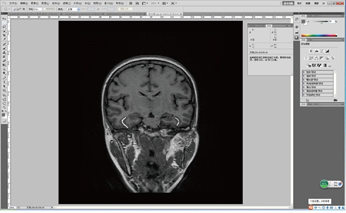
将已经扫描完成图像数据以Dicom 3.0格式导入到eFilm 3.1工作站中,在Study Manager页面中选择一例样本ID数据双击打开,选择“Screen Layout”,将格式调整为一行三列分别显示矢、横、冠状面图像(见图 1),再分别选中“Show First and Last Reference Lines ”“Show current Reference Line”“3D-Cursor”,以识别横断面层面的枕颞沟为依据,利用“基于连续断层的结构追踪法”按住右键,追踪冠、矢状面枕颞沟结构形态,并将冠状面图像导出,以JPEG格式保存。

图 1 eFilm 3.1工作站中枕颞沟影像断面的识别(横、冠状面黄色“+”表示枕颞沟所在位置, “+”随前者联动, 提示该取样点与前者对应)
-
在Adobe Photoshop CS 5.0软件中打开从eFilm 3.1工作站中导出,以JPEG格式保存的经过AP-PC线的横断面及所有冠状面图像。设置图像大小长宽为240 mm,像素54.186,放大图片后,将鼠标分别置于前连合、后连合处,读取坐标信息分别为(X0,Y1)、(X0,Y2),计算得出前、后连合线的中点Y0=(Y1+Y2)/2, 即为大脑原点坐标(X0,Y0)。在菜单栏选择“视图”,“新建参考线”,“垂直参考线”设定数值为X0,“水平参考线”设定数值为Y0。打开冠状面图像,在菜单栏“视图”,“标尺”中建立笛卡尔坐标系,用鼠标左键拖拽左上角十字坐标,至大脑原点,当信息板坐标显示(0, 0)时,松开鼠标。在笛卡尔坐标系中,横断面Z=0,冠状面X=0,矢状面Y=0,层厚2mm,其Y值为所在层面与零层面的层距乘以2mm层厚(见图 2)。

图 2 Adobe Photoshop Cs 5.0中冠状面枕颞沟图像(白线标注为枕颞沟, 鼠标放置枕颞沟内侧缘取样点上,右侧信息板上即出现此点坐标值)
-
构建三维立体定位数据集,将所得数据用Excel软件进行处理,绘制出枕颞沟内侧缘的投影散点图,再用SPSS 22.0软件分析出枕颞沟内侧缘的拟合曲线方程,及其相关系数。
-
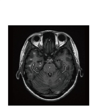
此横断面位于AC-PC层面以下第7层,与大脑原点的距离为14mm,此层面出现视交叉、海马、大脑脚及脚间池等结构,枕颞沟显示较清晰,位于海马后外侧方,且路径呈前后走行的波浪状,一直延伸入枕叶,右侧连续出现,左侧有间断。此时,位于枕颞沟内侧的侧副沟刚出现(见图 3)。

图 3 经视交叉及脚间池横断层面(枕颞沟呈“波浪”型,白色实线为枕颞沟)
-
此横断面位于AC-PC层面以下第9层,与大脑原点的距离为18mm,此层面出现侧脑室下角,第四脑室中央管等结构。此时的枕颞沟位于海马后外侧方,两侧均出现分支,呈现出镜面对称的数字“3”型,右侧侧副沟较短小,左侧侧副沟出现与枕颞沟平行走向的路径(见图 4)。

图 4 经第四脑室中央管横断层面(枕颞沟呈“3”型,白色实线为枕颞沟)
-
此横断面位于AC-PC层面以下第10层,与大脑原点的距离为20 mm。此层面出现鞍结节、鞍背等结构。此时的枕颞沟位于海马外侧,呈现左右对称的“()”型,走行路径相对较短且弧度弯曲直至小脑边缘,两侧未出现分支,左侧侧副沟走行与枕颞沟近似平行,右侧侧副沟较短小(见图 5)。

图 5 经鞍结节鞍背横断层面(枕颞沟呈“()”型,白色实线为枕颞沟)
-
此层面显示的枕颞沟呈现数字“1”型,左右对称。内侧红色实线为侧副沟,走行方向近似一致;侧脑室前角外侧为重要基底核区,依次为尾状核、内囊前肢,豆状核;蝶窦上方所示为视束及第三脑室,外侧为海马结构(见图 6)。

图 6 经颞下颌冠状面层面(枕颞沟呈“1”型,白色实线为枕颞沟,红色实线为侧副沟)
-
此层面显示的枕颞沟,右侧出现分支且走行方向相同,枕颞沟内侧红色实线所示的侧副沟,左右两侧均出现分支,且呈对称存在;侧脑室三角区下方为海马结构,紧邻小脑上方为四叠体的上丘;外侧为颞叶部分,颞上回、颞中回、颞下回清晰可见(见图 7)。

图 7 经下丘冠状面层面(枕颞沟呈“11”型,白色实线为枕颞沟,红色实线为侧副沟)
-
此层面所示的枕颞沟左右两侧均出现分支,走行大致相同,右侧呈数字“11”型,左侧呈“U”型;内侧红色实线所示的侧副沟左右两侧均出现分支,走行大致相同;位于枕颞沟和侧副沟之间的为梭状回;可见小脑内的第四脑室,及下方的延髓结构(见图 8)。

图 8 经侧脑室三角区冠状面层面(枕颞沟呈“U”型, 白色实线为枕颞沟,红色实线为侧副沟)
-
大脑枕颞沟在横断面的映射长度=枕颞沟在冠状面出现的层数×2mm;大脑枕颞沟在冠状面的映射长度=枕颞沟在横断面出现的层数×2mm。分别男15名,女15名,大脑枕颞沟左右两侧在横断面上的层数差异无统计学意义(P>0.05)(见表 1);男女大脑枕颞沟左右两侧在冠状面上的层数差异无统计学意义(P>0.05)(见表 2)。
性别 n 右侧 左侧 d±sd t P 男 15 18±3.85 18.8±4.31 0.8±2.37 -1.31 >0.05 女 15 17.67±3.98 16.53±3.83 1.14±2.95 1.49 >0.05 t — 0.25 1.59 — — — P — >0.05 >0.05 — — — 表 1 男女大脑枕颞沟横断面左右侧层数比较(x±s)
性别 n 右侧 左侧 d±sd t P 男 15 36.73±4.36 35.93±4.02 0.80±2.78 1.11 >0.05 女 15 35.00±3.46 34.33±2.75 0.67±2.97 0.87 >0.05 t — 1.67 1.42 — — — P — >0.05 >0.05 — — — 表 2 男女大脑枕颞沟冠状面左右侧层数比较(x±s)
-
男性大脑枕颞沟内侧缘左侧、右侧的坐标X值、Z值标准差进行两样本独立t检验,得出男性大脑左、右两侧枕颞沟于冠状面X值定位数据集差异具有统计学意义(P < 0.01),Z值立体定位数据集差异无统计学意义(P>0.05)(见表 3);女性大脑左、右两侧枕颞沟于冠状面X值定位数据集差异有统计学意义(P < 0.05),Z值立体定位数据集差异无统计学意义(P>0.05)(见表 4)。
Y/mm x±s/mm d±sd/mm 右侧 左侧 右侧 左侧 x±s/mm -27.876 7±4.555 7 35.371 6±3.730 6 — — d±sd/mm — — 2.95 2.95 t 4.93 0.00 P < 0.01 >0.05 表 3 男性枕颞沟内侧缘冠状面平均三维坐标值(n=15;x±s)
Y/mm x±s/mm d±sd/mm 右侧 左侧 右侧 左侧 x±s/mm -27.420 0±3.445 0 30.681 7±4.620 6 — — d±sd/mm — — -11.049 2±5.837 3 -9.834 3±6.510 8 t 2.19 0.54 P < 0.05 >0.05 表 4 女性枕颞沟内侧缘冠状面平均三维坐标值(n=15;x±s)
-
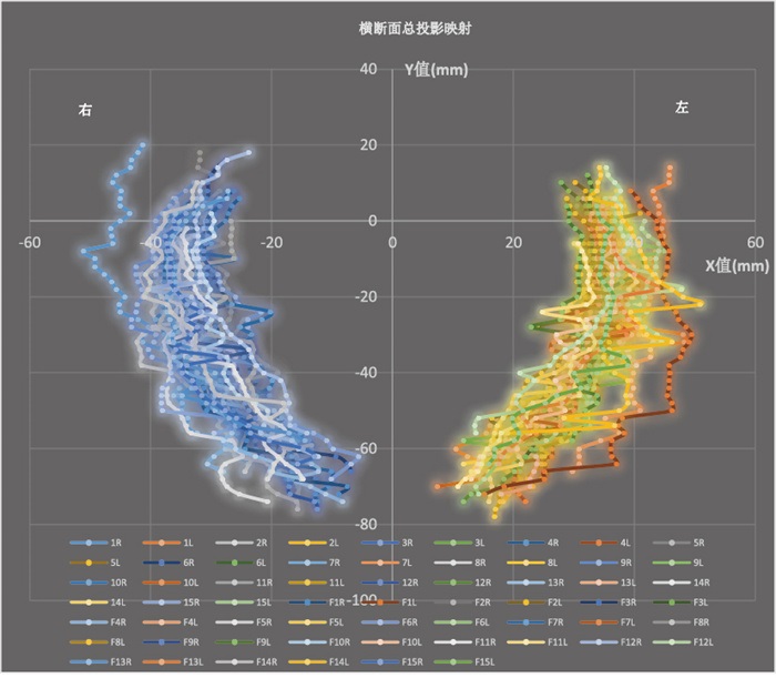
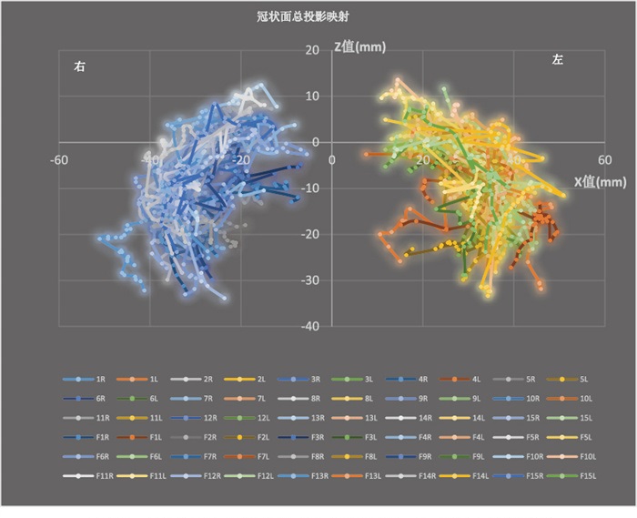
将30名大脑枕颞沟MRI图像数据集汇总整理,利用Excel软件,分别制作出男性及女性枕颞沟横断面(X-Y)、冠状面(X-Z)、的散点投影图,直观反映出枕颞沟空间走行位置变化及侧别变异情况(见图 9、10)。

图 9 枕颞沟内侧缘在横断面上的总投影映射

图 10 枕颞沟内侧缘在冠状面上的总投影映射
-
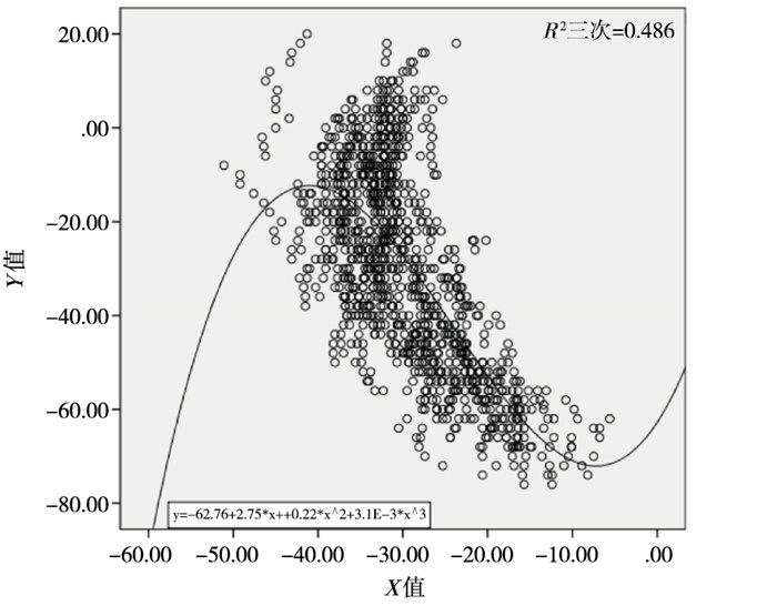
枕颞沟在横断面上Y值对X值的散点图及曲线回归方程分析:右侧$\mathop Y\limits^ \wedge$=-62.76+2.75×X+0.22×X2+3.1×10⁻³×X3(见图 11), 左侧$\mathop Y\limits^ \wedge$=3.84-10.83×X+0.5×X2-6.04×10⁻³×X3(见图 12);枕颞沟在冠状面上Z值对X值的散点图及曲线回归方程分析:右侧$\mathop Y\limits^ \wedge$=7.67-1.54×X-0.09×X2-1.06×10⁻³×X3, 左侧$\mathop Y\limits^ \wedge$=17.99+2.78×X-0.12×X2+1.42×10⁻³×X3;枕颞沟在矢状面上Z值对Y值的散点图及曲线回归方程分析:右侧$\mathop Y\limits^ \wedge$=20.75-0.35×X+2.62×10-3X2+5.49×10⁻⁵×X3, 左侧$\mathop Y\limits^ \wedge$=19.64-0.39×X+3.75×10-4X2+3.15×10⁻⁵×X3。

图 11 右侧枕颞沟横断面散点图及曲线回归方程分析

图 12 左侧枕颞沟横断面散点图及曲线回归方程分析
-
近几年来,人们对于脑磁共振成像技术的运用范围越发广泛,随着科学技术的突飞猛进,先进性和非侵入性的检测手段深受欢迎。此项技术为研究脑组织损伤后出现的行为、认知、情感等功能性障碍疾病的治疗提供技术支持。同时作为神经外科疾病的鉴别诊断标准之一,脑磁共振成像技术贯穿整个治疗及手术过程[4-5]。颞叶位于大脑外侧裂的下方,周围有重要的基底核及海马结构,当颞叶发生病变时,例如颅脑外伤、颞叶肿瘤、颞叶皮质支及颞叶深动脉闭塞,均可导致癫痫发作、失语症、瘫痪、听觉障碍、共济失调等症状。而颞叶癫痫是公认的网络化疾病,病人大脑中静息态网络连接遭到破坏,其发病机制可能与海马硬化导致颞叶白质病变有关系[6-7],除此之外,颞叶其他病变如局部脑皮质发育不良(FCD)、胚胎发育不良性神经上皮肿瘤(DNT)、节细胞胶质瘤等混合性神经胶质肿瘤(MNGTs)均为其发病机制[8]。研究[9]报道,治疗颞叶癫痫最好的方式是手术,且经证实手术的安全性和有效性可靠。而磁共振成像技术能够清晰显示病人大脑颞叶形态特征及功能学表现,为临床疾病鉴别诊断提供精准定位,同时也为颞叶癫痫手术入路路径提供解剖学基础[8, 10]。
本研究通过磁共振成像技术采集30名正常成人大脑数据,得到颞叶上枕颞沟在横断面及冠状面的影像学形态特征,经数据统计分析得出其立体定位数据集,有助于在三维立体空间中全方位识别枕颞沟的位置走行及形态规律。男性大脑左、右两侧枕颞沟于冠状面X值定位数据集存在差异,具有统计学意义(P < 0.05);女性大脑左、右两侧枕颞沟于冠状面X值定位数据集存在差异,具有统计学意义(P < 0.05),而对于大脑枕颞沟在冠状面出现的层数,男女左右两侧差异无统计学意义(P>0.05)。以此说明男、女大脑枕颞沟冠状面结构存在侧别差异性,这种左、右不对称的差异性可能与大脑功能侧化有关。颞叶对人的记忆起重要作用,国外研究[11]报道,其中嗅皮层主要负责记忆中新的关联,海马结构主要负责加固和回忆刺激之间的关联,而颞叶癫痫病人,会出现学习和记忆结合度明显下降,以及行为上的差异性。近些年研究发现,听觉功能障碍的人患认知障碍和痴呆的风险性更大,其主要与颞叶白质完整性相关[12]。AEBY等[13]对35~43周新生儿大脑右侧颞上沟及枕颞内侧回进行研究,发现这两者随着年龄呈现出非线性变化,这些结构对于大脑言语及非言语功能中起至关重要的作用。枕颞沟是位于颞叶最下方的一条脑沟,与侧副沟呈平行关系,将枕颞内侧回和枕颞外侧回划分开。横断面显示为前后走行,后方直接伸入枕叶,通过投影图及回归方程分析可看出,X、Y具有相关系数性,二者具有线性稳定性。
随着微创神经外科手术的广泛应用和不断发展,医学要求对颅内病变区域达到精准定位,本研究对大脑枕颞沟进行磁共振成像的横断面及冠状面形态学观察,揭示其规律性及差异性,对脑功能影像定位诊断及手术入路途径具有重要科学意义。
大脑枕颞沟形态学分析及投影回归方程
Morphological analysis and establishment of projection regression equation of occipital temporal sulcus
-
摘要:
目的构建以AC-PC线为扫描基线的活体大脑薄层MRI图像,研究枕颞沟在横、冠、矢状面的形态学规律及投影回归方程。 方法选定健康男、女各15名。以AC-PC为基准线,连续扫描MRI T1W1图像,所得横、冠、矢状面MRI成像数据,采用“3D-Cursor”技术观察枕颞沟在横、冠状面位置变化及连续形态学特征。将所测得的数据以横断面Z=0,冠状面X=0,矢状面Y=0,层厚2 mm,以及Y值为所在层面与零层面的层距乘以2 mm层厚为标准,建立笛卡尔三维立体坐标系,绘制枕颞沟内侧缘投影图,并利用SPSS 22.0统计分析出枕颞沟内侧缘的空间拟合曲线平面回归方程。 结果横断面的枕颞沟与大脑半球边缘平行,呈前后方向走行,后方一直延伸至枕叶,与侧副沟呈平行关系。分为“波浪”型,“()”型,“3”型;冠状面主要分为“1”型,“11”型,“U”型;成功构建大脑枕颞沟在横、冠、矢状面的投影回归方程。枕颞沟在横断面上Y值对X值的曲线回归方程右侧:$\mathop Y\limits^ \wedge$=-62.76+2.75×X+0.22×X2+3.1×10⁻³×X3,左侧:$\mathop Y\limits^ \wedge$=3.84-10.83×X+0.5×X2-6.04×10⁻³×X3;枕颞沟在冠状面上Z值对X值的曲线回归方程右侧:$\mathop Y\limits^ \wedge$=7.67-1.54×X-0.09×X2 -1.06×10⁻³×X3,左侧:$\mathop Y\limits^ \wedge$=17.99+2.78×X-0.12×X2+1.42×10⁻³×X3;枕颞沟在矢状面上Z值对Y值的曲线回归方程右侧:$\mathop Y\limits^ \wedge$=20.75-0.35×X+2.62×10-3X2+5.49×10⁻⁵×X3,左侧:$\mathop Y\limits^ \wedge$=19.64-0.39×X+3.75×10-4X2+3.15×10⁻⁵×X3。 结论利用“3D-Cursor”及“连续追踪”技术识别枕颞沟,其在横、冠面形态各异,为颞叶疾病定位及通过枕颞沟手术入路途径提供解剖学依据。 Abstract:ObjectiveTo investigate the thin layer MRI image of living brain with AC-PC line as the baseline, and study the morphology and projection regression equation of occipital temporal sulcus in cross section, coronal and sagittal plane. MethodsFifteen healthy male and 15 female were selected.With AC-PC as the baseline, the continuous scanning of MRI T1W1 images were performed to obtain MRI imaging data of cross, coronal and sagittal planes.The changes of occipital temporal sulci in cross and coronal plane, and continuous morphological characteristics were observed using 3D-cursor technology.The cross section Z=0, coronal plane X=0, sagittal plane Y=0, layer thickness for 2 mm, and Y value for the distance between the layer and zero layer multiplied by 2 mm layer thickness were set as the standard, the Cartesian three-dimensional coordinate system was established, the projection diagram of the medial margin of the occipital temporal sulcus was drawn, and the spatial fitting curve plane regression equation of the medial margin of occipital temporal sulcus was analyzed using SPSS 22.0. ResultsThe cross section of the occipital temporal sulcus was parallel to the edge of cerebral hemisphere, running forward and backward, extending to the occipital lobe, and parallel to the lateral accessory groove.The cross section was divided into the "wave" type, "()" type and "3" type, and the coronal plane was mainly divided into "1", "11" and "U" types.The projection regression equation of occipital temporal sulcus in cross section, coronal and sagittal plane were successfully constructed.The right and left sides of the cross-sectional Y-X curve regression equation of occipito-temporal sulcus were ($\mathop Y\limits^ \wedge$=62.76+2.75×X+0.22×X2+3.1×10⁻³×X3) and ($\mathop Y\limits^ \wedge$=3.84-10.83×X+0.5×X2-6.04×10⁻³×X3), respectively.The right and left sides of the coronal plane Z-X curve regression equation of occipito-temporal sulcus were ($\mathop Y\limits^ \wedge$= 7.67-1.54×X-0.09×X2 -1.06×10⁻³×X3) and ($\mathop Y\limits^ \wedge$=17.99+2.78×X-0.12×X2+1.42×10⁻³×X3), respectively.The right and left sides of the sagittal plane Z-Y curve regression equation of occipito-temporal sulcus were ($\mathop Y\limits^ \wedge$=20.75-0.35×X+2.62×10⁻³×X2+5.49×10⁻⁵×X3) and ($\mathop Y\limits^ \wedge$= 19.64-0.39×X+3.75×10⁻⁴×X2+3.15×10⁻⁵×X3), respectively. ConclusionsThe occipital temporal sulcus can be identified using 3D-Cursor and continuous tracking technology, has different shapes in the transverse and coronal planes, and can provide the anatomic basis for the location of temporal lobe diseases and approach of occipital temporal sulcus surgery. -
表 1 男女大脑枕颞沟横断面左右侧层数比较(x±s)
性别 n 右侧 左侧 d±sd t P 男 15 18±3.85 18.8±4.31 0.8±2.37 -1.31 >0.05 女 15 17.67±3.98 16.53±3.83 1.14±2.95 1.49 >0.05 t — 0.25 1.59 — — — P — >0.05 >0.05 — — —  下载: 导出CSV
表 2 男女大脑枕颞沟冠状面左右侧层数比较(x±s)
性别 n 右侧 左侧 d±sd t P 男 15 36.73±4.36 35.93±4.02 0.80±2.78 1.11 >0.05 女 15 35.00±3.46 34.33±2.75 0.67±2.97 0.87 >0.05 t — 1.67 1.42 — — — P — >0.05 >0.05 — — —
下载: 导出CSV
表 3 男性枕颞沟内侧缘冠状面平均三维坐标值(n=15;x±s)
Y/mm x±s/mm d±sd/mm 右侧 左侧 右侧 左侧 x±s/mm -27.876 7±4.555 7 35.371 6±3.730 6 — — d±sd/mm — — 2.95 2.95 t 4.93 0.00 P < 0.01 >0.05
下载: 导出CSV
表 4 女性枕颞沟内侧缘冠状面平均三维坐标值(n=15;x±s)
Y/mm x±s/mm d±sd/mm 右侧 左侧 右侧 左侧 x±s/mm -27.420 0±3.445 0 30.681 7±4.620 6 — — d±sd/mm — — -11.049 2±5.837 3 -9.834 3±6.510 8 t 2.19 0.54 P < 0.05 >0.05
下载: 导出CSV
-
[1] 黄子迅, 沈龙山, 王震寰, 等. 侧副沟形态学及分叉点的立体定位研究[J]. 蚌埠医学院学报, 2016, 41(1): 1. [2] KEIYA I, MASAFUMI H, TAKAAKI M, et al. Microrecording and image-guided stereotactic biopsy of deep-seated brain tumors[J]. J Neurosurgery, 2015, 123 (4), : 978 doi: 10.3171/2014.10.JNS14963 [3] ZHANG QJ, WANG WH, WEI XP, et al. Safety and efficacy of frameless stereotactic brain biopsy techniques[J]. Chin Med Sci J, 2013, 28(2): 113 doi: 10.1016/S1001-9294(13)60032-2 [4] MICHAEL JC, ANDREW JG, WOJTOWICZ M, et al. Task-related functional magnetic resonance imaging activations in patients with acute andsubacute mild traumatic brain injury: a coordinate-based meta-analysis[J]. NeuroImage Clin, 2019, 14(25): 102129. [5] OGAWA S, SUNG YW. Selected topics relating to functional MRI study of the brain[J]. Keio J Med, 2019, 68(4): 73 doi: 10.2302/kjm.2018-0011-OA [6] 王康. 基于磁共振指纹成像技术的颞叶内侧癫痫的颞叶白质改变[C]. 中国抗癫痫协会. 第八届CAAE国际癫痫论坛论文汇编. 中国抗癫痫协会: 中国抗癫痫协会, 2019: 168. [7] 周晓坤, 张众一, 郑彤, 等. 颞叶内侧病变相关性癫痫外科治疗研究进展[J]. 中国现代医药杂志, 2019, 21(11): 101. [8] 张笑娜, 王芳. MRI检查对癫痫病人海马硬化的应用价值[J]. 中国CT和MRI杂志, 2019, 17(5): 26. [9] GREWE P, NEU D, AENGENENDT J, et al. Rhinal and hippocampal contributions to spontaneous inter-item binding and verbal memory recall: evidence from temporal lobe epilepsy[J]. Cortex, 2020, 124: 204. doi: 10.1016/j.cortex.2019.11.015 [10] SPERLING MR, BARSHOW S, NEI M, et al. A reappraisal of mortality after epilepsysurgery[J]. Neurology, 2016, 86(21): 1938. doi: 10.1212/WNL.0000000000002700 [11] YU HT, CAI LH, WU XY, et al. Cross-frequency network analysis of functional brain connectivity in temporal lobe epilepsy[J]. Chinese Physics B, 2019, 28(4): 396. [12] GRAY DT, DE LA PEÑA NM, ENGLE JR, et al. Auditory processing deficits are selectively associated with medial temporal lobe mnemonic function and whitematter integrity in aging macaques[J]. Cereb Cortex, 2019, 13: 275. [13] AEBY A, VAN BOGAERT P, DAVID P, et al. Nonlinear microstructural changes in the right superior temporal sulcus and lateral occipitotemporal gyrus between 35 and 43 weeks in the preterm brain[J]. Neuroimage, 2012, 63(1): 104. -

 点击查看大图

图(12)表(4)
计量
- 文章访问数: 2104
- HTML全文浏览量: 1131
- PDF下载量: 23
- 被引次数: 0
