

-
随着医疗技术的不断发展,脑立体定向手术以安全、微创等优点近些年在临床应用广泛,是治疗难治性癫痫的有效方法。颞叶癫痫是难治性癫痫的常见类型,其传统外科手术治疗如颞前叶切除术及选择性海马、杏仁核切除术会大范围切除病灶,对颞叶皮质造成较大损伤从而导致病人可能出现语言、记忆功能障碍以及视野缺损等并发症[1-2],而脑立体定向技术的应用可以减小手术创伤面积,提高癫痫病灶损毁的精细度,从而保护病人的智力及情感等脑高级功能[3-4]。在痫性放电起于海马沟区域的颞叶内侧难治性癫痫外科手术治疗中, 脑立体定向电极植入术以及立体定向射频热凝阻滞疗法等的穿刺轨迹及靶点定位均基于海马沟立体定位数据集的获取。大脑海马沟位于海马本体(Ammon′s horn, 阿蒙角)、齿状回及下托之间,在MRI图像上对海马沟的识别有助于区分海马内部信号一致的灰质结构。本研究将基于MRI观察海马沟的形态学,同时在冠状面图像上测量海马沟的三维坐标值,构建出海马沟的立体定位数据集并进行回归分析,了解海马沟在横断面、冠状面及矢状面上坐标值间的相关性,揭示海马沟的形态学规律,为海马区域病灶的立体定向治疗, 如立体定向海马杏仁核射频损毁术、脑深部电刺激等的精准定位提供数字化解剖学参考。
-
选取30名健康成年人,男、女各15名,年龄18~34岁,均知情同意且无神经、精神系统疾病及家族史。选择T1 3D-BRAVO序列:TR 8.6 ms,TE 3.3 ms,以AC-PC线为扫描基线,获取层厚1 mm的横断面脑MRI图像。
-
Signa3.0T超导磁共振扫描仪及头颅正交线圈(美国GE公司);微型计算机(A8S,华硕公司);eFilm Workstation医学图像处理软件(版本4.2.2,美国Merge eMed公司); Adobe Photoshop CS6.0软件包。
-
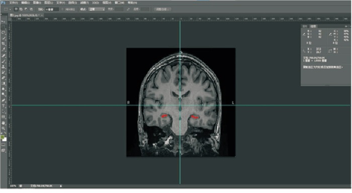
将采集的横断面颅脑MRI图像以DICOM3.0格式导入eFilm工作站中,利用其多面重建工具(Multip-lanar tools,MPR)重建出层厚为0.5 mm, 层间距为3 mm的冠状面及矢状面图像,在冠状层面上识别海马沟并以JPEG格式导出图像,将所保存的图像在Adobe Photoshop CS软件中打开,选择“图像”菜单栏里的“图像大小”,将文档大小的宽度和高度均设置为240 mm,分辨率为54.186像素/英寸,使其与实际参数一致,之后在新建的笛卡尔坐标系中测量海马沟外侧界三维坐标值的X值和Z值,二者可在软件中直接读取,Y值为当前图像层面距离AC-PC原点层面的层数与层间距的乘积, 所有取样点的坐标值构成海马沟的立体定位数据集(见图 1)。将取样点的坐标数据集导入SPSS25.0软件中,求解其在横切面、矢状面、冠状面上的拟合曲线和投影回归方程。

图 1 在photoshop中新建参考线, 将鼠标置于海马沟(红色线条标示)外侧缘, 右上方显示其X、Z坐标值
-
采用直线相关和回归分析。
-
在30名正常成年人脑T1 Bravo MRI图像中,海马沟在冠状面上共出现344层,其中左侧海马沟所在层面共164层(男性85层,女性79层),右侧180层(男性91层,女性89层)。海马沟90%出现在冠状面原点层面的后方,共出现315层,其中左侧153层(男性78层,女性75层),右侧162层(男性81层,女性81层)。各层面上海马沟X、Z坐标值的均差及标准差见表 1~2。
Y/mm x±s z±sd 右侧 左侧 右侧 左侧 -6 -30.600±4.101 36.300±0.000 -39.150±7.990 -35.400±0.000 -3 -30.500±3.057 34.067±1.849 -38.300±6.773 -37.283±8.804 0 -32.346±3.409 33.862±2.943 -36.654±6.860 -36.523±6.986 3 -33.093±3.217 34.513±3.176 -34.771±5.493 -33.780±6.236 6 -34.613±2.809 35.287±3.010 -33.140±6.069 -32.160±6.884 9 -35.093±3.136 35.880±3.127 -31.173±5.390 -31.100±6.551 12 -34.838±3.887 35.093±3.098 -29.285±5.351 -29.764±6.576 15 -35.000±4.725 34.967±3.199 -28.163±5.403 -26.267±9.373 18 -36.300±3.466 — -30.400±5.546 — 合计 -33.807±3.632 34.908±2.971 -33.076±6.522 -32.515±7.346 表 1 男性冠状面上海马沟外侧缘取样点的三维坐标值(n=15)
Y/mm x±s z±sd 右侧 左侧 右侧 左侧 -6 -38.350±5.445 — -41.250±2.051 — -3 -36.400±7.619 36.450±4.288 -42.483±8.011 -36.400±3.896 0 -36.140±7.168 34.625±6.449 -40.510±8.600 -38.663±9.289 3 -36.492±6.355 34.125±5.356 -40.346±8.598 -38.208±7.925 6 -36.813±5.221 35.300±6.017 -39.907±8.872 -37.871±7.970 9 -36.933±5.256 35.647±5.184 -37.193±9.469 -37.033±8.904 12 -36.079±5.457 35.971±5.390 -36.329±8.423 -36.229±8.508 15 -35.573±4.756 35.210±4.917 -35.909±7.394 -34.330±7.962 18 -39.000±2.921 34.950±0.354 -33.667±12.712 -30.650±6.859 合计 -36.522±5.548 35.276±5.235 -38.518±8.586 -36.847±8.042 表 2 女性冠状面上海马沟外侧缘取样点的三维坐标值(n=15)
-
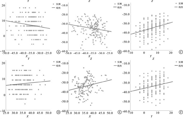
(1) 横断面上Y值对X值的回归分析,右侧:Y=-0.380-0.198X(r=0.161,P>0.05),左侧:Y=2.982+0.106X(r=0.083, P>0.05)。(2)冠状面上Z值对X值的回归分析,右侧:Z=-46.379-0.302X(r=0.182, P < 0.05), 左侧:Z=-49.512+0.425X(r=0.224, P < 0.05)。(3)矢状面上Z值对Y值的回归分析,右侧:Z=-38.501+0.416Y(r=0.308, P < 0.01),左侧:Z=-36.793+0.326Y(r=0.221, P < 0.01)。(4)海马沟在各个层面的回归方程散点图见图 2。

图 2 回归方程散点图
-
脑立体定向技术集合了神经病学、生物医学工程、影像学、定位学及计算机学于一体,在植入立体脑电图电极和引导精确切除癫痫灶方面具有重要价值[5],为精准定位的微创癫痫手术,包括立体定向放射治疗及激光诱导热疗法等[6]提供了坚实的临床基础。在癫痫病人海马区域的病灶损毁或阻断此处痫性放电的脑立体定向治疗中,海马沟是实施上述操作时颅内定位的有效参照结构。本研究在笛卡尔三维坐标系中构建的大脑海马沟立体定位数据集有利于海马沟的精确识别及精准定位,为海马沟区域病变的微创治疗提供影像数据支持。通过对海马沟坐标值的统计分析,求得其在横断面、冠状面及矢状面上的回归方程,利用此方程可以有效预测海马沟在各个层面上的坐标值,有助于确定其外侧缘在所设定的脑内坐标系中的空间位置。根据横断面上海马沟的回归方程可知,男、女性海马沟Y对X的相关性及回归分析结果均不显著,冠状面上Z对X及矢状面上Z对Y的回归方程虽具有统计学意义,但相关系数较小,表明海马沟坐标值在上述层面的相关关系不密切,变异性较高。通过回归方程散点图可以看出,海马沟外侧缘投影的离散程度较大,以上均可说明海马沟具有较大的个体差异性。
大脑海马沟是阿蒙角和齿状回在胚胎发育过程中不断卷曲折叠形成的,妊娠30周左右海马沟因二者之间的融合逐渐闭合[7-8]。BAJIC等[9]基于MRI研究了胎儿时期海马沟发育的不对称性,发现右侧海马沟较左侧发育速度快、闭合时间早,这可能会对成人左右侧海马沟形态产生影响。本研究获取的成人海马沟立体定位数据集显示,左右侧海马沟主要出现在Y=-3~15 mm的冠状面上(从经边缘内回的海马头的层面至经穹窿脚的海马尾层面),并且左右侧海马沟坐标值及男、女性的同侧坐标值均出现差异,提示健康成人的海马沟形态学存在性别及侧别上的非对称性。海马沟是区分海马内部结构——阿蒙角和齿状回的重要沟裂,由此推测二者的容积可能也存在个体差异性,而每个海马亚区在海马的记忆处理过程中都有其独特的功能如编码、巩固、检索等[10],是否海马沟的非对称性会对海马的记忆功能产生影响还需深度研究。
在健康成人的大脑中,典型的海马沟仅可见位于海马内侧的齿状回与前下托和旁下托间的小开口,T1WI图像上呈线性低信号且宽度小于1 mm[11]。当海马沟扩大或增宽时可能与海马区域的萎缩性病变有关。一项关于小鼠海马的研究[12]证实了在海马萎缩过程中海马沟的扩张,该研究表明海马沟的增宽与整个海马体积的减少高度相关,并且可以作为判断海马萎缩的简便指标。因此,对正常成人海马沟形态学的掌握是临床正确诊断海马相关病变的前提。有研究[13]表明颞叶癫痫病人海马区存在形态学改变,主要为海马萎缩,本研究分析的海马沟宽度指标对上述疾病及其他海马萎缩性疾病的筛查具有便捷、有效的意义。
综上所述,海马沟立体定位数据集的建立不仅说明了海马沟的形态学特点,而且为该区域及其周围结构病变的微创神经外科手术及立体定向放射治疗提供了影像学资料,对提高颞叶癫痫外科手术的安全性、准确性具有重要意义。
大脑海马沟立体定位数据集的构建及投影回归分析
Construction of stereotactic localization data set and projection regression analysis of hippocampal sulcus
-
摘要:
目的构建大脑海马沟基于连合间径(AC-PC)定位体系中的立体定位数据集及其平面投影回归方程。 方法将30名健康成年人颅脑横断层MRI数据经格式转化导入Photoshop CS软件包,经过严格的图像配准,建立以AC-PC中点为原点的三维立体坐标系,获取海马沟在脑MRI冠状面图像上的三维立体坐标值,以海马沟的最外侧点为起始点,记录该点坐标的X、Z值,其在软件的信息面板中直接显示,Y值为该图像所在层面与AC-PC层面间隔的层数乘以层间距,所有取样点坐标值构成大脑海马沟在三维坐标系中的立体定位数据集。利用SPSS25.0对所获取的取样点数据进行统计处理,求解其投影回归方程。 结果成功构建大脑海马沟在在三维坐标系中的立体定位数据集及其在横断、矢状、冠状面上的投影回归方程。冠状面上Z值对X值回归方程,右侧: Z=-46.379-0.302X(P < 0.05), 左侧: Z=-49.512+0.425X(P < 0.05);横断面上Y值对X值回归方程,右侧: Y=-0.380-0.198X(P>0.05), 左侧: Y=2.982+0.106X(P>0.05);矢状面上Z值对Y值回归方程,右侧Z=-38.501+0.416Y(P < 0.01), 左侧: Z=-36.793+0.326Y(P < 0.01)。 结论大脑海马沟横断、冠状及矢状面的三维坐标值间的相关关系较不密切。构建的海马沟立体定位数据集可为海马区域病灶定位及该区域脑立体定向手术的靶点确定、手术路径规划提供精确的影像解剖学数据。 Abstract:ObjectiveTo construct the stereotatic localization data set and its plane projection regression equation of the hippocampal sulcus based on the AC-PC localization system. MethodsThe transverse tomography data of 30 healthy adults were imported into Photoshop CS software package by format transformation.After strict image registration, a three-dimensional sitting system with AC-PC midpoint as the origin was established to obtain the three-dimensional coordinate values of hippocampal sulcus in the coronal plane images of brain.Taking the outermost point of the hippocampal sulcus as the starting point, record the X-value and Z-value, which were directly displayed in the information panel of the software, and the Y-value was the product of the number of layers between the image layer and AC-PC plane and interlayer spacing.The stereotactic data set of the hippocampal sulcus in a three-dimensional coordinate system were constituted by all the coordinate values of sample points.The SPSS25.0 software was used to calculate the obtained sampling point data, and solve the projection regression equation. ResultsThe stereotaxic dataset of the hippocampal sulcus in the three-dimensional coordinate system and its projection regression equation on the transverse, sagittal and coronal planes were successfully constructed.On the right side of the regression equation of Z-value to X-value on the coronal plane: Z=-46.379-0.302X(P < 0.05), on the left side: Z=-49.512+0.425X(P < 0.05).On the right side of the regression equation of Y-value to X-value on the transverse plane: Y=-0.380-0.198X(P>0.05), on the left side: Y=2.982+0.106X(P>0.05).On the right side of the regression equation of Z-value to Y-value on the sagittal plane: Z=-38.501+0.416Y(P < 0.01), on the left side: Z=-36.793+0.326Y(P < 0.01). ConclusionsThe correlation among the three-dimensional coordinate values of the transverse, coronal and sagittal plane of the hippocampal sulcus of the brain is not close.The stereotactic localization data set of the hippocampal sulcus provides accurate image anatomical data for the localization of lesions, the target determination and surgical path planning of stereotactic surgery in the hippocampal region. -
Key words:
- hippocampal sulcus /
- stereotatic localization /
- regression analysis
-
表 1 男性冠状面上海马沟外侧缘取样点的三维坐标值(n=15)
Y/mm x±s z±sd 右侧 左侧 右侧 左侧 -6 -30.600±4.101 36.300±0.000 -39.150±7.990 -35.400±0.000 -3 -30.500±3.057 34.067±1.849 -38.300±6.773 -37.283±8.804 0 -32.346±3.409 33.862±2.943 -36.654±6.860 -36.523±6.986 3 -33.093±3.217 34.513±3.176 -34.771±5.493 -33.780±6.236 6 -34.613±2.809 35.287±3.010 -33.140±6.069 -32.160±6.884 9 -35.093±3.136 35.880±3.127 -31.173±5.390 -31.100±6.551 12 -34.838±3.887 35.093±3.098 -29.285±5.351 -29.764±6.576 15 -35.000±4.725 34.967±3.199 -28.163±5.403 -26.267±9.373 18 -36.300±3.466 — -30.400±5.546 — 合计 -33.807±3.632 34.908±2.971 -33.076±6.522 -32.515±7.346  下载: 导出CSV
表 2 女性冠状面上海马沟外侧缘取样点的三维坐标值(n=15)
Y/mm x±s z±sd 右侧 左侧 右侧 左侧 -6 -38.350±5.445 — -41.250±2.051 — -3 -36.400±7.619 36.450±4.288 -42.483±8.011 -36.400±3.896 0 -36.140±7.168 34.625±6.449 -40.510±8.600 -38.663±9.289 3 -36.492±6.355 34.125±5.356 -40.346±8.598 -38.208±7.925 6 -36.813±5.221 35.300±6.017 -39.907±8.872 -37.871±7.970 9 -36.933±5.256 35.647±5.184 -37.193±9.469 -37.033±8.904 12 -36.079±5.457 35.971±5.390 -36.329±8.423 -36.229±8.508 15 -35.573±4.756 35.210±4.917 -35.909±7.394 -34.330±7.962 18 -39.000±2.921 34.950±0.354 -33.667±12.712 -30.650±6.859 合计 -36.522±5.548 35.276±5.235 -38.518±8.586 -36.847±8.042
下载: 导出CSV
-
[1] 杨子茵, 高安亮, 李佳宇, 等. 前颞叶切除术与颞下入路选择性海马杏仁核切除术治疗颞叶内侧癫痫疗效对比[J]. 西部医学, 2022, 34(4): 566. doi: 10.3969/j.issn.1672-3511.2022.04.019 [2] 李亚飞, 尹绍雅. 前颞叶切除术与选择性杏仁核海马切除术治疗颞叶癫痫的疗效对比[J]. 中国实用神经疾病杂志, 2020, 23(12): 1036. [3] 徐成伟, 周文静, 柏建军, 等. 立体定向脑电图引导射频热凝在难治性颞叶癫痫中的应用[J]. 中华医学杂志, 2018, 98(13): 1008. doi: 10.3760/cma.j.issn.0376-2491.2018.13.011 [4] MALIKOVA H, KRAMSKA L, VOJTECH Z, et al. Different surgical approaches for mesial temporal epilepsy: resection extent, seizure, and neuropsychological outcomes[J]. Stereotact Funct Neurosurg, 2014, 92(6): 372. doi: 10.1159/000366003 [5] KARAS PJ, GIRIDHARAN N, TREIBER JM, et al. Accuracy and workflow improvements for responsive neurostimulation hippocampal depth electrode placement using robotic stereotaxy[J]. Front Neurol, 2020, 11: 590825. doi: 10.3389/fneur.2020.590825 [6] QUIGG M, HARDEN C. Minimally invasive techniques for epilepsy surgery: stereotactic radiosurgery and other technologies[J]. J Neurosurg, 2014, 121 Suppl: 232. [7] KIER EL, KIM JH, FULBRIGHT RK, et al. Embryology of the human fetal hippocampus: MR imaging, anatomy, and histology[J]. AJNR Am J Neuroradiol, 1997, 18(3): 525. [8] ALLEOBONE J, KANAAN RA, MALLER JJ, et al. Enlarged hippocampal fissure in psychosis of epilepsy[J]. Epilepsy Behav, 2020, 111: 107290. doi: 10.1016/j.yebeh.2020.107290 [9] BAJIC D, CANTO MOREIRA N, WIKSTROM J, et al. Asymmetric development of the hippocampal region is common: a fetal MR imaging study[J]. AJNR Am J Neuroradiol, 2012, 33(3): 513. doi: 10.3174/ajnr.A2814 [10] ZAMMIT AR, EZZATI A, ZIMMERMAN ME, et al. Roles of hippocampal subfields in verbal and visual episodic memory[J]. Behav Brain Res, 2017, 317: 157. doi: 10.1016/j.bbr.2016.09.038 [11] BRONEN RA, CHEUNG G. MRI of the normal hippocampus[J]. Magn Reson Imaging, 1991, 9(4): 497. doi: 10.1016/0730-725X(91)90035-K [12] LI Y, YAN J, ZHU X, et al. Increased hippocampal fissure width is a sensitive indicator of rat hippocampal atrophy[J]. Brain Res Bull, 2018, 137: 91. [13] DUAN Y, LIN Y, ROSEN D, et al. Identifying morphological patterns of hippocampal atrophy in patients with mesial temporal lobe epilepsy and Alzheimer disease[J]. Front Neurol, 2020, 11: 21. -

 点击查看大图
图(2)表(2)
计量
- 文章访问数: 1976
- HTML全文浏览量: 906
- PDF下载量: 22
- 被引次数: 0
