


-
前列腺癌是一种泌尿生殖系统的恶性肿瘤, 是全世界发病率最高的男性恶性肿瘤之一。据估计,2020年全球新增约141万前列腺癌确诊病例,约37万人因前列腺癌死亡[1]。中国的前列腺癌发病率仍低于西方发达国家,但是近年来随着生活方式的改变和筛查方法的普及,我国前列腺癌的发病率和死亡率都在持续上升[2]。因此对前列腺癌侵袭和转移机制的分子水平研究仍有十分重要的意义。线粒体亚甲基四氢叶酸脱氢酶2(mitochondrial methylenetetrahydrofolate dehydrogenase 2, MTHFD2),于1960年在艾氏腹水瘤细胞中首次被发现,它是线粒体中一种参与细胞活动的重要酶[3]。本研究通过RNA干扰技术下调MTHFD2基因在前列腺癌PC3细胞中的表达,探讨其对前列腺癌细胞侵袭和转移能力的影响。
-
人前列腺癌细胞系PC3购自中国科学院上海生命科学研究院中心,于含10%胎牛血清的RPMI-1640液体培养基(Sigma)中培养,培养条件为37 ℃、5% CO2的恒温培养箱。细胞贴壁生长,用0.25%的胰酶消化传代。将5×10⁵个PC3细胞接种于6孔板中,待细胞生长密度至80%左右时,使用LipofectamineTM2000转染试剂,按照试剂说明书进行转染。实验分为空白对照组(ctrl组)、阴性对照组(si-NC组)及转染siRNA-MTHFD2组(si-MTHFD2组)。
-
利用Trizol试剂盒提取细胞总RNA,利用分光光度计检测RNA纯度和浓度,检测合格后将总RNA逆转录为cDNA;MTHFD2正向引物:5′-AGC CGG AGA CTT ACC CAA TTG-3′,反向引物: 5′-GTC TCT CGA CAG TTA CTC-3′;GAPDH正向引物: 5′-GCT CAT CGG CCT CCA TTC A-3′, 反向引物: 5′-TAC GCG ACG TCG GCG AGC TT-3′。以cDNA为模板进行qRT-PCR反应,参照试剂盒配置反应体系,反应条件: 95 ℃预热5 min,95 ℃ 30 s,60 ℃ 30 s,72 ℃ 30 s(循环40次)。采用2-ΔΔCT方法计算MTHFD2的相对表达水平。
-
取对数生长期PC3细胞,胰蛋白酶消化,用经预冷的磷酸盐缓冲液(PBS)洗涤细胞,调整细胞密度为105个/毫升,按照Annexin V-FITC/ PI凋亡试剂盒说明书要求,通过流式细胞仪检测细胞凋亡情况。
-
细胞转染后使用无胎牛血清培养基培养24 h,用20 μL枪头形成划痕,洗去脱落细胞。显微镜下拍照并记录划痕宽度。培养箱培养24 h后再次显微镜下拍照并记录划痕宽度。依据细胞愈合相对距离判断细胞运动能力。
-
收集对数生长期PC3细胞,0.25% 胰蛋白酶消化,加入1 mL不含血清的培养液制备单细胞悬液,并将100 μL(1×10⁵) 细胞接种到Transwell小室的上室,在下室中加入400 μL含有10% 胎牛血清的RPMI 1640培养基。放入5% CO2、37 ℃培养箱内培养24 h后,0.2%结晶紫染液染色30 min,染色结束后洗脱残余结晶紫溶液,用棉签擦去基质胶和小室内的细胞并拍照计数。
-
取对数生长期PC3细胞裂解提取总蛋白,通过BCA试剂盒检测蛋白质含量,取等量蛋白质样品,用10% SDS-PAGE凝胶电泳法分离蛋白,转移蛋白至PVDF膜,5%脱脂牛奶室温封闭2 h。封闭完成后加入适宜浓度E-cadherin、Vimentin和N-cadherin一抗,4 ℃孵育过夜,24 h后TBST洗涤,室温条件下二抗孵育1 h,TBST洗涤,最后加入ECL发光液曝光拍照,对蛋白质表达量进行定量分析,以GAPDH为内参。
-
采用t检验、方差分析和q检验。
-
采用qRT-PCR法检测人正常前列腺上皮细胞RWPE-1以及前列腺癌PC3细胞中MTHFD2的表达。结果显示,前列腺癌PC3细胞中MTHFD2表达水平(3.86±0.38),较正常前列腺上皮细胞表达量(1.00±0.24)升高(t=11.02,P < 0.05)。
-
qT-PCR结果显示,ctrl组、si-NC组以及si-MTHFD2组MTHFD2水平差异有统计学意义(P < 0.01);si-MTHFD2组MTHFD2 mRNA水平较ctrl组以及si-NC组表达均降低(P < 0.05)(见表 1)。
分组 n MTHFD2 F P MS组内 ctrl组 3 1.00±0.08 52.02 < 0.01 0.005 si-NC组 3 0.99±0.08 si-MTHFD2组 3 0.48±0.05*# q检验: 与ctrl组比较*P < 0.05;与si-NC组比较#P < 0.05 表 1 下调MTHFD2抑制PC3细胞中MTHFD2水平的表达(x±s)
-
细胞凋亡实验结果显示,ctrl组、si-NC组以及si-MTHFD2组凋亡率差异有统计学意义(P < 0.01);si-MTHFD2组凋亡能力较ctrl组以及si-NC组表达均增加(P < 0.05)(见图 1、表 2)。

图 1 下调MTHFD2对前列腺癌PC3细胞凋亡能力的影响
分组 n 凋亡率/% F P MS组内 ctrl组 3 17.91±1.26 54.10 < 0.01 2.239 si-NC组 3 19.70±1.32 si-MTHFD2组 3 29.13±1.84*# q检验: 与ctrl组比较*P < 0.05;与si-NC组比较#P < 0.05 表 2 下调MTHFD2对前列腺癌PC3细胞凋亡能力的影响(x±s)
-
划痕实验结果显示,si-MTHFD2组细胞迁移能力较ctrl组、si-NC组明显减弱,表明si-MTHFD2显著降低了PC3细胞迁移能力(见图 2)。

图 2 下调MTHFD2对前列腺癌PC3细胞迁移能力的影响
-
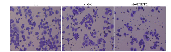
Transwell细胞侵袭实验结果显示,si-MTHFD2组细胞侵袭能力较ctrl组、si-NC组均减弱(P < 0.05)(见图 3、表 3)。

图 3 下调MTHFD2对前列腺癌PC3细胞侵袭能力的影响
分组 n 侵袭数/个 F P MS组内 ctrl组 3 53.21 ± 3.46 111.10 < 0.05 9.311 si-NC组 3 52.36 ± 3.52 si-MTHFD2组 3 20.63± 1.89*# q检验: 与ctrl组比较*P < 0.05;与si-NC组比较#P < 0.05 表 3 下调MTHFD2对前列腺癌PC3细胞侵袭能力的影响(x±s)
-
Western blotting结果显示, 与ctrl组和si-NC组相比,si-MTHFD2组中E-cadherin蛋白水平升高(P < 0.05);与ctrl组和si-NC组相比,si-MTHFD2组中Vimentin和N-cadherin蛋白水平降低(P < 0.05)(见图 4、表 4)。

图 4 下调MTHFD2对前列腺癌PC3细胞EMT相关蛋白表达的影响
分组 n E-cadherin Vimentin N-cadherin ctrl组 3 0.40±0.04 0.42±0.03 0.48±0.03 si-NC组 3 0.43±0.04 0.40±0.03 0.45±0.03 si-MTHFD2组 3 0.82±0.05*# 0.23±0.02*# 0.26±0.02*# F — 86.68 44.59 58.23 P — < 0.01 < 0.01 < 0.01 MS组内 — 0.002 0.001 0.001 q检验: 与ctrl组比较*P < 0.05;与si-NC组比较#P < 0.05 表 4 下调MTHFD2对前列腺癌PC3细胞上皮-间充质转化(EMT)相关蛋白表达的影响(x±s)
-
前列腺癌病人早期无明显的症状,因此病人往往在癌症晚期才发现,虽然初期肿瘤生长缓慢,但进展迅速,易引起全身转移, 并进一步发展为去势抵抗性前列腺癌[4-5]。血清前列腺特异性抗原(PSA)是诊断前列腺癌常用的生物标志物,但由于PSA缺乏特异性,常常导致过度诊断[6]。因此,寻找新的分子标志物及探明机制对提升前列腺癌诊疗水平及改善前列腺癌病人的预后至关重要。
MTHFD2具有甲酰四氢叶酸合成酶和环水解酶的双重功能,其产生的NADPH能够对抗氧化应激,维持氧化还原动态平衡,为肿瘤细胞快速增殖创造了条件[7-8]。MTHFD2已经在蛋白水平上被反复证实与肿瘤的发生、进展和病人的预后有关。最近研究[9-10]表明MTHFD2在肝癌、乳腺癌和肾癌中高表达,并与病人的预后不良有关,同时MTHFD2的高表达已被证明与肿瘤的发生、发展密切相关。本研究通过体外转染前列腺癌细胞系PC3获得了MTHFD2瞬转,发现si-MTHFD2转染组细胞中凋亡能力增加,细胞迁移、侵袭能力明显减弱,表明MTHFD2在前列腺癌中可能作为促癌因子促进肿瘤的发生与发展。
肿瘤转移是肿瘤病人死亡的主要原因,其是一个多步骤的过程,包括附着、降解和脱离细胞外基质,最后从原发肿瘤主动迁移[11]。上皮起源的肿瘤细胞会重新激活一种被称为EMT的程序,其特征是上皮标志物如E-cadherin的表达减少,间质标志物如Vimentin和N-cadherin的表达增加。EMT引起的表型变化包括细胞间黏附丧失、细胞极性丧失以及细胞获得迁移和侵袭[12]。具有间充质表型的细胞具有侵袭和转移潜能,并且能获得干细胞状态(自我更新和分化的能力)或具有抗药性。越来越多的证据表明EMT与前列腺癌的侵袭、转移和治疗耐药密切相关[13]。本研究中通过检测EMT相关蛋白的表达水平, 探讨下调MTHFD2对前列腺癌细胞侵袭和转移的影响,结果显示,si-MTHFD2转染组E-cadherin表达明显升高,而Vimentin和N-cadherin表达明显降低,表明si-MTHFD2可明显降低PC3细胞的侵袭和转移能力。
综上所述,下调MTHFD2对前列腺癌细胞侵袭和转移能力具有明显抑制的作用,其作用机制还需要深入研究。本研究进一步加深了MTHFD2在前列腺癌中的功能和分子机制的认识,为前列腺癌在临床的诊疗提供了一定的理论依据。
下调MTHFD2对前列腺癌PC3细胞的侵袭和转移的作用及影响机制
Down-regulation of MTHFD2 inhibits invasion and metastasis in prostate cancer PC3 cells
-
摘要:
目的观察线粒体亚甲基四氢叶酸脱氢酶2(mitochondrial methylenetetrahydrofolate dehydrogenase 2, MTHFD2)基因表达对人前列腺癌PC3细胞侵袭和转移能力的影响。 方法人前列腺癌PC3细胞分为3组: 空白对照组(ctrl组)、阴性对照组(si-NC组)及转染siRNA-MTHFD2组(si-MTHFD2组)。采用qRT-PCR法检测人正常前列腺上皮细胞RWPE-1以及前列腺癌PC3细胞中MTHFD2的mRNA表达,通过流式细胞术检测MTHFD2对PC3细胞凋亡的影响,通过划痕实验检测细胞迁移能力的变化,通过Transwell实验检测细胞侵袭能力的变化,采用Western blotting检测E-cadherin、Vimentin和N-cadherin蛋白表达变化。 结果前列腺癌PC3细胞较正常前列腺上皮细胞中MTHFD2表达水平升高(P < 0.05),si-MTHFD2组MTHFD2 mRNA表达水平较ctrl组和si-NC组降低(P < 0.05);si-MTHFD2组细胞迁移和侵袭能力较ctrl组和si-NC组降低(P < 0.05);si-MTHFD2组E-candherin蛋白表达水平较ctrl组和si-NC组升高(P < 0.05),Vimentin和N-cadherin蛋白表达水平较ctrl组和si-NC组降低(P < 0.05)。 结论下调MTHFD2表达可增加前列腺癌细胞凋亡能力,抑制前列腺癌细胞迁移和侵袭能力。 -
关键词:
- 前列腺肿瘤 /
- 线粒体亚甲基四氢叶酸脱氢酶2 /
- 侵袭 /
- 转移
Abstract:ObjectiveTo investigate the effect of mitochondrial methylenetetrahydrofolate dehydrogenase 2 (MTHFD2) on cell invasion and metastasis in prostate cancer PC3 cells. MethodsThe prostate cancer PC3 cells were divided into 3 groups: blank control group (ctrl group), negative control group (si-NC group) and transfected siRNA-MTHFD2 group (si-MTHFD2 group).The mRNA expression of MTHFD2 in human normal prostate epithelial cells RWPE-1 and prostate cancer PC3 cells was detected by qRT-PCR.The apoptosis rate in different groups was detected by flow cytometry, the changes of cell migration ability were detected by scratch test, the changes of cell invasion ability were detected by Transwell test, and the changes of E-cadherin, Vimentin and N-cadherin protein expressions were detected by Western blotting. ResultsThe expression level of MTHFD2 in prostate cancer PC3 cells was significantly higher than that in normal prostate epithelial cells(P < 0.05), and the mRNA expression level of MTHFD2 in si-MTHFD2 group was significantly lower than that in ctrl and si-NC groups(P < 0.05).The results of cell scratch and invasion assay showed that the migration and invasion abilities in si-MTHFD2 group was significantly lower than in ctrl and si-NC groups(P < 0.05).E-candherin protein expression level in si-MTHFD2 group was significantly higher than that in ctrl and si-NC groups(P < 0.05), and the protein expression levels of vimentin and N-cadherin were significantly lower than those in ctrl and si-NC groups. ConclusionsDown-regulation of MTHFD2 expression can increase the apoptosis of prostate cancer cells and inhibit the migration and invasion of prostate cancer cells. -
表 1 下调MTHFD2抑制PC3细胞中MTHFD2水平的表达(x±s)
分组 n MTHFD2 F P MS组内 ctrl组 3 1.00±0.08 52.02 < 0.01 0.005 si-NC组 3 0.99±0.08 si-MTHFD2组 3 0.48±0.05*# q检验: 与ctrl组比较*P < 0.05;与si-NC组比较#P < 0.05  下载: 导出CSV
表 2 下调MTHFD2对前列腺癌PC3细胞凋亡能力的影响(x±s)
分组 n 凋亡率/% F P MS组内 ctrl组 3 17.91±1.26 54.10 < 0.01 2.239 si-NC组 3 19.70±1.32 si-MTHFD2组 3 29.13±1.84*# q检验: 与ctrl组比较*P < 0.05;与si-NC组比较#P < 0.05
下载: 导出CSV
表 3 下调MTHFD2对前列腺癌PC3细胞侵袭能力的影响(x±s)
分组 n 侵袭数/个 F P MS组内 ctrl组 3 53.21 ± 3.46 111.10 < 0.05 9.311 si-NC组 3 52.36 ± 3.52 si-MTHFD2组 3 20.63± 1.89*# q检验: 与ctrl组比较*P < 0.05;与si-NC组比较#P < 0.05
下载: 导出CSV
表 4 下调MTHFD2对前列腺癌PC3细胞上皮-间充质转化(EMT)相关蛋白表达的影响(x±s)
分组 n E-cadherin Vimentin N-cadherin ctrl组 3 0.40±0.04 0.42±0.03 0.48±0.03 si-NC组 3 0.43±0.04 0.40±0.03 0.45±0.03 si-MTHFD2组 3 0.82±0.05*# 0.23±0.02*# 0.26±0.02*# F — 86.68 44.59 58.23 P — < 0.01 < 0.01 < 0.01 MS组内 — 0.002 0.001 0.001 q检验: 与ctrl组比较*P < 0.05;与si-NC组比较#P < 0.05
下载: 导出CSV
-
[1] SUNG H, FERLAY J, SIEGEL RL, et al. Global cancer statistics 2020: GLOBOCAN estimates of incidence and mortality worldwide for 36 cancers in 185 countries[J]. CA Cancer J Clin, 2021, 71(3): 209. doi: 10.3322/caac.21660 [2] YE DW, ZHU Y. Prostate cancer and prostatic diseases Best of China, 2018[J]. Prostate Cancer Prostatic Dis, 2019, 22(1): 1. doi: 10.1038/s41391-018-0117-y [3] HE H, LI PC, JIA W, et al. High expression of methylenetetrahydrofolate dehydrogenase 2 (MTHFD2) in esophageal squamous cell carcinoma and its clinical prognostic significance[J]. Med Sci Monit, 2020, 26: e920259. [4] GILLESSEN S, ATTARD G, BEER TM, et al. Management of patients with advanced prostate cancer: report of the advanced prostate cancer consensus conference 2019[J]. Eur Urol, 2020, 77(4): 508. doi: 10.1016/j.eururo.2020.01.012 [5] WANG S, YANG Y, CAO YD, et al. Androgen downregulation of miR-760 promotes prostate cancer cell growth by regulating IL6[J]. Asian J Androl, 2021, 23(1): 85. doi: 10.4103/aja.aja_20_20 [6] LOMAS DJ, AHMED HU. All change in the prostate cancer diagnostic pathway[J]. Nat Rev Clin Oncol, 2020, 17(6): 372. doi: 10.1038/s41571-020-0332-z [7] NILSSON R, JAIN M, MADHUSUDHAN N, et al. Metabolic enzyme expression highlights a key role for MTHFD2 and the mitochondrial folate pathway in cancer[J]. Nat Commun, 2014, 5: 3128. doi: 10.1038/ncomms4128 [8] LIU X, HUANG Y, JIANG C, et al. Methylenetetrahydrofolate dehydrogenase 2 overexpression is associated with tumor aggressiveness and poor prognosis in hepatocellular carcinoma[J]. Dig Liver Dis, 2016, 48(8): 953. doi: 10.1016/j.dld.2016.04.015 [9] LIU F, LIU Y, HE C, et al. Increased MTHFD2 expression is associated with poor prognosis in breast cancer[J]. Tumour Biol, 2014, 35(9): 8685. doi: 10.1007/s13277-014-2111-x [10] LIN H, HUANG B, WANG H, et al. MTHFD2 overexpression predicts poor prognosis in renal cell carcinoma and is associated with cell proliferation and vimentin-modulated migration and invasion[J]. Cell Physiol Biochem, 2018, 51(2): 991. doi: 10.1159/000495402 [11] GONG R, LIN W, GAO A, et al. Forkhead box C1 promotes metastasis and invasion of non-small cell lung cancer by binding directly to the lysyl oxidase promoter[J]. Cancer Sci, 2019, 110(12): 3663. doi: 10.1111/cas.14213 [12] THIERY JP, ACLOQUE H, HUANG RY, et al. Epithelial-mesenchymal transitions in development and disease[J]. Cell, 2009, 139(5): 871. doi: 10.1016/j.cell.2009.11.007 [13] NAKAZAWA M, KYPRIANOU N. Epithelial-mesenchymal-transition regulators in prostate cancer: androgens and beyond[J]. J Steroid Biochem Mol Biol, 2017, 166: 84. doi: 10.1016/j.jsbmb.2016.05.007 -

 点击查看大图

图(4)表(4)
计量
- 文章访问数: 2539
- HTML全文浏览量: 1238
- PDF下载量: 28
- 被引次数: 0
