

-
流行病学数据显示,急性心肌梗死仍然是当前世界上主要的致死性疾病之一。及时的血管内溶栓和支架成形术等再灌注手段来开通罪犯血管对于缓解心肌缺血、减少梗死面积是非常重要的[1]。然而,过往有研究[2]表明冠脉再通后的再灌注过程可引起缺血区域心肌细胞的二次打击,这种现象被称为“缺血/再灌注损伤”。因此,对于急性心肌梗死的治疗而言,缓解缺血/再灌注损伤是必不可少的[3]。
研究[4]显示,缺血/再灌注损伤的发生发展与多种病理生理机制有关,内质网应激在其中发挥着重要作用。内质网应激相关通路活化可诱导下游的凋亡途径激活,进而促进心肌组织中的缺血/再灌注损伤进展[5-6]。以往研究[7-8]证实心肌细胞在缺血再灌注环境中高表达内质网应激相关的信号蛋白,如活化转录因子6(ATF6)、C/EBP同源蛋白(CHOP)、葡萄糖调节蛋白78(GRP78)等,且抑制内质网应激信号转导可有效降低细胞凋亡率。
在生理条件下,Nrf2通过与Keap1结合而锚定于胞质中。一旦受到应激作用,Nrf2与Keap1分离,转入核内并调控下游靶基因如HO-1的表达,它们可显著抑制细胞内凋亡级联反应的活化[9-10]。有研究[11]表明,Nrf2/HO-1通路表达上调对缺血再灌注环境中的心肌细胞有明显的保护作用,但是其中的潜在机制尚未完全阐明。本研究探讨Nrf2/HO-1抗心肌缺血再灌注损伤的可能机制。
-
细胞培养用PBS、胰酶及DMEM/F12培养基购于美国HyClone公司;5-溴-2-脱氧尿苷购自美国Sigma公司;心肌肌钙蛋白Ⅰ(cTnⅠ)和α-横纹肌肌动蛋白抗体购于美国Abcam公司;Opti-MEM和Lipofectamine 2000试剂购自美国Thermo Fisher公司;重组腺病毒购于上海吉凯基因有限公司;MTT试剂盒购于美国Sigma公司;TUNEL试剂盒购于德国Roche公司;检测cTnⅠ和肌酸激酶同工酶(CK-MB)含量的ELISA试剂盒购于上海酶联生物科技有限公司;逆转录及荧光定量PCR试剂盒购于大连Takara公司;PCR引物购于上海生工生物工程有限公司。
-
新生乳鼠(1~3 d)由三峡大学实验动物中心提供。本研究已通过华中科技大学同济医学院附属武汉市中心医院伦理委员会批准。在本实验中,乳鼠心肌细胞通过酶解法提取。首先,小鼠由75%乙醇消毒处理并给予安乐死。然后,快速取出心脏并将心室组织切为约1 mm3组织块。将组织块用冰PBS洗涤后置于0.125%胰酶中进行消化。随后加入PBS终止消化,收集消化液,300 g离心5 min。将细胞沉淀重悬于DMEM/F12完全培养基中并接种于细胞培养皿中,利用5-溴-2-脱氧尿苷将心肌细胞与成纤维细胞分离。随后将提取的心肌细胞进行免疫荧光染色,观察cTnⅠ和α-横纹肌肌动蛋白的表达情况。
-
心肌细胞饥饿培养24 h,使用Opti-MEM和Lipofectamine 2000转染试剂将HO-1-siRNA、control-siRNA转入细胞内。首先用Opti-MEM分别稀释siRNA和Lipofectamine 2000,放置5 min。随即将两者充分混匀,室温放置20 min,将其加入细胞培养基中。37 ℃共孵育6 h,更换为完全培养基进行后续实验。siRNA序列: HO-1-siRNA,5′-GCC ACA CAG CAC UAU GUA ATT-3′;control-siRNA,5′-UUG UCC GAA CGU GUC ACG UTT-3′。
-
原代心肌细胞密度达到70%~80%转染分别载有Nrf2片段、HO-1片段或空载的腺病毒。首先,心肌细胞密度达到70%~80%时更换新鲜DMEM培养基。随后将滴度为5×10¹² VG/mL的病毒液加入培养孔中,37 ℃、5%CO2饱和湿度培养过夜,后更换为完全培养基进行后续实验。应用重组腺相关病毒9型(rAAV9)作为体外特异性调控靶基因表达的工具。
-
与siRNA或腺病毒共孵育后,心肌细胞培养于无糖无血清培养基,置于37 ℃缺氧(1% O2、5% CO2、94% N2)环境中60 min。随之更换为完全培养基,细胞转入正常条件(5% CO2、95%空气)培养24 h,随后收集细胞和上清液。分组如下: 正常对照,空载-rAAV9转染后缺氧/复氧处理,Nrf2-rAAV9转染后缺氧/复氧处理,HO-1-rAAV9转染后缺氧/复氧处理,HO-1-siRNA共孵育后Nrf2-rAAV9转染、随之缺氧/复氧处理。
-
采用MTT试验检测细胞活性。首先,提取的原代心肌细胞接种于96孔板中,密度为1.0×10⁴个/孔。过夜培养后,将细胞与siRNA或腺病毒共孵育后,进行缺氧/复氧处理。处理结束后,向每孔中加入10 μL MTT试剂,37 ℃孵育4 h。随后将MTT试剂从孔中吸出,加入150 μL二甲基亚砜,放置10 min后使用酶标仪测定570 nm波长处吸光度(OD)值。
-
用TUNEL试剂盒检测细胞凋亡情况。将各组处理后的细胞爬片固定于4%多聚甲醛25 min,然后PBS洗涤3次。随之将细胞爬片浸没于0.1% TritonX-100中2 min。PBS洗涤,然后加入TUNEL试剂,避光37 ℃孵育60 min。随后PBS洗涤,加入DAB显色剂,在显微镜下观察记录细胞凋亡情况。
-
收集细胞培养上清,2 000 r/min离心10 min,收集上层液体并检测其cTnⅠ和CK-MB含量,使用南京建成生化试剂盒进行测定。
-
使用Trizol试剂提取心肌细胞中的总RNA,然后采用逆转录试剂盒将提取的RNA逆转录为cDNA。随之,荧光定量PCR检测使用SYBR Green Master试剂以及ABI QuantStudio 6 real-time PCR系统。mRNA相对表达量采用2-△△Ct方法计算,GAPDH设为内参基因,引物序列: GRP78,正向5′-CCA TCC CGT GGC ATA AAC-3′,反向5′-TGT CTT TTG TTA GGG GTC GTT-3′;ATF6,正向5′-ACC ACT AGT AGT ATC AGC AGG A-3′,反向5′-AAT GTG TCT CCC CTT CTG CG-3′;CHOP,正向5′-TCA CTA CTC TTG ACC CTG CG-3′,反向5′-ACT GAC CAC TCT GTT TCC GT-3′;Caspase-3,正向5′-GAG CTT GGA ACG GTA CGC TAA-3′,反向5′-CGT CCA CAT CCG TAC CAG AG-3′;GAPDH,正向5′-ATG GGT GTG AAC CAC GAG A-3′,反向5′-CAG GGA TGA TGT TCT GGG CA-3′。
-
采用t(或t′)检验、方差分析和q检验。
-
cTnⅠ和α-横纹肌肌动蛋白均在所提取的细胞中高表达(见图 1)。Nrf-2 rAAV9和HO-1 rAAV9处理细胞后,胞内Nrf-2和HO-1 mRNA表达量上升(P < 0.01和P < 0.05);HO-1 siRNA处理细胞后,胞内HO-1 mRNA表达量下调(P < 0.05)(见表 1)。

图 1 c-TnⅠ和α -横纹肌肌动蛋白在提取的心肌细胞上的表达情况(红色荧光为cTnⅠ,绿色荧光为α-横纹肌肌动蛋白,蓝色荧光为细胞核)
分组 n Nrf2 mRNA HO-1 mRNA 对照组 3 0.95±0.04 1.04±0.05 过表达组 3 2.63±0.44 3.25±0.31* 沉默组 3 — 0.38±0.08* F — 6.59△ 193.67 P — < 0.01 < 0.01 MS组内 — — 0.035 △示t′值; q检验: 与对照组比较*P < 0.05 表 1 各组目的基因表达情况(x±s)
-
Nrf2或HO-1高表达可抑制缺氧/复氧诱导的心肌细胞活性的下降(P < 0.05);HO-1基因沉默后,Nrf2过表达对心肌细胞活性无明显影响(P>0.05)(见表 2)。
分组 n OD值 F P MS组内 对照组 5 1.01±0.09 27.75 < 0.01 0.007 模型组 5 0.51±0.09* Nrf2过表达组 5 0.71±0.06*# HO-1过表达组 5 0.72±0.08*# HO-1沉默+Nrf2过表达组 5 0.56±0.09*▲△ q检验: 与对照组比较*P < 0.05;与模型组比较#P < 0.05;与Nrf2过表达组比较▲P < 0.05;与HO-1过表达组比较△P < 0.05 表 2 各组心肌细胞活性比较(x±s)
-
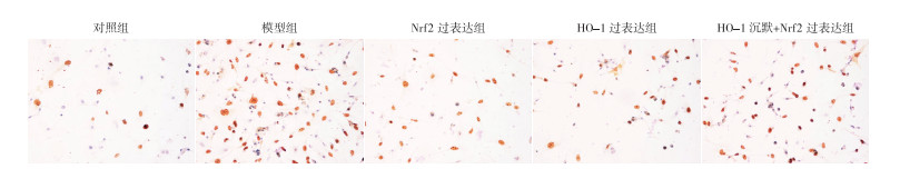
TUNEL染色结果显示,缺氧/复氧组心肌细胞凋亡率高于对照组(P < 0.05),Nrf2或HO-1过表达可有效抑制缺氧/复氧处理诱导的细胞凋亡(P < 0.05),HO-1表达下调减弱了Nrf2上调的抗凋亡效果(P < 0.05)(见图 2、表 3)。Nrf2或HO-1过表达降低了缺氧/复氧引起的心肌细胞cTnⅠ及CK-MB释放量(P < 0.05)(见表 3)。

图 2 各组心肌细胞凋亡情况
分组 n 细胞凋亡率/% cTnⅠ /(pg/mL) CK-MB /(U/L) 对照组 4 10.13±3.11 42.98±13.52 20.89±11.06 模型组 4 59.78±9.16* 452.30± 47.44* 93.16±7.56* Nrf2过表达组 4 41.78±4.1*# 344.50±52.93*# 64.94±13.80*# HO-1过表达组 4 40.21±4.98*# 312.20±46.07*# 61.52±9.05*# HO-1沉默+Nrf2过表达组 4 58.15±8.01*▲△ 450.00± 43.69*▲△ 89.14±8.06*▲△ F — 40.03 45.05 24.16 P — < 0.01 < 0.01 < 0.01 MS组内 — 39.870 2 022.489 103.321 q检验: 与对照组比较*P < 0.05;与模型组比较#P < 0.05;与Nrf2过表达组比较▲P < 0.05;与HO-1过表达组比较△P < 0.05 表 3 各组细胞凋亡率及细胞培养上清中cTnⅠ和CK-MB含量比较(x±s)
-
缺氧/复氧组心肌细胞的GRP78和ATF6的mRNA表达量高于对照组(P < 0.05);Nrf2或HO-1过表达降低了GRP78和ATF6的表达量(P < 0.05);当HO-1表达受抑时,Nrf2过表达对缺氧/复氧诱导的GRP78和ATF6表达无明显影响(P>0.05);Nrf2或HO-1过表达可抑制缺氧/复氧诱导的CHOP及Caspase-3表达(P < 0.05)(见表 4)。
分组 n GRP78 ATF6 CHOP Caspase-3 对照组 3 1.06±0.21 0.98±0.12 1.11±0.14 1.09±0.20 模型组 3 2.51±0.30* 2.14±0.28* 3.28±0.26* 2.56±0.20* Nrf2过表达组 3 1.72±0.14*# 1.57±0.15*# 2.10±0.29*# 1.80±0.11*# HO-1过表达组 3 1.79±0.22*# 1.51±0.17*# 1.80±0.21*# 1.64±0.09*# HO-1沉默+Nrf2过表达组 3 2.30±0.20*▲△ 2.00±0.1*▲△ 2.99±0.23*▲△ 2.24±0.17*▲△ F — 19.82 20.55 43.75 37.68 P — < 0.01 < 0.01 < 0.01 < 0.01 MS组内 — 0.048 0.031 0.053 0.026 q检验: 与对照组比较*P < 0.05;与模型组比较#P < 0.05;与Nrf2过表达组比较▲P < 0.05;与HO-1过表达组比较△P < 0.05 表 4 各组心肌细胞内GRP78、ATF6、CHOP及Caspase-3 mRNA的相对表达水平比较(x±s)
-
尽管溶栓药物以及血管成形术等再灌注手段的广泛应用,急性心肌梗死仍然是当前世界上主要的致死原因之一[12]。造成这一现象最重要的原因之一是梗死后的缺血再灌注过程加重心肌细胞凋亡、扩大心肌缺血损伤面积[13]。本研究使用缺氧/复氧处理心肌细胞作为缺血/再灌注损伤的体外模型,发现Nrf2/HO-1通路表达上调可有效缓解缺氧/复氧过程诱导的心肌细胞损伤,其机制可能与抑制内质网应激相关的信号转导有关。
既往研究[14-15]表明, 在缺血/再灌注过程中,细胞凋亡相关通路激活,进而导致线粒体功能失调、凋亡蛋白释放、胞膜结构紊乱以及心肌酶漏出。与以往研究相似,本研究结果显示,缺氧/复氧处理后,心肌细胞活性降低、细胞凋亡增强、cTnⅠ及CK-MB释放增加。核转录因子Nrf2在维持细胞稳态方面发挥着重要作用,其分子机制主要是通过增加下游HO-1表达,发挥HO-1相关的效应。研究[16]表明, HO-1表达上调可有效抑制细胞凋亡,促进细胞存活。当Nrf2表达量增加,可使下游HO-1转录活性增强,进而使HO-1表达量上升,细胞凋亡率明显降低。而一旦HO-1表达受抑制时,Nrf2不能有效发挥抗凋亡的作用,表明Nrf2/HO-1级联反应对细胞存活有明显的促进作用[17]。鉴于此,我们研究了Nrf2/HO-1通路是否影响缺氧/复氧诱导的心肌细胞损伤。结果显示,与缺氧/复氧处理组相比,Nrf2或HO-1过表达组均可提高心肌细胞活性、抑制心肌细胞凋亡、降低cTnⅠ及CK-MB含量。这些结果表明上调Nrf2/HO-1表达可有效改善缺氧/复氧诱导的心肌细胞损伤。
大量研究[18-19]报道,内质网应激参与缺血/再灌注损伤的发生发展。以往实验[20-21]显示,内质网应激相关信号分子GRP78与ATF6高表达于遭受缺血/再灌注的心、肝及脑组织。与这些结果相似,本研究结果显示,缺氧/复氧处理引起GRP78和ATF6表达量增高,而Nrf2或HO-1过表达可减少GRP78和ATF6的表达,说明Nrf2/HO-1表达增强可抑制缺氧/复氧诱导的内质网应激信号转导。当上游的应激信号持续出现,CHOP表达增加,导致Caspase-3表达上调,进而引起凋亡通路激活。既往研究[22-23]表明,抑制CHOP/Caspase-3信号流能够缓解内质网应激诱导的细胞凋亡,从而改善缺血/再灌注损伤。我们进而推测Nrf2/HO-1缓解心肌细胞损伤的机制可能是通过抑制内质网应激相关的促凋亡信号转导。本研究结果显示,Nrf2/HO-1上调降低了CHOP和Caspase-3的表达量,而HO-1基因沉默可引起Nrf2过表达对CHOP/Caspase-3抑制的失效。
综上所述,上调Nrf2/HO-1通路可有效改善缺氧/复氧处理诱导的心肌细胞损伤,其机制可能在于抑制内质网应激以及下游的凋亡通路信号转导。本研究为Nrf2/HO-1作为靶点治疗缺血/再灌注损伤提供了新的研究证据。
Nrf2/HO-1通路通过调控内质网应激改善小鼠心肌细胞缺氧/复氧损伤的作用研究
Nrf2/HO-1 pathway improves anoxia/reoxygenation-induced injury in murine cardiomyocytes via regulating endoplasmic reticulum stress
-
摘要:
目的探讨Nrf2/HO-1信号上调对小鼠心肌细胞缺氧/复氧损伤的改善作用及相关机制。 方法小鼠原代心肌细胞分为正常培养处理和缺氧/复氧处理,其中缺氧/复氧处理的心肌细胞分为模型组、Nrf2过表达组、HO-1过表达组、HO-1沉默+Nrf2过表达组。采用MTT法检测细胞活性,TUNEL染色检测细胞凋亡,ELISA法检测细胞培养上清中心肌肌钙蛋白Ⅰ(cTnⅠ)及肌酸激酶同工酶(CK-MB)含量,荧光定量PCR检测内质网应激相关分子水平。 结果小鼠心肌细胞缺氧/复氧处理后,心肌细胞活性降低、细胞凋亡增加及细胞培养上清中cTnⅠ和CK-MB含量升高(P < 0.05)。Nrf2或HO-1过表达可有效减轻缺氧/复氧诱导的心肌细胞损伤(P < 0.05)。给予HO-1基因沉默预处理后,Nrf2过表达产生的细胞保护作用明显减弱(P < 0.05)。此外,缺氧/复氧处理后,心肌细胞内GRP78、ATF6、CHOP和Caspase-3的mRNA表达量升高(P < 0.05)。Nrf2或HO-1过表达引起GRP78、ATF6、CHOP和Caspase-3表达水平下调(P < 0.05),而下调HO-1水平可使Nrf2过表达对内质网应激信号转导的抑制作用减弱(P < 0.05)。 结论Nrf2/HO-1通路表达上调可有效改善缺氧/复氧诱导的心肌细胞损伤,其机制可能在于抑制内质网应激以及下游的细胞凋亡通路信号转导。 -
关键词:
- 急性心肌梗死 /
- 缺氧/复氧损伤 /
- 内质网应激 /
- Nrf2/HO-1通路
Abstract:ObjectiveTo discuss the effect of Nrf2/HO-1 up-regulation on improving anoxia/reoxygenation-induced injury in murine cardiomyocytes and its related mechanism. MethodsThe primary murine cardiomyocytes were divided into normal culture treatment and anoxia/reoxygenation treatment, and the anoxia/reoxygenation treated cardiomyocytes were divided into model group, Nrf2 overexpression group, HO-1 overexpression group, HO-1 silence+Nrf2 overexpression group.Cell viability was detected by MTT assay, apoptosis was analyzed by TUNEL staining, cardiac troponin Ⅰ(cTnⅠ) and creatine kinase isoenzyme MB(CK-MB) contents in cell culture supernatant were determined by ELISA, and levels of endoplasmic reticulum stress-related molecules were measured by fluorescence quantitative PCR. ResultsAfter anoxia/reoxygenation treatment, the activity decreased, apoptosis increased, and the contents of cTnⅠ and CK-MB in cell culture supernatant of murine cardiomyocytes increased(P < 0.05).Overexpression of Nrf2 or HO-1 could effectively alleviate anoxia/reoxygenation-induced injury in murine cardiomyocytes(P < 0.05).After pretreatment with HO-1 gene silencing, the protection effect of Nrf2 overexpression was significantly weakened(P < 0.05).In addition, after anoxia/reoxygenation treatment, the mRNA expressions of GRP78, ATF6, CHOP and Caspase-3 in cardiomyocytes increased(P < 0.05).Overexpression of Nrf2 or HO-1 resulted in down-regulation of the expression levels of GRP78, ATF6, CHOP and Caspase-3(P < 0.05), while down-regulation of HO-1 level reduced the inhibitory effect of Nrf2 overexpression on endoplasmic reticulum stress signal transduction(P < 0.05). ConclusionsThe up-regulation of Nrf2/HO-1 pathway can effectively improve anoxia/reoxygenation-induced injury in cardiomyocytes, which may be related to the suppression of endoplasmic reticulum stress and its downstream apoptosis signaling transduction. -
表 1 各组目的基因表达情况(x±s)
分组 n Nrf2 mRNA HO-1 mRNA 对照组 3 0.95±0.04 1.04±0.05 过表达组 3 2.63±0.44 3.25±0.31* 沉默组 3 — 0.38±0.08* F — 6.59△ 193.67 P — < 0.01 < 0.01 MS组内 — — 0.035 △示t′值; q检验: 与对照组比较*P < 0.05  下载: 导出CSV
表 2 各组心肌细胞活性比较(x±s)
分组 n OD值 F P MS组内 对照组 5 1.01±0.09 27.75 < 0.01 0.007 模型组 5 0.51±0.09* Nrf2过表达组 5 0.71±0.06*# HO-1过表达组 5 0.72±0.08*# HO-1沉默+Nrf2过表达组 5 0.56±0.09*▲△ q检验: 与对照组比较*P < 0.05;与模型组比较#P < 0.05;与Nrf2过表达组比较▲P < 0.05;与HO-1过表达组比较△P < 0.05
下载: 导出CSV
表 3 各组细胞凋亡率及细胞培养上清中cTnⅠ和CK-MB含量比较(x±s)
分组 n 细胞凋亡率/% cTnⅠ /(pg/mL) CK-MB /(U/L) 对照组 4 10.13±3.11 42.98±13.52 20.89±11.06 模型组 4 59.78±9.16* 452.30± 47.44* 93.16±7.56* Nrf2过表达组 4 41.78±4.1*# 344.50±52.93*# 64.94±13.80*# HO-1过表达组 4 40.21±4.98*# 312.20±46.07*# 61.52±9.05*# HO-1沉默+Nrf2过表达组 4 58.15±8.01*▲△ 450.00± 43.69*▲△ 89.14±8.06*▲△ F — 40.03 45.05 24.16 P — < 0.01 < 0.01 < 0.01 MS组内 — 39.870 2 022.489 103.321 q检验: 与对照组比较*P < 0.05;与模型组比较#P < 0.05;与Nrf2过表达组比较▲P < 0.05;与HO-1过表达组比较△P < 0.05
下载: 导出CSV
表 4 各组心肌细胞内GRP78、ATF6、CHOP及Caspase-3 mRNA的相对表达水平比较(x±s)
分组 n GRP78 ATF6 CHOP Caspase-3 对照组 3 1.06±0.21 0.98±0.12 1.11±0.14 1.09±0.20 模型组 3 2.51±0.30* 2.14±0.28* 3.28±0.26* 2.56±0.20* Nrf2过表达组 3 1.72±0.14*# 1.57±0.15*# 2.10±0.29*# 1.80±0.11*# HO-1过表达组 3 1.79±0.22*# 1.51±0.17*# 1.80±0.21*# 1.64±0.09*# HO-1沉默+Nrf2过表达组 3 2.30±0.20*▲△ 2.00±0.1*▲△ 2.99±0.23*▲△ 2.24±0.17*▲△ F — 19.82 20.55 43.75 37.68 P — < 0.01 < 0.01 < 0.01 < 0.01 MS组内 — 0.048 0.031 0.053 0.026 q检验: 与对照组比较*P < 0.05;与模型组比较#P < 0.05;与Nrf2过表达组比较▲P < 0.05;与HO-1过表达组比较△P < 0.05
下载: 导出CSV
-
[1] SMIT M, COETZEE AR, LOCHNER A. The pathophysiology of myocardial ischemia and perioperative myocardial infarction[J]. J Cardiothorac Vasc Anesth, 2020, 34(9): 2501. doi: 10.1053/j.jvca.2019.10.005 [2] HUANG J, LI R, WANG C. The role of mitochondrial quality control in cardiac ischemia/reperfusion injury[J]. Oxid Med Cell Longev, 2021, 2021: 5543452. [3] SZUMMER K, JERNBERG T, WALLENTIN L. From early pharmacology to recent pharmacology interventions in acute coronary syndromes: JACC state-of-the-art review[J]. J Am Coll Cardiol, 2019, 74(12): 1618. doi: 10.1016/j.jacc.2019.03.531 [4] JIN J, BLACKWOOD E, AZIZI K, et al. ATF6 decreases myocardial ischemia/reperfusion damage and links ER stress and oxidative stress signaling pathways in the heart[J]. Cric Res, 2017, 120(5): 862. doi: 10.1161/CIRCRESAHA.116.310266 [5] CACCIOPPO A, FRANCHIN L, GROSSO A, et al. Ischemia reperfusion injury: mechanisms of damage/protection and novel strategies for cardiac recovery/regeneration[J]. Int J Mol Sci, 2019, 20(20): 5024. doi: 10.3390/ijms20205024 [6] ZHANG T, GUO J, GU J, et al. Protective role of mtor in liver ischemia/reperfusion injury: involvement of inflammation and autophagy[J]. Oxid Med Cell Longev, 2019, 2019: 7861290. [7] LI J, ZHAO Y, ZHOU N, et al. Dexmedetomidine attenuates myocardial ischemia-reperfusion injury in diabetes mellitus by inhibiting endoplasmic reticulum stress[J]. J Diabetes Res, 2019, 2019: 7869318. [8] WANG R, WANG M, ZHOU J, et al. Shuxuening injection protects against myocardial ischemia-reperfusion injury through reducing oxidative stress, inflammation and thrombosis[J]. Ann Transl Med, 2019, 7(20): 562. doi: 10.21037/atm.2019.09.40 [9] NDISANG JF. Synergistic interaction between heme oxygenase(HO) and nuclear-factor e2-related factor-2(Nrf2) against oxidative stress in cardiovascular related diseases[J]. Curr Pharm Des, 2017, 23(10): 1465. doi: 10.2174/1381612823666170113153818 [10] LOBODA A, DAMULEWICZ M, PYZA E, et al. Role of Nrf2/HO-1 system in development, oxidative stress response and diseases: an evolutionarily conserved mechanism[J]. Cell Mol Life Sci, 2016, 73(17): 3221. doi: 10.1007/s00018-016-2223-0 [11] ZHANG X, YU Y, LEI H, et al. The Nrf2/HO-1 signaling axis: a ray of hope in cardiovascular diseases[J]. Cardio Res Pract, 2020, 2020: 5695723. [12] VOGEL B, MEHTA SR, MEHRAN R. Reperfusion strategies in acute myocardial infarction and multivessel disease[J]. Nat Rev Cardiol, 2017, 14(11): 665. doi: 10.1038/nrcardio.2017.88 [13] DAVIDSON SM, FERDINANDY P, ANDREADOU I, et al. Multitarget strategies to reduce myocardial ischemia/reperfusion injury: JACC review topic of the week[J]. J Am Coll Cardiol, 2019, 73(1): 89. doi: 10.1016/j.jacc.2018.09.086 [14] DU Y, MA X, MA L, et al. Inhibition of microRNA-148b-3p alleviates oxygen-glucose deprivation/reoxygenation-induced apoptosis and oxidative stress in HT22 hippocampal neuron via reinforcing Sestrin2/Nrf2 signaling[J]. Clin Exp Pharmacol Physiol, 2020, 47(4): 561. doi: 10.1111/1440-1681.13231 [15] CAI HA, TAO X, ZHENG LJ, et al. Ozone alleviates ischemia-reperfusion injury by inhibiting mitochondrion-mediated apoptosis pathway in SH-SY5Y cells[J]. Cell Biol Int, 2020, 44(4): 975. doi: 10.1002/cbin.11294 [16] DURANTE W. Targeting heme oxygenase-1 in the arterial response to injury and disease[J]. Antioxidants, 2020, 9(9): 829. doi: 10.3390/antiox9090829 [17] SHOPIT A, NIU M, WANG H, et al. Protection of diabetes-induced kidney injury by phosphocreatine via the regulation of ERK/Nrf2/HO-1 signaling pathway[J]. Life Sci, 2019, 242: 117248. [18] 宋乐乐, 刘浩, 蒋琛琛. 内质网应激与自噬信号通路研究进展[J]. 蚌埠医学院学报, 2014, 39(5): 687. doi: 10.13898/j.cnki.issn.1000-2200.2014.05.005 [19] ZHAO L, ZHAI M, YANG X, et al. Dexmedetomidine attenuates neuronal injury after spinal cord ischaemia-reperfusion injury by targeting the CNPY2-endoplasmic reticulum stress signalling[J]. J Cell Mol Med, 2019, 23(12): 8173. doi: 10.1111/jcmm.14688 [20] ZHONG W, WANG X, RAO Z, et al. Aging aggravated liver ischemia and reperfusion by promoting hepatocyte necroptosis in an endoplasmic reticulum stress-dependent manner[J]. Ann Transl Med, 2020, 8(14): 869. doi: 10.21037/atm-20-2822 [21] YAN B, LIU S, LI X, et al. Preconditioning with endoplasmic reticulum stress alleviated heart ischemia/reperfusion injury via modulating IRE1/ATF6/RACK1/PERK and PGC-1α in diabetes mellitus[J]. Biomed Pharmacother, 2019, 118: 109407. doi: 10.1016/j.biopha.2019.109407 [22] DENG T, WANG Y, WANG C, et al. FABP4 silencing ameliorates hypoxia reoxygenation injury through the attenuation of endoplasmic reticulum stress-mediated apoptosis by activating PI3K/Akt pathway[J]. Life Sci, 2019, 224: 149. doi: 10.1016/j.lfs.2019.03.046 [23] ZHANG BF, JIANG H, CHEN J, et al. Nobiletin ameliorates myocardial ischemia and reperfusion injury by attenuating en doplasmic reticulum stress-associated apoptosis through regulation of the PI3K/Akt signal pathway[J]. Int Immunopharmacol, 2019, 73: 98. doi: 10.1016/j.intimp.2019.04.060 -

 点击查看大图

图(2)表(4)
计量
- 文章访问数: 2903
- HTML全文浏览量: 2103
- PDF下载量: 15
- 被引次数: 0
