

-
再生障碍性贫血(再障)是一种因不同原因引起的骨髓造血功能衰竭疾病,临床以贫血、出血和感染等为主要症状[1]。雄激素是临床治疗再障的常用药物,但长期使用可导致肝占位病变,影响病人预后[2]。早期发现病灶、明确病灶范围及类型是肝占位病变治疗的重要环节,CT和MRI作为肝占位定性诊断的主要手段,在病灶定位及定性方面具有良好效果,但单一成像模式在多发病灶的识别中效果有限,多模融合影像技术通过联合多种成像,实现影像信息的互补与完善,可为疾病的诊断与治疗提供更全面的辅助信息[3-4]。当前,有关再障雄激素治疗相关肝占位的影像学研究报道较少,本研究在再障雄激素治疗相关肝占位的评估中应用多模融合影像技术,旨在评估螺旋CT扫描和MRI扫描融合影像在再障雄激素治疗相关肝占位中的应用价值。现作报道。
-
选取2016年4月至2019年10月经超声检查提示肝占位的雄激素治疗再障病人49例为观察组。纳入标准: (1)临床检查符合《再生障碍性贫血的诊断与治疗指南》中有关再障的诊断标准[5],且均接受长期雄激素治疗;(2)雄激素治疗前无肝占位病变,治疗后超声检查提示肝占位;(3)病人性别不限,年龄≥18岁,病程≥2年;(4)病人无相关禁忌证,均接受螺旋CT和MRI检查;(5)病人及其家属知情同意,均签署知情同意书。排除标准: (1)合并其他恶性肿瘤病人;(2)临床及影像学资料不完整病人;(3)存在严重认知功能障碍,难以配合研究病人。另选取同期在我院接受治疗的非再障雄激素治疗相关肝占位病变病人78例为对照组。其中,观察组男29例,女20例,年龄32~60岁;再障病程2~10年。对照组男45例,女33例,年龄30~65岁。2组病人性别、年龄等一般临床资料均具有可比性。
-
采用64排螺旋CT扫描仪(德国西门子公司)对病人行多层螺旋CT检查,管电压: 120~140 kV,管电流: 250~280 mA,层厚1.5 mm,矩阵512×512,旋转速度0.27 s/r。病人取仰卧位,先行常规平扫,扫描范围为膈顶至肝下缘;经肘静脉以3.0~4.0 mL/s的速率高压注射碘帕醇(370 mg/mL),待造影剂注射完成且主动脉增强达到150 HU时,采集动脉期图像,并于动脉期开始后25 s、门静脉期开始后60 s及延迟期180 s进行扫描。
-
采用1.5 T超导磁共振扫描仪(美国GE公司)和体部相控阵线圈对病人行MRI检查,层厚3 mm,层间距0.3 mm,矩阵256×256,FOV 200×200 mm。病人取仰卧位,对肝脏依次行常规矢状位T1WI、T2WI、轴位T2WI和增强扫描,扫描参数如下: (1)矢状位T1WI序列: TE=40 ms,TR=400 ms;(2)矢状位T2WI序列: TE=100 ms,TR=3 000 ms;(3)轴位T2WI序列: TE=120 ms,TR=3 000 ms。(4)动态增强扫描: TE=2.2 ms,TR=3.7 ms,扫描时间51 s;于第2个动态增强时相开始前使用双筒高压注射器于肘静脉注射对比剂(Gd-DTPA-BMA,0.01 mmol/kg,注射速度2.5 mL/s),完成后以相同速率注射20 mL 0.9%氯化钠溶液。分别于对比剂注射后10 s、60 s和180 s采集动脉期、门静脉期和延迟期扫描数据。
-
所有图像由2名影像学医生进行双盲分析,双方结论一致为最终结果,意见不统一时,由第3名医生进行分析,获得最终结果。(1)CT图像分析: 图像传输至ADW 4.6T工作站,采用标准软组织重建算法对各期相图像进行处理,重建层厚为1~3 mm,记录肝占位病灶数目及图像特征。(2)MRI图像分析: 图像传输至ADW 4.6T工作站,采用Functool软件对图像进行处理,在对比剂强化区画出感兴趣区(ROI),绘制时间信号强度曲线(TIC),记录肝占位病灶数目,分析病灶强化特点。(3)多模融合图像分析: 将螺旋CT扫描图像与MRI扫描图像以Dicom格式传输到Syngo Leonardo XWP工作站,使用Syngo Inspace 3D/3D融合软件,采用基于强度值自动配准或基于2组三维血管造影像素数据相似度的手动配准方法,将螺旋CT扫描的动脉期、门静脉期图像与MRI扫描的动脉期、门静脉期图像分别进行融合,待肝脏部位图像精准叠加后获取融合图像[6]。
-
比较2组病人的临床病理学资料;分析肝占位病人的CT及MRI图像特征,比较CT、MRI及融合图像的病灶检出情况;评估不同影像学检查方法对肝占位的诊断价值。
-
采用t检验、χ2检验和χ2分割检验。
-
经血清生物学检查、穿刺检查、术后组织病理学检查或临床及影像随访>6个月,最终证实观察组肝占位45例,病灶68个;对照组肝占位75例,病灶92个。2组病人的病变类型比较,观察组良性病变占比高于对照组(P < 0.05);2组病人的病灶直径和病灶类型差异无统计学意义(P>0.05)(见表 1)。典型病例图片见图 1。
分组 n 病灶直径/cm 病灶类型 病变类型 单发病灶 多发病灶 肝细胞癌/肝内胆管癌 良性病变 观察组 45 2.25±0.76 32(71.11) 13(28.89) 19(42.22) 26(57.78) 对照组 75 2.31±0.82 49(65.33) 26(34.67) 52(69.33) 23(30.67) χ2 — 0.40* 0.43 8.56 P — >0.05 >0.05 < 0.01 *示t值 表 1 2组病人病理诊断结果比较[n;百分率(%)]

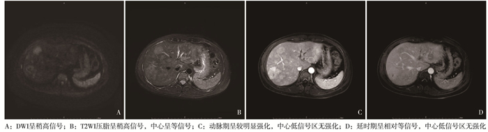
图 1 病人女,53岁
-
不同影像学检查的肝占位病灶检出率差异有统计学意义(P < 0.05);其中,观察组融合图像的病灶检出率高于CT检查,对照组融合图像的病灶检出率高于CT和MRI检查,差异均有统计学意义(P < 0.05),但观察组融合图像的病灶检出率与MRI检查差异无统计学意义(P>0.05)(见表 2)。
分组 n CT MRI 融合图像 χ2 P 观察组 68 49(72.04) 53(77.94) 62(91.18)* 8.27 < 0.05 对照组 92 43(66.15) 47(72.31) 59(90.77)*△ 6.07 < 0.05 χ2分割检验: 与CT检查比较*P < 0.05;与MRI检查比较△P < 0.05 表 2 CT、MRI及融合图像的肝占位病灶检出情况分析[n;百分率(%)]

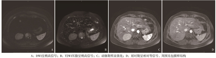
图 2 病人男,56岁
-
以手术、病理穿刺及随访后的最终结果为“金标准”,螺旋CT检查诊断再障雄激素治疗相关肝占位的灵敏度为71.43%,特异度为87.50%,准确率为80.00%;MRI检查诊断再障雄激素治疗相关肝占位的灵敏度为85.00%,特异度为88.00%,准确率为86.67%;融合图像诊断再障雄激素治疗相关肝占位的灵敏度为100.00%,特异度为94.74%,准确率为97.78%。融合图像检查对再障雄激素治疗相关肝占位的诊断准确率高于CT(χ2=7.41,P < 0.01)和MRI(χ2=5.24,P < 0.05)(见表 3)。
螺旋CT检查 手术、病理穿刺及随访结果 合计 良性 恶性 良性 15 3 18 恶性 6 21 27 MRI检查 良性 17 3 20 恶性 3 22 25 融合图像 良性 26 1 27 恶性 0 18 18 表 3 CT、MRI及融合图像检查对再障雄激素治疗相关肝占位的诊断价值分析
-
雄激素可直接作用于造血细胞,通过增加红细胞生成浓度,刺激骨髓造血,达到改善机体造血功能的作用,是临床再障治疗的常用药物。但临床研究发现,长期服用雄激素易引发肝脏病变,增加病人肝脏相关肿瘤的发生风险[7-8]。尽早明确再障雄激素治疗相关肝占位病变的范围及性质,并据此选择合适的治疗方案,已成为当前的研究热点。CT、MRI被认为是肝占位定性诊断的主要手段,被广泛应用于肝脏相关疾病的临床诊断中。但值得注意的是,再障雄激素相关肝占位病人多数为多发病灶,多发病灶在不同成像模式的图像上不尽相同,单一成像模式已无法满足多发病灶的定性诊断[9]。多模融合影像技术可融合不同模态的图像信息,弥补病灶影像信息的不足,实现疾病的精确诊断与治疗,在肝脏疾病、甲状腺疾病等的诊断及手术治疗方面均有所应用[10]。
本研究发现,再障雄激素治疗相关肝占位病人的良性病变占比明显高于对照组,与既往研究[11]结果类似,提示接受雄激素治疗的再障病人,其肝占位病变以良性病变为主。雄激素和雄激素受体(AR)信号通路一方面可能参与肝细胞癌变过程,另一方面雄激素和AR可调节B淋巴细胞瘤-2基因(Bcl-2)水平,使肝细胞受到炎症损伤、凋亡和氧化应激的影响,最终出现肝占位病变[12-13]。因此,临床应密切关注再障病人接受雄激素治疗期间的肝脏局部性病变发生情况,对病人肝占位病情进行及时的诊断与评估,为指导后续治疗提供支持。
本研究在再障雄激素治疗相关肝占位的诊断中应用多模融合影像技术,研究结果显示,观察组、对照组融合图像的病灶检出率均高于CT和MRI检查,提示多模融合影像技术可提高肝占位病灶的检出率。螺旋CT检查可利用病灶组织与周围组织间较大的密度差,清晰提供肝脏占位性病变信息,但对微小病灶及分化程度较低的肿瘤,难以准确显示其边界。MRI检查具有检测微小病灶及准确显示病灶边界的优点,但图像质量易受病人呼吸影响,且成像时间较长,会影响最终诊断。多模融合图像技术将CT与MRI图像融合,弥补了以上2种检查手段的不足,不仅可清晰显示病灶边界、病灶与周围组织的空间位置关系,还可增强肝占位病灶的减出效果,从而提高了判断占位病灶特征的可靠性[14]。曾帅等[15]比较了CT/MRI单一影像技术的图像和CT/MRI与实时双期C臂锥形束CT融合图像对肝占位病灶检测能力,发现多模融合图像技术可明显改善单一影像技术对肝占位病灶的检测能力,帮助施术者更精确地定位病灶,优化手术治疗方案。以手术病理检查结果为“金标准”,进一步分析不同影像学检查方法对再障雄激素治疗相关肝占位的诊断价值发现,融合图像诊断再障雄激素治疗相关肝占位的灵敏度、特异度和准确率分别为100.00%、94.74%和97.78%,诊断准确率明显高于CT和MRI,提示多模融合图像技术在评估再障雄激素治疗相关肝占位的病变类型方面具有较高的诊断价值。本研究中,有1例恶性病人经多模融合图像技术诊断后误诊为良性,考虑与病灶直径太小,病灶密度与周围组织差异不大,病变组织微血管状态无典型变化等有关。
综上所述,多模融合影像在评估再障雄激素治疗相关肝占位方面具有良好的诊断价值,可提高肝占位病灶的检出率,为再障雄激素治疗相关肝占位的后续治疗提供可靠依据,且诊断效能优于CT和MRI,值得临床推广与应用。
多模融合影像评估再障雄激素治疗相关肝占位的临床研究
Clinical study on the evaluation of hepatic space-occupying lesions related to androgen treatment for aplastic anemia by multi-modality image fusion
-
摘要:
目的探究多模融合影像评估再生障碍性贫血(再障)雄激素治疗相关肝占位的临床价值。 方法选取经超声检查提示肝占位的雄激素治疗再障病人49例为观察组,另选取同期接受治疗的其他肝占位病变病人78例为对照组,所有病人均接受螺旋CT和MRI扫描,将CT与MRI双期图像进行融合,分析多模融合影像在再障雄激素治疗相关肝占位中的评估价值。 结果49例观察组病人经血清生物学检查、穿刺检查、术后组织病理学检查或临床及影像随访>6个月,最终证实观察组肝占位45例,病灶68个,对照组肝占位75例,病灶92个。观察组良性病变占比高于对照组(P < 0.05),观察组融合图像的病灶检出率高于CT检查,对照组融合图像的病灶检出率高于CT和MRI检查,差异均有统计学意义(P < 0.05),但观察组融合图像的病灶检出率与MRI检查差异无统计学意义(P>0.05)。以手术、病理穿刺及随访后的最终结果为“金标准”,螺旋CT检查诊断再障雄激素治疗相关肝占位的灵敏度、特异度和准确率分别为71.43%、87.50%和80.00%;MRI检查诊断的灵敏度、特异度和准确率分别为85.00%、88.00%和86.67%;融合图像诊断的灵敏度、特异度和准确率分别为100.00%、94.74%和97.78%,融合图像检查对再障雄激素治疗相关肝占位的诊断价值高于CT和MRI(P < 0.05)。 结论多模融合影像在评估再障雄激素治疗相关肝占位方面具有良好的诊断价值,可提高肝占位病灶的检出率,且诊断效能优于CT和MRI,值得临床推广与应用。 Abstract:ObjectiveTo explore the clinical value of multi-modality image fusion in evaluating hepatic space-occupying lesions related to androgen treatment for aplastic anemia (AA). MethodsTotal 49 patients with AA who had undergone androgen treatment and had hepatic space-occupying lesions detected by ultrasound were selected as the observation group.Meanwhile, 78 patients with the other hepatic space-occupying lesions were selected as the control group.All patients were subjected to spiral CT and MRI scanning.CT and MRI dual-phase images were fused, and the value of multi-modality image fusion in evaluating hepatic space-occupying lesions related to androgen treatment for AA was analyzed. ResultsSerum biological examination, puncture, postoperative histopathological examination or more than 6 months of clinical and imaging follow-up found that there were 45 cases with 68 hepatic space-occupying lesions in the observation group and 75 cases with 92 hepatic space-occupying lesions in the control group.The proportion of benign lesions in the observation group was higher than that in the control group (P < 0.05).The proportion of lesions detected by fusion images was higher than that by CT in the observation group (P < 0.05), and the proportion of lesions detected by fusion images was higher than that by CT or MRI in the control group (P < 0.05), but the proportion of lesions detected by fusion images in the observation group was not significantly different from that in MRI examination (P>0.05).Taking surgical results, pathological results or follow-up results as the golden standard, the sensitivity, specificity and accuracy of spiral CT, MRI and fusion images to diagnose hepatic space-occupying lesions related to androgen treatment for AA were (71.43%, 87.50%, 80.00%), (85.00%, 88.00%, 86.67%) and (100.00%, 94.74%, 97.78%), respectively.The diagnostic value of fusion images was higher than that of CT or MRI (P < 0.05). ConclusionsMulti-modality image fusion has good diagnostic value in evaluating the hepatic space-occupying lesions related to androgen therapy in aplastic anemia.It can improve the detection rate of hepatic space-occupying lesions, and its diagnostic efficiency is better than that of CT or MRI.It is worthy of clinical promotion and application. -
表 1 2组病人病理诊断结果比较[n;百分率(%)]
分组 n 病灶直径/cm 病灶类型 病变类型 单发病灶 多发病灶 肝细胞癌/肝内胆管癌 良性病变 观察组 45 2.25±0.76 32(71.11) 13(28.89) 19(42.22) 26(57.78) 对照组 75 2.31±0.82 49(65.33) 26(34.67) 52(69.33) 23(30.67) χ2 — 0.40* 0.43 8.56 P — >0.05 >0.05 < 0.01 *示t值  下载: 导出CSV
表 2 CT、MRI及融合图像的肝占位病灶检出情况分析[n;百分率(%)]
分组 n CT MRI 融合图像 χ2 P 观察组 68 49(72.04) 53(77.94) 62(91.18)* 8.27 < 0.05 对照组 92 43(66.15) 47(72.31) 59(90.77)*△ 6.07 < 0.05 χ2分割检验: 与CT检查比较*P < 0.05;与MRI检查比较△P < 0.05
下载: 导出CSV
表 3 CT、MRI及融合图像检查对再障雄激素治疗相关肝占位的诊断价值分析
螺旋CT检查 手术、病理穿刺及随访结果 合计 良性 恶性 良性 15 3 18 恶性 6 21 27 MRI检查 良性 17 3 20 恶性 3 22 25 融合图像 良性 26 1 27 恶性 0 18 18
下载: 导出CSV
-
[1] 庄顺红, 胡慧仙, 魏斌, 等. 重型再生障碍性贫血患者免疫抑制疗法后血流感染的研究[J]. 中华医院感染学杂志, 2018, 3(28): 12. [2] 张慧敏, 袁炜, 白明明, 等. 雄激素对再生障碍性贫血患者调节性T细胞的影响[J]. 河北医药, 2019, 11(21): 126. [3] 翟亚娟, 上官建伟, 王丹阳. CT与MRI鉴别肝脏良恶性病灶的临床应用价值[J]. 医学影像学杂志, 2020, 15(7): 123. [4] 王祥芝, 张书海, 徐敏, 等. 多模态影像组学预测肿块型乳腺癌术前淋巴结转移的价值[J]. 蚌埠医学院学报, 2021, 46(5): 652. doi: 10.13898/j.cnki.issn.1000-2200.2021.05.024 [5] MARSH JCW, BALL SE, CAVENAGH J, et al. Guidelines for the diagnosis and management of aplastic anaemia[J]. Br J Haematol, 2009, 147(1): 43. doi: 10.1111/j.1365-2141.2009.07842.x [6] GOLITZ P, STRUFFERT T, GANSLANDT O, et al. Optimized angiographic computed tomography with intravenous contrast injection: an alternative to conventional angiography in the follow-up of clipped aneurysms[J]. J Neurosurg, 2012, 117(1): 29. doi: 10.3171/2012.3.JNS111895 [7] 宋琳, 叶蕾, 李建平, 等. R-ATG联合环孢素与环孢素联合雄激素一线治疗输血依赖非重型再生障碍性贫血的疗效比较: 单中心回顾性研究[J]. 临床血液学杂志, 2019, 32(5): 358. [8] 刘璐, 姜雨佑, 李书书. 性激素及其受体在肝脏脂类代谢中的作用机制研究进展[J]. 中华内分泌代谢杂志, 2020, 36(3): 267. doi: 10.3760/cma.j.cn311282-20190520-00190 [9] 李政良, 徐亚明, 李凤菊, 等. 多模态影像融合技术在肝内胆管癌诊断与治疗中的应用价值[J]. 影像研究与医学应用, 2020, 4(11): 33. [10] 隋洋,孙医学,李阳,等.ACR TI-RADS危险分级结合多模态超声影像技术对甲状腺良恶性病变的诊断价值[J].蚌埠医学院学报,2021,46(8):1099. [11] 马华林,隗功华,张哲.多模融合影像评估慢性再生障碍性贫血雄激素治疗肝脏占位病灶的价值[J].临床和实验医学杂志,2020,19(22):108. [12] 李虹,刘维新.雄激素受体对肝细胞癌发生发展作用的研究进展[J].山东医药,2020,60(10):95. [13] 王开宇,李文心,马作红,等.雄激素受体与三磷酸腺苷酶家族蛋白2在肝细胞癌中的表达及二氢睾酮对其影响[J].中华肝胆外科杂志,2018,24(11):742. [14] 李小娟,黄品同,徐永远,等.超声造影肝影像报告和数据系统与磁共振肝影像报告和数据系统对肝细胞癌高危患者肝占位性病变的一致性评价研究[J].中华超声影像学杂志,2020,29(6):522. [15] 曾帅,王嵇,程杰军,等.肝动脉化学治疗栓塞术中实时多模影像融合和双期C臂锥形束CT对改善肝脏占位病灶检测能力的研究[J].生物医学工程与临床,2019,23(4):86. -

 点击查看大图
图(2)表(3)
计量
- 文章访问数: 1494
- HTML全文浏览量: 658
- PDF下载量: 7
- 被引次数: 0
